
1. Introduction
Pre-eruptive intra coronal caries are often an accidental lesion as a radiolucent finding in the coronal dentin of a tooth which did not erupted into the oral space (1). However, treatment modality is still not reported systematically (2).
Pre-eruptive resorption in crown of tooth (PEIR) is depicted as a Well-bounded, radiolucent and irregular range inside the coronal part of tooth near to the junction of dentin and enamel which spread into different profundities of dentin in unerupted tooth. This condition could be a uncommon peculiarity that happens in both dentitions. Within the past, these abandons were not being seen and named “pre-eruptive caries” or “hidden caries”. These injuries are frequently recognized inadvertently during routine dental radiographic examination (3), and nearly 61% of dentists are aware of PEIR, which is possible to be misdiagnosed (4).
The prevalence of teeth with pre-eruptive caries is 0.2 - 3.5%, results from these variables: The type of radiograph utilized for examination, sex, age, and type of dentition (5).
Pre-eruptive caries lesions have been commonly seen in molars and premolars, but incidence in canines, have been seen. Involvement in one tooth is more often, but cases with several teeth have also been reported (3).
Seow classification for the PEIR defects involves three class. In first class, the lesion extends up to one-third of the dentin. Second class is the involvement of between one-third and two-thirds of the dentin layer. In third class, the lesion extends more than two-thirds of the width of dentin layer (3). Treatment plans were based on the nature of the defect which have a desire for progression in size. Clinicians must consider the status of tooth eruption, progression of lesion, the size of the it and the severity of pulp penetration (2).
Untreated caries, especially deep caries, can result in pulpitis, pulp necrosis, periapical abscess, and tooth loss. Although there are yet doubts about the different methods used to treat deep carious lesions, it is agreed that an exact assessment of the depth of the carious lesions and the status of the pulp tissue is essential for timely and appropriate treatment (6).
Apexogenesis keeps the nerve of the tooth alive to develop the root tooth and increase its thickness, which can fortify the it stand up to break. Since the pulp is vital and capable of mineralizing root dentin, there is no coherent defense for giving a regenerative endodontic or routine endodontic treatment. The long-term overall success rate of apexogenesis was 82.5% and 96.4% (7).
Stainless-steel crowns (SSC) performed on permanent teeth are used as an restoration to restore severely broken-down molars until placement of the final restoration. Stainless-steel crowns has many advantages compared with other filling materials, such as durability, full coverage, being cheap, and minimal technique sensitivity during crown placement (8).
The reason of PEIR remains hazy, and different clarifications have been mentioned (1). the foremost affirmed speculation is an acquired imperfection, developmental in source, coming from a resorption in tooth crown (3).
The hypothesis of lytic activity involves apical inflammatory cells in primary teeth (in spite of the fact that not all PEIR teeth have a progenitor), an idea that sets a formative beginning that results in hypoplastic or hypomineralized enamel and dentin (but these lesions are ordinarily beginning from full crown development), or internal or external resorption hypotheses. The less particular hypothesis is pre-eruptive caries without oral cavity exposure. At the same time, the foremost worthy is resorption by resorptive cells, either by attacking through enamel ruptures or via communication near the junction of cement and enamel (1).
2. Case Presentation
A 6-year-old boy was referred to the pediatric faculty of Hamadan University with a main complaint of inconvenience and torment on the left side of the maxilla. His medical history was not related, and no history of dental trauma was detailed. Extra-oral survey uncovered non-significant discoveries.
Intra-oral examination appeared a mixed dentition with many dental caries, and we ordered a panoramic view radiography. The maxillary left second primary molar presented pulpal involvement. An intracoronal radiolucency was detected on the unerupted mandibular left first molar, limited to the occlusal surface without mesial or distal tooth involvement (Figure 1).
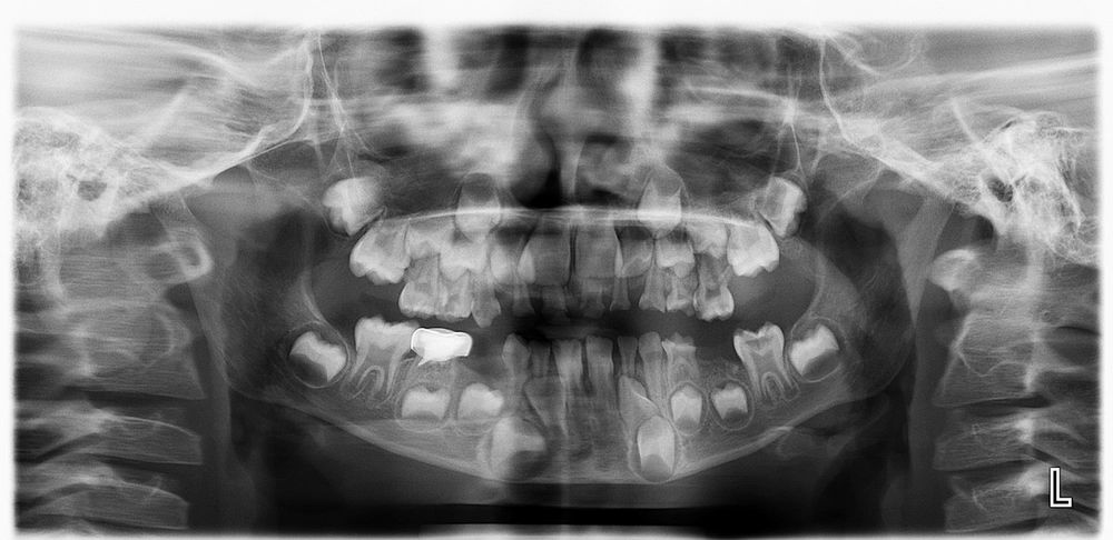
Figure 1.
Panoramic view of the patient notice; the shadow of soft tissue above the left mandibular first molar.
For specific diagnoses, we ordered a periapical radiography of that tooth (Figure 2).
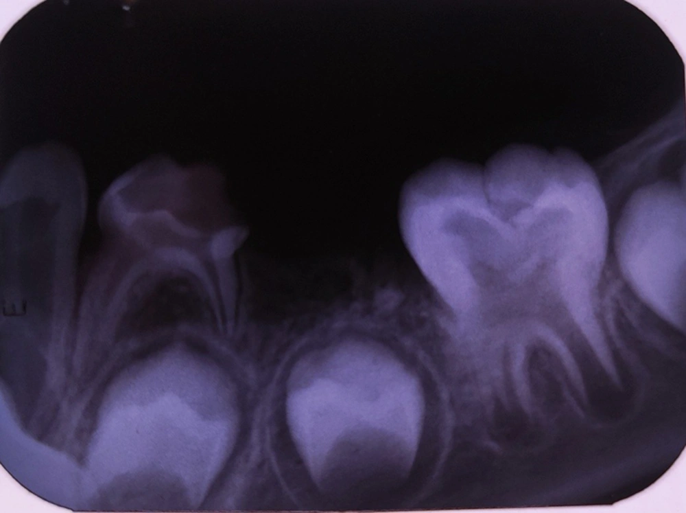
Figure 2.
Pre-eruptive caries located in the central aspect of the unerupted left mandibular first molar crown.
The lesion was presumed to be a PEIR with pulpal involvement based on clinical and radiographic aspects. The patient had no related symptoms in that area, and we informed his parent about the necessity of surgical exposure of the tooth for access to treatment. Parents’ informed consent was taken for treatment and publication of the treatment result as an article.
At the first session, treatment of the painful tooth and consultation was performed for dental exposure surgery.
Access was granted after the gum was removed, and pre-eruptive caries could be treated. According to pulp involvement in an immature tooth with no symptoms, we choose apexogenesis as his treatment. The treatment was accomplished by using MTA, and bleeding control during treatment was difficult according to the short length of roots and large size of the pulp chamber. A light-cured Glass Inomer dressing was applied as a dressing after seven days, and the access cavity was filled with amalgam. The tooth walls were weak, so they were covered with a stainless-steel crown.
Six months later, a radiographic examination was done. Pulp was survival, so root developementand thickening were continued, which can reinforce the tooth to stand up to break (Figure 3).
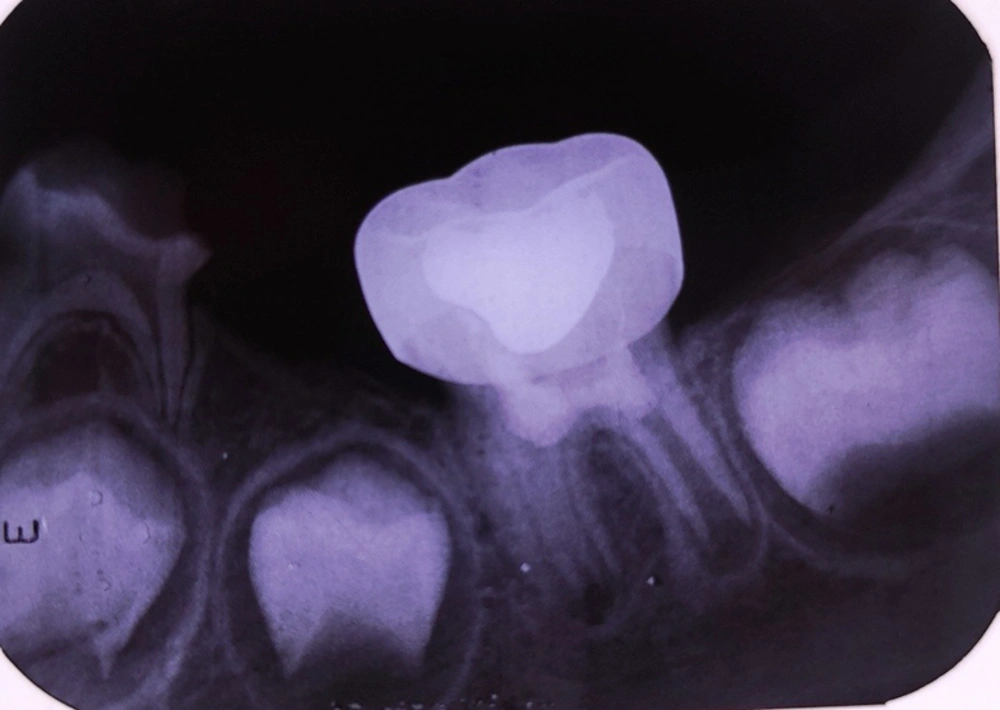
Figure 3.
Radiograph of follow-up session after six months.
3. Discussion
In tooth eruption, the tooth germ emerges harmoniously through the space to the oral cavity. The tooth growth mechanism results from the interaction between osteoblasts, osteoclasts, and dental follicle, along with genetic factors. Hence, there are various systemic, local, and genetic factors responsible for tooth eruption, and any disturbances in these factors might lead to abnormalities in eruption (9).
Dental caries can happen during the eruption process under the title of pre-eruptive caries. Caries lesions are frequently found within the mesial or central measurement of the tooth crown, close the intersection of enamel and dentin, and can penetrate the dentin to a variable extent but rarely involve the pulp, unlike our case, which was extended to the pulp.
Tooth decay is still the most common oral disease in the world. The incidence of dental caries is highly associated with the microbial properties in the oral cavity (10), but the microbiota is not the single etiology in cases with pre-eruptive caries, and intracoronal resorption is also responsible for the attack of resorptive cells into the dentine through breakdowns within the crown arrangement (3).
Detection of caries by visual sense is adequate for most patients in routine clinical practice without paying attention to tooth type or its surface (11), but it is impossible for pre-eruptive caries to be diagnosed with this method, and only radiographic examination can appear it. However, this is not usually performed in children during routine dental tests (12).
Millions of patients have their immature permanent teeth extracted every year. However, most dental specialists of endodontics and pediatric have done regenerative approach of endodontic treatment accessible to do nearly everything conceivable to save these teeth (7). In our case, the pre-eruptive caries were extended to pulp, and pulp therapy was necessary. We scheduled regenerative endodontic treatment because of his immature roots, and the outcome was satisfactory.
The clinical significance of these lesions is that they account for the majority of potential caries and may be associated with dysgenesis, ectopic position and supernumerary teeth, and delayed tooth development (5). In our case, dental development or several teeth was not affected, and its expansion was up to two-thirds of the dentin thickness, called type III in the classification of Seow (3).
Seow stated that the misfortune of the integrity of the Nasmyth layer is triggered by abnormal local pressure caused by affected tooth germ or by the ectopic position of the adjacent tooth. Therefore, the ectopic location was cited as a trigger for initiation of intracoronary resorption. The affected and adjacent teeth described in this report are in normal position (3).
3.1. Conclusions
Accurate radiographic analysis of unerupted teeth is strongly recommended because rapid diagnosis and timely treatment of caries before an eruption can save the tooth.