
1. Context
Anatomy is a foundational discipline in medical science and plays a significant role in understanding the structure and function of the body’s systems. Textbooks and atlases provide 2-dimensional (2D) images of organs, and anatomical models are used as supplementary resources; however, none can replace real CD. CD has been regarded as the gold standard in anatomy education because it helps students understand structures in three dimensions (1, 2). In recent years, owing to reduced cadaver availability and restrictions related to the COVID-19 pandemic, a significant decrease in medical students’ memorization and understanding of anatomical concepts has been observed. This issue is considered one of the major challenges in medical education and has prompted a review of educational methods and the adoption of new technologies (1, 3). This study aimed to investigate and compare new tools with traditional teaching methods in anatomy and to present a combined approach to improve the learning process and the quality of medical education.
2. Evidence Acquisition
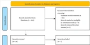
To identify sources for this study, a search was conducted on October 8, 2025, in databases including PubMed and Scopus. The search used the keywords "virtual reality," "cadaver dissection," and "medical anatomy education." A total of 18,000 articles were identified without time limits. Initially, 34 articles were selected based on their titles and then reviewed. Subsequently, 22 articles were selected based on the inclusion criteria, including thematic relevance, full-text availability, publication in English, and a focus on human anatomy education, and the exclusion criteria, including the removal of duplicate studies, abstracts, and conference proceedings. These articles were examined in detail. The findings of the selected studies were categorized and analyzed across 3 main axes: 1) teaching anatomy with CD, 2) teaching anatomy with VR, and 3) the combined approach. A comparison of CD, VR, and combined methods is presented in Table 1, and the variables extracted from the selected articles are presented in Table 2.
Table 1.
Comparison of Cadaver Dissection, Virtual Reality, and Combined Methods in Anatomy Education
| Variables | Cadaver Dissection | Virtual Reality | Combined Approach |
|---|---|---|---|
| Benefits | Accurate understanding of spatial relationships between structures; professional skills such as communication and respect for human life; critical thinking; careful observation and touch; enhanced teamwork skills; study of simpler body parts; enhanced manual skills and motor coordination; practical and deeper learning of concepts; introduction to anatomical diversity among humans; increased emotional resilience; increased pathological and tissue understanding | Provides immediate feedback to students; better spatial understanding of complex structures; interactive and safe learning; ability to visualize spatially, solve problems, and think analytically; reduced teacher workload; increased student engagement; reduced learning time; more engaging instruction; ability to practice and review concepts without physical limitations; reduced technical errors; reduced cost and reusability | Consolidates knowledge and improves skills; longer-lasting learning; a more complete and interactive learning experience |
| Disadvantages | Time-consuming dissection; requirement of basic training; shortage of cadavers and a decreasing number of donated cadavers; unpleasant odor; inability to see all details; difficulty in identifying structures; ethical considerations | Nausea and dizziness (cybersickness); headache; blurred vision | More expensive |
| Cost | High | Average | High |
| Access | Low | High | Average |
| Application | Strengthening surgical and clinical skills | Repetition and review of concepts and better learning | Comprehensive and complete learning |
| Effectiveness | Good | Good | Excellent |
Table 2.
Variables Extracted from Articles About Cadaver Dissection, Virtual Reality, and Combined Methods in Anatomy Education
| Authors and Ref. | Year | Aim | Application Type | Target Population | Domain | Outcome |
|---|---|---|---|---|---|---|
| Elbeshbeishy et al. (16) | 2025 | Assessing the opinions of medical and health science students about integrating learning with digital tools and CD | CD, 3D models, animations, and interactive apps | 454 students from various health disciplines at Ras Al Khaimah Medical and Health Sciences University | CD (thorax, abdomen, pelvis, limbs, and head and neck); Complete Anatomy and 3D organ visualization tools | Medical students were more likely to support autopsies than allied health students. |
| Ragland et al. (13) | 2025 | Assessing students' satisfaction and confidence after teaching inner ear anatomy with 3D and 2D models | 2D and 3D models | 30 first-year medical students | Inner ear | 3D models of the inner ear increased student satisfaction and confidence in learning the material. |
| Abundez Toledo et al. (10) | 2024 | Assessing the use and understanding of VR | VR | A group at a traditional 4-year medical school in the United States | Body systems, including cardiovascular, musculoskeletal, lymphatic, and nervous systems | Most students said VR was effective in learning anatomy topics. |
| Tahir et al. (3) | 2020 | Evaluating the effect of 3D-VR on the retention of human anatomy content | 3D-VR | 182 third-year medical students of the foundation block (ie, 3rd year, Phase II) | Human anatomy | Short-term and long-term grades were higher for a group of male students who used 3D-VR. |
| Chen et al. (14) | 2020 | Evaluation by objective questionnaires and perceptual surveys regarding teaching with VR and traditional teaching methods | 3D skull model, CD skull, and atlas | 73 medical students | Skull | Students in all 3 atlas, CD, and VR instruction groups had higher scores after the intervention. However, students in the VR and CD groups had better feedback than those in the atlas group. |
| Kalthur et al. (8) | 2018 - 2019 | Assessing students' opinions on the usefulness of anatomy in modern education | CD | 145 medical undergraduate students exposed to CD in anatomy during their first year (2018 - 19) | Human anatomy | Most students agreed that CD of the body is effective in providing a deep and 3D understanding of structures. |
| Jeyakumar et al. (5) | 2018 | Evaluating students' opinions on the effectiveness of explanation | Musculoskeletal dissection | 174 medical students of Griffith University in their second-year anatomy course | Musculoskeletal anatomy | CD is effective in retaining applied anatomical concepts and improving clinical skills. |
| Flack et al. (6) | 2018 | Assessing medical students' learning, coping, and effects of CD sessions | Musculoskeletal dissection | 267 second-year medical students at Otago Medical School | Musculoskeletal anatomy | Students stated that CD is a useful and valuable educational tool that is effective in understanding the 3-dimensional relationships of the body, teamwork, and coping with death. |
| Izard et al. (12) | 2017 | Evaluating the effect of VR in teaching human anatomy | 3D model | - | The cranium | It was shown that VR creates a sense of presence in a virtual environment, as well as an understanding of the skull and its foramina. |
3. Results
3.1. Teaching Anatomy with CD
Despite the availability of advanced tools, CD remains the most important method for teaching anatomy. CD not only helps students learn 3-dimensional (3D) anatomy but also strengthens critical thinking, careful observation, and teamwork. This experience also fosters professional skills, such as respect for human life. Many researchers believe that education and the development of medical attitudes are shaped in the dissection room. Recent findings indicate that students’ learning experiences in CD courses are not uniform. In addition to strengthening the scientific foundation and consolidating knowledge, this method plays a vital role in developing clinical skills and ensuring basic medical competencies. In recent years, many universities have reduced anatomy teaching hours to devote more time to clinical skills training. This reduction should not compromise the quality of physician education or patient safety (2, 4).
The results of a study on anatomy education with CD showed that most participants believed that working with a corpse provides a deeper understanding of body structure and anatomical complexities and that no digital method can fully replace it. CD not only promotes active learning but also fosters respect and responsibility toward the human body and the concept of death, and it can prepare students emotionally and ethically for dealing with patients. The researchers emphasized that CD should remain in the medical education curriculum; however, to adapt to current needs, a procedure-based dissection model can be used. In this model, body regions are examined through exercises resembling clinical and surgical procedures, such as cutting the carpal tunnel or saphenous vein, so that students become familiar with the clinical application of basic surgical structures and skills (5).
Another study was conducted to clarify students’ perspectives on the CD experience. In this study, researchers used a qualitative approach and inductive analysis to examine students’ responses and determine what they learn from CD and how the experience affects their personal and professional development. The results showed that most medical students found the CD experience positive and valuable and that they coped well with it. Most viewed the activity as a unique opportunity for hands-on learning and direct observation of the human body and expressed gratitude to the donors. Although some initially felt anxious, nauseous, or uncomfortable, they gradually adapted to the dissection room environment (6).
In contrast, in some areas, anatomy education is not based on CD. Instead, education is provided through medical imaging, live human models, and training sessions with experts. The educational philosophy of this approach is "outside-in" learning, meaning that students first learn surface anatomy and then deeper structures to gain a more clinical and practical understanding of anatomy. After learning the basics of anatomy in the preclinical years, students have the opportunity to apply their knowledge in a real patient environment in the operating room. However, this approach has limitations, including limited access to the operating room, variability in educational quality, and the inability to view areas outside the surgical field. Researchers emphasize that eliminating CD from medical programs does not necessarily reduce educational quality. They also acknowledge that many students still want to reinforce their knowledge by observing or working with cadavers (7). Another study found that digital-generation students, despite access to technological tools, consider CD the most effective way to learn anatomy. Researchers emphasized that educational technologies such as simulators should be used alongside CD to achieve optimal learning (8).
Research has shown that CD is suitable for studying simpler body parts, such as abdominal organs, whereas more complex structures, such as the skull and brain tissue, can be better taught using virtual models. Overall, there is general agreement that anatomical knowledge is essential to medicine and that education would be incomplete without CD. However, CD alone cannot meet all the needs of modern medical curricula and must be complemented by modern educational methods. This collaboration between traditional and modern methods, with CD as the central focus, is likely to shape the future direction of anatomical sciences education (4).
3.2. Teaching Anatomy with VR
Simulation is a newer educational method that provides students with an interactive and engaging environment, real-time feedback, and ease of deployment, enabling effective learning and skill development. VR is a high-level technology that offers interactive learning opportunities and enables the simulation of real-world anatomical or clinical scenarios. By providing active and risk-free learning conditions, these technologies improve spatial understanding, reduce errors, and improve student performance (1, 2, 9, 10). They allow interaction with 3-dimensional structures and observation from different angles, helping students visually and spatially understand complex anatomical concepts. Augmented reality provides an interactive experience by overlaying digital images onto the real environment, whereas VR immerses the student in a fully virtual environment. In addition to teaching anatomical structures, these methods enhance spatial visualization, problem-solving, and analytical thinking and provide opportunities to practice and review concepts without physical limitations (3, 11).
VR is usually delivered using a headset equipped with a monitor and headphones connected to a computer or mobile phone. Several studies have shown that VR can improve learning as a supplementary teaching tool compared with books or pamphlets alone, although it cannot replace the experience of actual CD. The most important advantage of VR is that it provides students with immediate feedback, allowing them to promptly identify and correct their weaknesses while reducing the need for professors to provide correction and assessment (2). Some VR simulators, including skull simulators, allow placement of bone fragments in the correct position. These technologies enable users to interact easily with the virtual environment and gain an accurate 3-dimensional understanding of the spaces and structures of the skull, including the anterior, middle, and posterior cranial cavities (12).
A study comparing traditional anatomy teaching using plastinated models with 3D-VR was conducted. The results showed that this technology improved concept comprehension, increased learning, and improved short- and long-term knowledge retention; this effect was more pronounced among male students. Qualitative analysis also indicated that VR made learning easier, more interactive, and more accessible (3). In another study, to assess the effect of 3D models on anatomy learning, 2D and 3D models of the inner ear were presented to students for viewing. The results showed that self-confidence and satisfaction were higher in the 3D group, indicating active learning skills among the students. However, in this study, the students’ academic progress in the 2 groups was not objectively studied and measured (13).
Another study evaluated the effect of VR training on learning anatomy, including the skeletal, cardiovascular, lymphatic, and nervous systems. The results showed that most students found the learning environment safe and supportive and reported feeling comfortable, with the headset being easy to use. Most stated that VR-based training helped them learn better than traditional methods, although most believed that VR should be used as a supplement to, not a complete replacement for, in-person training (10). However, this technology is also associated with challenges such as eye fatigue and technical difficulties (3), cost, physical discomfort during prolonged headset use, and the need for initial training (10).
Comparative studies have also shown that VR skull models are as effective as dissected skulls and atlases in teaching anatomical structures. Although no significant difference in student performance was observed between groups, students in the VR and CD groups provided more positive feedback than those in the atlas group (14). Given these findings, VR technologies, as complementary tools in anatomy education, can provide a more dynamic and interactive learning experience and facilitate understanding of complex anatomical structures (15, 16).
VR simulators for teaching cardiac anatomy (17) and neuroanatomy (18) provide better spatial understanding of complex structures and increase students’ motivation and mental engagement to understand the material. The only negative aspect is the occurrence of nausea and dizziness (cybersickness) in some students, although these side effects did not significantly affect the final scores. The findings indicate that the use of VR and stereoscopic technologies can be effective in improving anatomy teaching and 3D understanding of neural structures (19).
Some studies have not reported a significant difference between 3D and traditional methods; however, the use of VR in some universities has had a significant impact on improving the retention of anatomical knowledge among medical students. Accordingly, the use of 3D technologies can be an important step toward innovation in medical education (3).
3.3. Combined Approach
Teaching human anatomy has always required methods that balance accuracy, accessibility, and meaningful student engagement. Current comparisons between CD and VR show that both approaches support learning in different ways. They should be regarded as complementary rather than competitive. CD provides students with the most authentic experience of human structure. Working with real tissue helps students appreciate color, texture, and natural variation, details that are difficult to replicate digitally. This hands-on experience also fosters teamwork, patience, and respect for the human body. These qualities have long been linked to the ethical training of medical students. However, maintaining cadaver facilities requires substantial financial and logistical support. Issues related to body donation, storage, and chemical safety also persist (20).
In contrast, VR offers a flexible and interactive space in which students can explore anatomy repeatedly without material limitations. Research shows that well-designed VR modules can significantly improve spatial reasoning and short-term recall (21). Students can rotate structures, isolate systems, and view areas that are difficult to access in traditional CD. However, despite these benefits, the lack of tactile feedback and the emotional weight of real CD remain important limitations. The study showed that learning outcomes were statistically similar between the VR and CD groups, suggesting that digital tools alone cannot fully replace the experience of physical exploration (14).
A growing number of studies support a combined model that integrates the cognitive clarity of VR with the sensory reality of CD. In such programs, students can begin with digital simulations to build mental maps and then apply that knowledge in the dissection laboratory. Evidence suggests that this sequence improves both confidence and understanding (22).
3.4. Limitations
In this debate article, despite extensive and careful database searches, the possibility of bias cannot be completely ruled out because the authors’ opinions and perspectives may have influenced the search and writing of the article. In addition, during the initial search, access to some sources was not possible; however, an effort was made to ensure that the search was as complete and comprehensive as possible despite this limitation.
4. Conclusions
Although CD is the cornerstone of anatomy education and prepares students for a 3D understanding of body parts through realism, hands-on experience, and interaction with real patients, dissection does not meet many students’ scientific learning needs despite these advantages because, in topics such as neuroanatomy, adequate spatial understanding is not possible. At the same time, VR, as a new method, makes learning in these cases more accessible and easier. However, a well-designed combined model offers the best approach by enabling practice, repetition, and an understanding of spaces that are not possible with a cadaver, providing students with real organs to touch, and facilitating long-term knowledge retention and skills transfer.