
1. Background
Chronic endometritis (CE) is a persistent and continuous inflammatory disease of the endometrium associated with the presence of plasma cells in endometrial stroma. It is often asymptomatic or presents with mild symptoms, like pelvic pain or discomfort, vaginal discharge, abnormal uterine bleeding (AUB), dyspareunia, and vaginitis (1). The prevalence of CE in premenopausal women in Iran is about 31% (2) compared to wide varied rate in general population (2.8 to 72%) worldwide (3). High prevalence of CE has been reported in infertile women particularly in those with unexplained infertility (4).
Chronic endometritis contributes to infertility and adverse, poor reproductive outcomes, including recurrent implantation failure (RIF) and recurrent pregnancy loss (RPL) after in vitro fertilization (IVF) (5-12). CE as a negative factor for IVF outcome is highly prevalent in infertile women (7), but it is not evaluated routinely in these women.
Clinical diagnosis of CE is complex and may be neglected by gynecologists due to the absence or minimal signs and symptoms and normal vaginal ultrasound. The gold standard for diagnosing CE is the hysteroscopic-guided endometrial biopsy and histological diagnosis, particularly with immunohistochemical staining for CD138, a glycoprotein antigen on the surface of plasma cells (13). However, as a minimally invasive procedure, hysteroscopy can also be a method of CE diagnosis. Nevertheless, there is no consensus on the value of hysteroscopy in diagnosing CE, which remains controversial (14).
Hysteroscopy findings, including micro-polyp ≤ 1 mm, endometrial hyperemia, and mucosal edema flushed with strawberry aspect or a white central point, are considered the reliable diagnostic features of CE (15-17).
In a study on 2190 women with various indications for diagnostic hysteroscopy the prevalence of CE was reported 20% which among them 37% were infertile that is considerable. Previous studies found out a moderate sensitivity and higher specificity of hysteroscopy in diagnosis of CE (7, 18).
As CE contributes in infertility (19) and affects the outcome of assisted reproduction technology (ART) and endometer receptivity (5-12), a strategy to treat infertility and improve ART outcomes is easy minimally invasive diagnosis and treatment of CE (as a modifiable risk factor) in infertile women and those candidate for ART. However, there is lack of information about the prevalence of CE in infertile women and diagnostic value of hysteroscopy in CE in infertile women.
2. Objectives
This study was conducted to assess the CE prevalence and the sensitivity, specificity, and diagnostic value of hysteroscopy in its diagnosis in female candidates for IVF undergoing hysteroscopy.
3. Methods
This cross-sectional study was done in the infertility ward, Vali-e-Asr Hospital, affiliated with Imam Khomeini Hospital Complex (an educational third-level and referral hospital), Tehran, Iran, from October 2019 to February 2020. Ninety infertile women referred to the clinic for IVF (both primary and secondary infertility) undergoing hysteroscopy due to any cause were consecutively included in the study.
Women with menstruation at examination time, positive Beta-hCG test, uterine anatomical abnormalities disorder (myoma, malformation, and endometrial polyp in sonography), previous ART, and using hormone replacement therapy or hormonal therapy within the last three months were excluded from the research.
Demographic and clinical information, including age, duration of infertility, type and cause of infertility, and detailed history, were taken from all eligible individuals. Symptoms of abnormal vaginal bleeding (abnormal cycle duration and bleeding volume), dyspareunia (painful intercourse), lower abdominal pain in history, abnormal vaginal discharge, and findings in favor of pelvic inflammatory disease (such as cervical motion tenderness and abnormal vaginal discharge) were recorded during the pelvic examination, by speculum and bimanual examination.
3.1. Hysteroscopy and Endometrial Biopsy
Endometrial biopsy and hysteroscopy were performed as outpatient procedures with sedation. Chronic endometritis in hysteroscopy was defined as the presence of one or more criteria, including stromal edema, focal or diffuse hyperemia, and a micro-polyp ≤ 1 mm, as reported by the same expert physician (15-17).
Hysteroscopy was performed using a compact hysteroscope (HOPKINS® telescope 30°, size 4 mm) with a channel for semirigid 5 Fr (Karl Storz). Normal saline solution distended the uterine cavity at the pressure of 100 mmHg.
In the operating room, an infertility assistant and recorded evidence of CE in hysteroscopy (including stromal edema and defused or local hyperemia).
During hysteroscopy, an endometrial biopsy sample was taken randomly from uterine cavity walls, immediately placed in 10% formalin solution, and sent to the pathology lab within 24 - 48 hours. Endometrial specimens were taken during the follicular phase of the menstrual cycle (days 5 - 12) by an expert infertility sub-specialist.
3.2. Histopathology
In the pathology lab, formalin-fixed endometrial specimens were embedded in paraffin before histological analysis. Conventional hematoxylin-eosin staining was done to detect positive plasma cells in the endometrial stroma.
The samples were transferred to the same hospital laboratory and examined by the same pathologist, unaware of the patient’s clinical and hysteroscopic findings. In histological examination, positive CE diagnosis was considered as ≥ 5 plasma cells in 10 non-overlapping high-power fields (HPFs) (400X magnifications) in the endometrial stroma. For < 5 plasma cells in the field, it was considered normal or negative CE (1). Histopathology examination of endometrial sample was considered as the gold standard method for calculating sensitivity and specificity of hysteroscopy.
Ethics committee at Tehran University of Medical Sciences approved the study protocol (IR.TUMS.IKHC.REC.1396.4849), and all participants signed informed consent before enrollment. The research was done according to the Helsinki declaration (20).
Considering the estimated prevalence of chronic endometritis in women referring to the infertility department about 50% and the confidence level of 0.95% and margin of error 10%, the sample size of 90 people was obtained.
3.3. Statistical Analysis
Quantitative and qualitative data were presented as the mean and standard deviation (SD) and the number and percentage, respectively. Categorical data were compared using chi-square test.
Specificity, sensitivity, negative and positive predictive value (NPV and PPV), accuracy, and Kappa statistics also was determined. A P-value less than 0.05 was regarded significant. Data analyses were done by SPSS software (ver. 20.00 for Windows).
4. Results
In this study, hysteroscopy was performed for 90 infertile women candidates for IVF. The mean age of women was 35.6 ± 5.1 years (26 - 46 years). Table 1 shows the clinical characteristics of patients.
Table 1.
Clinical Characteristics of the Infertile Women
| Variables | No. (%) |
|---|---|
| Duration of infertility (y) | |
| < 5 | 27 (30) |
| 5 - 10 | 38 (38.5) |
| 10 - 15 | 16 (17.8) |
| > 15 | 12 (13.3) |
| Infertility type | |
| Primary | 77 (85.6) |
| Secondary | 13 (14.4) |
| Cause of infertility | |
| Male factor | 28 (31.1) |
| Female factor | 42 (46.7) |
| Both male and female factors | 4 (4.4) |
| Unexplained | 16 (17.8) |
| Symptoms and disorders | |
| AUB | 8 (8.9) |
| PID | 6 (6.7) |
| Dyspareunia | 7 (7.8) |
| Vaginal discharge | 11 (12.2) |
| Hysteroscopy findings | |
| Normal | 68 (75.6) |
| Hyperemic | 14 (15.6) |
| Edema | 8 (8.9) |
| Micro polyp | 0 (0) |
Abbreviations: AUB, abnormal uterine bleeding; PID, pelvic inflammatory disease.
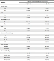
According to the pathological report, CE prevalence was present in 19 (21.1%) (95% CI: 12.67 - 29.53) women candidates for IVF. Chronic endometritis prevalence in women with primary and secondary infertility is shown in Table 2. Although the CE prevalence was higher in secondary infertility compared to primary infertility, but the difference was not significant. The frequency of hysteroscopy findings of CE in women with histologically confirmed CE is shown in Table 3.
Table 2.
Relationship Between Chronic Endometritis and Different Clinical Factors and Gynecological Disorders
| Findings | Chronic Endometritis by Histology; No. (%) | P-Value | |
|---|---|---|---|
| Yes (N = 19) | No (N = 71) | ||
| Dyspareunia | 0.6 | ||
| Yes | 2 (28.6) | 5 (71.4) | |
| No | 17 (20.5) | 66 (79.5) | |
| AUB | 0.001 | ||
| Yes | 6 (75) | 2 (25) | |
| No | 13 (15.9) | 69 (84.1) | |
| Vaginal discharge | 0.03 | ||
| Yes | 5 (45.5.3) | 6 (54.5) | |
| No | 14 (17.7) | 65 (82.3) | |
| PID | 0.01 | ||
| Yes | 4 (66.7) | 2 (33.3) | |
| No | 15 (17.9) | 69 (82.1) | |
| Duration of infertility (y) | 0.1 | ||
| < 5 | 4 (14.8) | 23 (85.2) | |
| 5 - 10 | 12 (34.3) | 23 (65.7) | |
| 10 - 15 | 2 (12.5) | 14 (87.5) | |
| > 15 | 1 (8.3) | 11 (91.7) | |
| Infertility type | 0.09 | ||
| Primary | 14 (18.2) | 63 (81.8) | |
| Secondary | 5 (38.5) | 8 (61.5) | |
| Cause of infertility | 0.7 | ||
| Male factor | 5 (17.9) | 23 (82.1) | |
| Female factor | 8 (19) | 34 (81) | |
| Both factors | 1 (25) | 3 (75) | |
| Unexplained | 5 (31.3) | 11 (68.8) | |
Abbreviations: AUB, abnormal uterine bleeding; PID, pelvic inflammatory disease.
Table 3.
Frequency of Hysteroscopic Findings in Women with Histologically Confirmed Chronic Endometritis
| Hysteroscopy Findings of CE | Histology Result for CE Positive and Negative; No. (%) | P-Value | Kappa | |
|---|---|---|---|---|
| Normal | 6 (31.6) | 62 (87.3) | < 0.001 | 0.5 |
| Edema | 6 (31.6) | 7 (9.9) | ||
| Hyperemia | 7 (36.8) | 2 (2.8) | ||
| Total | 19/90 (21) | 71/90 (78) | ||
False-positive and negative hysteroscopic diagnoses of CE were 10% (9 of 90) and 6.6% (6 of 90), respectively. The relationships between CE prevalence and AUB, PID, dyspareunia, and vaginal discharge are shown in Table 2. Although the CE prevalence was higher in women with a history of dyspareunia than in those without, the difference was not significant. Abnormal uterine bleeding, vaginal discharge, and PID were significantly higher in patients with CE compared to those without it (Table 2). Abnormal uterine bleeding was significantly higher in women who had secondary infertility compared to those with primary infertility. The relationship between clinical and hysteroscopy findings of CE is shown in Table 4.
Table 4.
Frequency of Different Hysteroscopy Findings of Chronic Endometritis in Infertile Women
| Factors | Hysteroscopy Findings of Chronic Endometritis; No. (%) | P-Value | ||
|---|---|---|---|---|
| Hyperemia | Edema | Normal Hysteroscopy | ||
| Dyspareunia | 0.08 | |||
| Yes | 3 (42.9) | 1 (14.3) | 3 (42.9) | |
| No | 11 (13.3) | 7 (8.4) | 65 (78.3) | |
| AUB | 0.03 | |||
| Yes | 3 (37.5) | 2 (25) | 3 (37.5) | |
| No | 11 (13.4) | 6 (7.3) | 65 (79.3) | |
| Vaginal discharge | 0.004 | |||
| Yes | 5 (45.5) | 2 (18.2) | 4 (36.4) | |
| No | 9 (11.4) | 6 (7.6) | 64 (81) | |
| PID | 0.09 | |||
| Yes | 1 (16.7) | 2 (33.3) | 3 (50) | |
| No | 13 (15.5) | 6 (7.1) | 65 (77.4) | |
| Duration of infertility (y) | 0.1 | |||
| < 5 | 4 (14.8) | 1 (3.7) | 22 (81.5) | |
| 5 - 10 | 4 (11.4) | 5 (14.3) | 26 (74.3) | |
| 10 - 15 | 6 (37.5) | 1 (6.3) | 9 (56.3) | |
| > 15 | 0 (0) | 1 (8.3) | 11 (91.7) | |
| Infertility type | 0.1 | |||
| Primary | 13 (16.9) | 5 (6.5) | 59(76.6) | |
| Secondary | 1 (7.7) | 3 (23.1) | 9 (69.2) | |
| Cause of infertility | 0.07 | |||
| Male factor | 3 (10.7) | 2 (7.1) | 23 (82.1) | |
| Female factor | 6 (14.3) | 1 (2.4) | 35 (83.3) | |
| Both factors | 1 (25) | 1 (25) | 2 (50) | |
| Unexplained | 4 (25) | 4 (25) | 8 (50) | |
Abbreviations: AUB, abnormal uterine bleeding; PID, pelvic inflammatory disease.
The NPV, PPV, specificity, sensitivity, and accuracy of hysteroscopy for diagnosing CE are shown in Table 5. Hysteroscopy had low sensitivity (68.4%) and good specificity (87.3%) in diagnosing CE. Nonetheless, the NPV of hysteroscopy for CE diagnosis was high (91.2%) and higher than PPV (59%). The accuracy of hysteroscopy for CE diagnosis was 83.3%. There was a moderate agreement between hysteroscopy and histology in diagnosing CE (Kappa = 0.5, P < 0.001) (Table 3).
Table 5.
Sensitivity, Specificity, Negative and Positive Predictive Value (NPV and PPV) of Hysteroscopy in Diagnosis of Chronic Endometritis in Infertile Women a
| Variables | Sensitivity | Specificity | NPV | PPV | Accuracy |
|---|---|---|---|---|---|
| Hyperemia | 36.8 (16.3 - 61.6) | 90.1 (80.7 - 95.9) | 84.3 (79 - 88.4) | 49.8 (28.4 - 71.3) | 78.9 (69 - 86.8) |
| Edema | 31.6 (12.6 - 56.5) | 97.2 (90.2 - 99.6) | 84.2 (79.7 - 87.9) | 74.9 (39.5 - 93.1) | 83.4 (74.1 - 90.4) |
| Hysteroscopy (by each sign/ hyperemia or edema) | 68.4 (43.4 - 87.4) | 87.3 (77.3 - 94) | 91.2 (84.2 - 95.3) | 59 (42 - 73.9) | 83.3 (74 - 90.4) |
aValues are expressed as No. (%).
5. Discussion
Chronic endometritis is an inflammatory condition in endometrial mucosa with a challenging clinical diagnosis. In this study, histology detected CE in 21.1% of infertile women candidates for IVF. Recent studies have revealed an association between CE and fertility failure and the negative effect of CE on reproductive outcomes (21, 22). Increasing evidence indicates an increased prevalence of CE in RIF patients (23).
The prevalence of CE was reported 3 - 67.6% in women with RPL (24-26), and 14 - 67.5% in RIF (12, 23, 27-30), which is different based on patient population and diagnostic technique.
Different studies estimated the CE prevalence at 2.8 - 56.8% in infertile females (4, 10, 15, 27, 31). The specificity and NPV of hysteroscopy were high for the diagnosis of CE. There was moderate agreement (k = 0.52) between histology and hysteroscopy findings in the diagnosis of CE. Sensitivity, specificity, PPV, NPV, and diagnostic accuracy of hysteroscopy in diagnosis of CE in our study was 68.4%, 87.3%, 91.2%, 59% and 83.3%, respectively which is similar to the results of other studies with higher specificity than sensitivity and higher NPV than PPV (7, 18). The hysteroscopy sensitivity and specificity in diagnosing CE in a previous study were reported at 40% and 80%, respectively (7). In another study, sensitivity, specificity, PPV, NPV, and diagnostic accuracy of one or more hysteroscopy features were reported at 59.3%, 69.7%, 42.1%, 82.8%, and 66.9%, respectively (18). These indices were higher in our study (7, 18) this may be due to different study population.
Also, AUB, vaginal discharge, and PID were significantly higher in CE patients compared to those without it.
A previous study identified CE in 30% of RIF, 28% of unexplained infertility, and 12% of recurrent pregnancy loss (32). A study revealed that the incidence of CE was higher in infertile women than in fertile women (2206 vs. 806) and also reported that the infertility history was significantly linked to CE diagnosis (33). Although CE in infertile women was high in our study, the comparison with fertile women was impossible, due to lack of control group in our study. The CE prevalence in infertile women without symptoms was reported at 2.8% by a study, and reproductive outcome following IVF/ICSI was not affected negatively by CE (10). In-vitro fertilization outcome was not evaluated in the current study.
The prevalence of CE in unexplained infertility in our study was 31.1% which is lower than that of a study reported CE in unexplained infertility at 40.7% (4). The mentioned study included only patients with unexplained infertility, but we studied all infertile women with any causes of infertility that of them only 17.8% had unexplained infertility.
In the current study, the prevalence of endometrial hyperemia in infertile women was 15.6%. The prevalence of focal or diffuse hyperemia was 65.5% in histologically confirmed CE in women with RIF and RPL in another study (7). In our study interstitial edema was 8.9% in infertile women which is similar to a study reported it 8.4% (18). In contrast, in a study the prevalence of hyperemia was 52.5% and 35.23% in all premenopausal women with RIF and RPL undergoing hysteroscopy, respectively (18).
As it was mentioned CE usually is asymptomatic or presents with mild symptoms (1). This was confirmed in another study (21), showing that most patients were asymptomatic, while symptomatic patients presented with abnormal uterine bleeding with or without abdominal pain and leucorrhea. A study reported abnormal uterine bleeding as the most common symptom in women with CE (34). However, in our study the most common symptoms in histologically confirmed CE were vaginal discharge, AUB and PID, respectively.
The current study’s limitations included using HE staining for diagnostic histopathology (instead of immunohistochemical analysis for CD138+), not recoding the indication of hysteroscopy, small sample size, and lack of a control group.
5.1. Conclusions
Due to the good specificity and accuracy, hysteroscopy and biopsy should be considered to diagnose CE in infertile women candidate for IVF without risk factors for CE, especially in patients with dyspareunia or history of vaginal discharge and PID. Considering good NPV of hysteroscopy for CE diagnosis, in patients who underwent hysteroscopy and have no CE finding (edema and hyperemia in hysteroscopy), endometrial biopsy can be avoided, and CE could be excluded, but in those with positive findings (presence of edema and hyperemia) endometrial biopsy should be performed to confirm CE diagnosis.
Further prospective studies with larger samples of unexplained infertility and IVF considering outcomes are required to confirm the results and make a firm conclusion.