
1. Introduction
Radical trachelectomy is a surgical procedure to remove the cervix, the upper part of the vagina, surrounding supporting tissues, and lymph nodes. It is designed and performed to preserve fertility in young women with early stages of cervical cancer (1). According to the Global Cancer Incidence, Mortality and Prevalence (GLOBOCAN) 2020 database, cervical cancer ranks fourth for both incidence and mortality in women worldwide and is reported as the second cause of cancer-related death in women below 40 years of age in the United States. While about half of the women with early stages of the disease undergo hysterectomy as the definitive surgical treatment, 40% expressed a desire for fertility preservation (2, 3).
Previous case reports show that the safety of trachelectomy is comparable with that of hysterectomy (3-5). Quality of life, including emotional and sexual well-being, significantly declined 6 weeks after trachelectomy but returned to baseline in 6 months (6). Trachelectomy can be done even when the cancer is diagnosed during pregnancy (7, 8).
Complications of trachelectomy, including cervical stenosis, result in menstrual or fertility problems, cervical insufficiency, first or second-trimester miscarriage, preterm rupture of membrane, and preterm delivery (9). In addition, lack of cervical mucus for sperm migration, subclinical endometritis, vascular compromise, and infertility should be explained in patient consultation before surgery (2, 10). Strategies to prevent preterm labor and rupture of the membrane include bed rest, tocolytic agents, vaginal administration of antifungal agents, and disinfectants (11).
This case report aims to introduce a successful pregnancy in a woman with a history of cervical cancer and total removal of the uterine cervix in the context of trachelectomy as the first case in Iran.
2. Case Presentation
A 28-year-old nulligravid (G0P0Ab0) woman was referred to the physician in Isfahan, Iran, complaining of abnormal vaginal bleeding and dyspareunia shortly after marriage in 2017. She had no specific past medical history (PMH) or family history (FH). Her body mass index was 23.18 kg/m2. She also had two previous failed in vitro fertilization cycles using embryo transfer methods different from the method we describe in this study. She received medical treatment for three months, including antibacterial and antifungal agents for her erosive cervicitis, but her contact bleeding and cervical erosion did not recover. The cervical smear showed a high-grade squamous intraepithelial lesion (ASC-H)(+).
A cervical biopsy was carried out, revealing invasive squamous cell carcinoma of the cervix (non-keratinizing). She was referred to the gyno-oncologists. During her first examination, they described the cervix as a cauliflower-like mass measuring 5 cm, with free parametrial tissue. The anterior lip was firmer compared to the posterior lip. Abdominal MRI was normal, and pelvic MRI assessment revealed no macroscopic tumoral mass in the cervix, and only minimal irregularities and contrast enhancement were noted in the endocervical canal, suggesting cervical cancer stage T1a according to the International Federation of Gynecology and Obstetrics TNM/FIGO classification that indicated invasive carcinoma diagnosed only by microscopy (12, 13). Based on the early stage of the disease, she was consulted about the treatment options, and because she decided to preserve her fertility, she became a candidate for Radical Trachelectomy (RT) and adjuvant chemotherapy. Before surgery, she received three courses of Neoadjuvant Chemotherapy (Taxol 150cc + Cisplatin 100 mg).
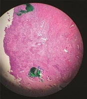
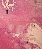
Pathology report of uterine endocervix resection indicated Residual Poorly Differentiated Invasive Squamous Cell Carcinoma measured 1x1 cm in greatest dimensions. All surgical margins were free of tumors. The distance between the tumor and the deep surgical margin (the closest one) was 0.2 cm in one focus. The lymph-vascular invasion was present, but no perineural invasion was observed. All the lymph nodes were free of tumor. After the trachelectomy, she received four more Cisplatin and Taxol courses, and her pelvic MRI was normal again.
One year after the trachelectomy, her oncologist allowed her to plan for pregnancy, which remained unsuccessful after a few months. Finally, she requested assisted reproductive procedures. Ovarian reserve based on the antral follicle count (>9) and anti-Müllerian hormone (2.2 ng/mL) was reasonable. Because of the absence of the cervix and difficulty finding the orifice in the vaginal cuff, she became a candidate for in vitro fertilization (IVF) and zygote intra-fallopian tube transfer at the first infertility clinic she visited. Induction of ovulation was initiated using the Gonadotropin-releasing hormone (GnRH) antagonist protocol. Five mature oocytes were retrieved, resulting in the acquisition of five high-quality cleavage-stage embryos.
Laparoscopy was carried out, and due to the absence of the left tube and the right tube adhesion, it was impossible to perform zygote intra-fallopian tube transfer. Therefore, they tried to transfer two embryos through a vaginal cuff which failed to result in pregnancy. The remaining three embryos were vitrified. In the first frozen embryo transfer cycle, one of the vitrified embryos was thawed and cultured to reach the blastocyst stage. The blastocyte was transferred without ultrasound guidance but failed to result in pregnancy. The next frozen embryo transfer cycle was canceled as the thawed embryos did not reach the blastocyst stage. She was finally offered to use the advantage of a surrogate uterus, but she did not agree.
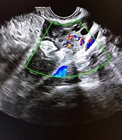
In the second IVF cycle (2019), which is related to this study, per the antagonist protocol, we retrieved 11 oocytes and vitrified 7 embryos on day 3. At the end of ovum pickup, in order to find the right path to the endometrial cavity through the vaginal cuff, an ultrasound-guided mock transfer was carried out using an IVF embryo transfer catheter (Cook Medical Incorporated, USA). In the next cycle, endometrial preparation using 3 × 2 mg oral estradiol valerate (Aburaihan, Iran) and 400 mg daily vaginal progesterone (Cyclogest, Actoverco, Iran) led to three laminar endometrium of 8.4 mm thickness and a single day 3 embryo was transferred under the guide of abdominal ultrasound, resulting in pregnancy.
In the first trimester screening, nuchal translucency was measured at 4.5 mm. Because of the time limitation for abdominal cerclage before 14 weeks of pregnancy, after consultation with a perinatologist and genetic specialist, chorionic villous sampling was carried out and was reported to be normal for chromosomes 13, 18, 21, and XX. Finally, she underwent a laparotomy for abdominal cerclage, and according to the request of the oncologist at the same time, a thin prep smear of the vaginal calf was sent for pathology that was reported free of malignant cells.
Second-trimester ultrasound and fetal echocardiography at the 18th week were normal. She received Hydroxyprogesterone Caproate 250 mg monthly IM (ProlutonDepot, BAYER SCHERING PHARMA, Germany) and 2 × 10 mg daily oral tablet beta-adrenergic agonist (Isoprin Tolidaru, Iran) as tocolytic. Prenatal care continued, and she remained in close touch with the clinic for every sign and symptom.
At 37 weeks, a female baby weighing 2840 g was born by cesarean section following uterine contractions.
3. Discussion
To the best of our knowledge, this is the first case report of successful pregnancy following embryo transfer in a woman with a history of cervical cancer in Iran. We find the right path to the endometrial cavity through the vaginal cuff, an ultrasound-guided embryo mock transfer. The outcome of pregnancy after radical trachelectomy was reviewed (2005). This review of 153 patients, with a follow-up period ranging from 1 to 44 months after RT, showed that 70% of the patients who attempted to conceive became pregnant at least once. A total of 161 pregnancies occurred, with 49% resulting in term deliveries (9). In a meta-analysis in 2019 to evaluate the short and long-term outcomes of abdominal trachelectomy (ART) versus radical hysterectomy for early-stage cervical cancer, they concluded that ART is a more complex and time-consuming technique but equally safe to radical hysterectomy in terms of oncological outcomes in selected patients and provided them with childbearing (4). In a study conducted in 2021, trans myometrial embryo transfer (TMET) was performed after trachelectomy. A successful pregnancy was achieved (14) when performing TMET endometrial trauma, and myometrial contraction can happen compared to a vaginal cuff. The main difference between this case report and the previous studies is that the operation for this young woman was total trachelectomy, while most of the previous pregnancies were reported in patients with less invasive operations.
On the other hand, our patient had experienced two previous failed IVFs, neither of which included a mock transfer to determine the optimal route for transfer. Additionally, there was no ultrasound-guided embryo transfer, which is crucial for patients without a normal cervix.
This study is important from two points of view. First, in young women with early stages of cervical cancer, there is a chance of preserving the uterus and getting pregnant, although it is considered a high-risk pregnancy and needs close observation, including abdominal cerclage. Second, this woman had two previously failed embryo transfer cycles that were not ultrasound-guided. It is especially important in patients with abnormal cervix to follow the true pathway at the time of embryo transfer.
The limitation of this study is that it only reports one case.
3.1. Conclusions
In young women with early stages of cervical cancer, there is a chance of preserving the uterus and getting pregnant, and special attention should be paid to evaluating the right path for embryo transfer. At the end of ovum pickup, in order to find the right path to the endometrial cavity through the vaginal cuff, an ultrasound-guided mock transfer was carried out using an IVF embryo transfer catheter. In the next cycle, a single day 3 embryo was transferred under the guide of abdominal ultrasound, resulting in pregnancy. Based on our experience, we suggest the mock transfer and ultrasound-guided ET in these women.