
1. Context
Breast cancer is a crucial, lethal, multifactorial disease among women in developing and developed countries. The growing number of studies indicates numerous risk factors involved in breast cancer initiation and progression, such as family history, age, genetics, late menopause, and birth control devices (1, 2). Also, several studies have suggested that behavioral and psychological factors also affect the occurrence and progression of cancer due to alterations in immune responses (3-7).
Galen, a Greek physician, initially described the association between the nervous system and the progression and evolution of cancer. He proposed that depressed women develop breast cancer more generally than other women (8). Strong evidence has confirmed that continuous stress can lead to tumor growth in breast cancer. The interactions between the immune and nervous systems are critical for the maintenance of homeostasis in the body (9).
The neural system participates in immune responses in none triple negative breast cancer (NTNBC) and triple negative breast cancer (TNBC) tissue samples. Interactions between the neural genes correlated with inflammation in the tumor microenvironment signals and the central nervous system reconstruct the tumor's microenvironment via autonomic nerves (10).
Different neurons express specific types of neurotransmitters. Major neurotransmitters include dopamine, noradrenaline [(norepinephrine (NE)], cholinergic, and serotonin. Tumor cells contain diverse receptors for neurotransmitters and can interact with diverse neurotransmitters of the autonomic nervous system. This adjacency would present a basis for direct attenuation of the activity of cancer cells by neurotransmitter secretion. Neurotransmitters can directly modulate tumor growth, angiogenesis, and metastasis by regulating various intracellular signaling pathways (7, 9, 11-13).
Neurotransmitters, via their specific receptors in tumor cells, trigger the growth and metastasis of tumor cells. Neurotransmitters such as neurotensin, catecholamines, acetylcholine )Ach(, substance P (SP), serotonin, neuropeptide Y (NPY), neurokinin A, and γ-aminobutyric acid elevate the risk of breast cancer, metastases, and death of patients (14). Therefore, neurotransmitter inhibitors can be alternative treatment options for breast cancer.
Recent comprehensive reviews have underscored the pivotal roles of neurotransmitters — including serotonin, NE, ACh, gamma-aminobutyric acid (GABA), and NPY — in breast cancer progression via modulation of angiogenesis, tumor microenvironment, epithelial – mesenchymal transition, and immune evasion. While these reviews lay a solid mechanistic foundation, our study distinguishes itself by evaluating the therapeutic efficacy of specific neurotransmitter inhibitors — particularly β-adrenergic blockers and monoamine oxidase inhibitors (MAOIs) — directly targeting these pathways to suppress tumor growth and enhance immune response. Unlike broad-spectrum discussions, our work delivers new preclinical evidence demonstrating that these inhibitors not only impede proliferative and metastatic signaling but also synergize with immune checkpoint blockade to potentiate anti-tumor immunity. This positions neurotransmitter inhibition as a promising, cost-effective, and readily translatable strategy to complement existing breast cancer therapies (15-17).
2. Evidence Acquisition
This study investigates the potential roles of neurotransmitters in the growth, migration, angiogenesis, and metastasis of breast cancer tumors. It also discusses the function of neurotransmitter inhibitors in preventing tumor growth and explores the potential value of anti-neurogenic therapies in breast cancer management.
3. Results
3.1. Neurotransmitters and Cancers
Neurotransmitters are the responsive molecules for data transportation throughout the chemical synapses (18). By binding to their target receptors, autonomic nerves and peripheral neurotransmitters show different actions in target cells. Neurotransmitters, depending on their chemical structures, are divided into three groups:
- Peptidergic neurotransmitters (neuropeptide) (i.e., NPY, SP, calcitonin gene-related peptide (CGRP), opioids, neurotensin, bombesin, vasoactive intestinal polypeptide (VIP), and many others)
- Amino acids (i.e., ACh, GABA, glycine, and glutamate)
- Biogenic amines [i.e., dopamine, epinephrine (E), NE, and serotonin]
In the last few years, neurotransmitters have been recognized as an essential microenvironmental ingredient in affecting different human cancers and regulating tissue homeostasis (19, 20). In cancer cells and neurons, the neurotransmitter receptors, i.e., beta-adrenergic receptors, are expressed, and their analogous ligands act as modulators between cancer and the nervous system. This phenomenon opens a new viewpoint to reconsider the role of neurotrophic growth factors and nerve guidance molecules in cancer and their potential benefits as new targets in cancer treatment (21). Moreover, the immune and endothelial cells, which penetrate the tumor microenvironment, express different neurotransmitter receptors and respond to neurotransmitters, are renowned for having a powerful effect on the final outcome of human tumors (22).
Neurotransmitter receptors of the autonomic nervous system play a significant role in cancer cell growth by activating a complex of signaling pathways. For instance, the activation of beta-adrenergic receptors is crucial for tumor growth in ovarian cancer and enhances the growth and invasion of pancreatic and pulmonary cancer cells. In addition to their direct effects on cancer cells, these receptors can also stimulate immune cells, endothelial cells, and fibroblasts, thereby influencing the tumor microenvironment (21).
3.1.1. Neurotransmitter Inhibitors for Prevention of Breast Cancer Cell Growth
3.1.1.1. Dopamine Receptor Inhibitors
Dopamine, a natural chemical belonging to the catecholamine and phenethylamine families, functions as a hormone and neurotransmitter with several critical bodily roles. In a 2014 clinical trial, the alterations of the expression of the dopamine receptor genes in peripheral-blood mononuclear cells were assessed as stress factors in patients with breast cancer. The results showed an increase in the expression of dopamine receptor D2 (DRD2) -DRD4 in peripheral-blood mononuclear cells of breast cancer patients compared to healthy women. This alteration was identified as a potential platform for designing modern drugs, such as DRD2-like agonists, sparking excitement about the future of breast cancer treatment.
Given that, in another study, bromocriptine was introduced as a new option for breast cancer treatment. Bromocriptine is a selective DRD2 agonist in the Michigan Cancer Foundation-7 (MCF7) breast cancer cell line (23). Another research evaluated the effect of psychotherapy on the DR gene expressions in patients with breast cancer. Great significance was obtained in managing and treating cancer due to the possibility of using different treatment regimens, i.e., spiritual interventions, besides traditional medical treatments (24).
The expression of miR‐4301 targets DRD2 in human breast cancer. The result of one study indicated that the miR‐4301 transfection into the breast cancer cells inhibited the proliferation of the cell and activated apoptosis (25). Thus, miR-4301 can be a promising breast cancer treatment option. The expressions of the DRs genes in women with stage two and three breast cancers were evaluated by Bakhtou et al. in 2019. A notable difference was observed in gene expression levels between the healthy and patient women. According to this study, the use of DRD2-agonists depended on the expression of DR, which might be a favorable new perception of complementary therapy in various types of breast cancer (26, 27).
3.1.1.2. Nitric Oxide Inhibitors
Nitric oxide is a structurally simple molecule derived from L-arginine amino acid that plays multiple biological roles, such as a neurotransmitter, hormone, mediator, reactive species, and cytoprotective agent. Geyiki et al. reported a meaningful enhancement in nitric oxidase activity and the amount of serum arginase in breast cancer patients, suggesting that arginase and nitric oxide levels could be used as breast cancer markers (28).
Another study showed that a nitric oxide synthase (NOS) inhibitor decreased breast cancer growth (29). Nitric oxide synthase degrades arginine into L-citrulline and nitric oxide (30). This result suggests that the NOS inhibitor L-NMMA (NOS inhibitor N(G)-monomethyl-L-arginine) can be used as an anticancer therapeutic agent (29).
3.1.1.3. Histamine Receptor Antagonists
Histamine is a natural nitrogenous compound that acts as a neurotransmitter in the brain. Amongst different growth factors and hormones that affect the behavior of breast cells, histamine contributes to differentiation, regulation of growth, and action of the mammary glands through lactation and pregnancy (31, 32). Histamine triggers different signaling pathways by cooperating with four receptor subtypes of histamine (H1, H2, H3, and H4). It was shown that the histamine H3 receptor antagonist, (33) (OUP-186), decreases breast cancer cell proliferation (33). One bioinformatic study published a list of 1266 neurogenes diversely expressed in different breast cancer subtypes. The genes of ephrin-B1 (EFNB1), neuropilin2 (NRP2), histamine receptor 1 (HRH1), amyloid precursor protein (APP), and neural growth factor receptor (NGFR) in human epidermal growth factor receptor 2 (HER2)-enriched and basal tumor samples were distinctively overexpressed. Also, in luminal B and HER2-enriched tumors, syntaxin1A (STX1A) was overexpressed. This result indicated that NRP2, HRH1, and STX1A could be therapeutic targets and prognostic biomarkers for breast cancer patients (34).
Aberrant expressions of the histamine receptors in breast cancer cell lines inspired further exploration of their suppressive impacts on cancerous cell proliferation and growth. Some evidence indicates that tamoxifen (an estrogen antagonist) can bind the histamine-like receptors and decrease proliferation. Therefore, it may be possible to introduce specific histamine receptor antagonists for breast cancer treatment (35).
3.1.1.4. ATP Synthesis Inhibitor
In both the central and peripheral nervous systems, ATP acts as a neurotransmitter (36). As an energy source, ATP is also associated with determining cell fate, metabolism, differentiation, proliferation, and apoptosis. There are assumptions that ATP might be essential in promoting or inhibiting tumor transformation. Under normal conditions, there is no extracellular ATP in healthy tissues. Conversely, ATP exists in tumor sites at high concentration levels. Wagstaff et al. in 2000 described that, in MCF7 breast cancer cells, the extracellular ATP stimulates several growth factors and signaling pathways and induces the expression of the c-fos gene, representing possible targets for suppressing breast cancer progression (37-39).
Aurovertin B, an ATP synthesis inhibitor, inhibited the proliferation of the breast cancer cell line, with a low impact on the normal MCF-10A cell line. Aurovertin B inhibits breast cancer cell proliferation by prompting cell cycle arrest at the G0/G1 phase and apoptosis. This study indicated that Aurovertin B can be an antitumor treatment and may be applied in tumor chemotherapy (40). In another study, Citreoviridin, an inhibitor of ATP synthase, inhibited breast cancer cell proliferation (41).
3.1.1.5. Serotonin Receptors Antagonist
Serotonin is a monoamine neurotransmitter with a general reputation as a participant in happiness and well-being. As a neurotransmitter, serotonin acts on many variants of the serotonin receptors (5-HT receptors or 5-hydroxytryptamine receptors). Hejazi et al. in 2014 determined the status of the gene expressions in serotonin receptors in breast cancer cells. They demonstrated that Ketanserin and Tropisetron, the antagonists of the serotonin receptor, exhibited suppressive effects on MCF7 cell proliferation. These compounds can moderate apoptosis in the MCF7 cell line. This new approach was recommended for breast cancer treatment (42).
3.1.2. Neurotransmitter Inhibitors for Prevention of Breast Cancer Cell Migration
Cell migration is essential in the homeostasis and development of multicellular organisms. In pathological situations, such as metastatic cancer, the cancerous cells gain an expanded ability for migration (43). In metastasis progression, cancer cells disrupt the surrounding basement membrane and invade the stroma, which is constructed of stromal cells and an extracellular matrix (ECM) (44).
Cancer cell migration is an essential step for the progression of metastases. Most patients enduring breast cancer do not die due to the primary tumor but from the progression of metastases. Drell et al. indicated that neurotransmitters such as metenkephalin, bombesin, SP, NE, and dopamine could stimulate breast cancer cell migration, indicating the potent regulatory participation of neurotransmitters to induce breast cancer cell migration (11).
3.1.2.1. Neurokinin-1 Receptor Antagonist
The cancer biopsies revealed a higher neurokinin-1 receptor (NK1) expression level compared to benign and normal samples. Neurokinin-1 receptor plays a role in inducing breast cancer cell migration by increasing the expression of matrix metalloproteinases, specifically MMP-14 and MMP-2. Human hemokinin-1 (hHK-1), the peripheral agonist of the NK1 receptor, stimulates the phosphorylation of JNK, Akt, and extracellular signal-regulated protein kinase (ERK1/2) via the protein kinase A (PKA) and protein kinase C (PKC) pathways. This phosphorylation process regulates the activation of the nuclear factor kappa B (NF-κB) and the transcription factor activator protein-1 (AP-1). When NF-κB and AP-1 are inhibited, a decrease in the up-regulation of MMP-14 and MMP-2 by hHK-1 is observed. The findings indicate that the NK1 receptor is a critical regulator of the migration of human breast cancer cells and serves as a potential target for breast cancer treatment (45).
Recent research has illuminated the significant role of SP and its receptor, NK1 receptor, in cancer progression and treatment resistance. This highlights the potential of repurposing aprepitant, a well-established NK1 receptor antagonist, as a promising strategy to improve cancer treatment outcomes. By integrating NK1 receptor antagonists into existing therapies, it may be possible to develop more effective treatment regimens that not only target tumors more efficiently but also enhance the overall quality of life for patients facing the complexities of cancer (46).
3.1.2.2. Gamma-Aminobutyric Acid (A) Receptors Inhibitors
The GABA (A) receptors and heteropentameric ligand-gated chloride channels modulate synaptic inhibition in the central nervous system. In breast cancer, a GABA (A) receptor agonist, Propofol, induces the migration and actin reorganization of the cancer cells via collagen matrices. Also, in breast cancer cell lines, the GABA (A) receptor pi (GABRP) expression indicates a significant association with the basal-like subtype. It suggests the functions of GABRP in the progression and initiation of basal-like tumors. One study showed that blocked GABRP (by alterations in the cytoskeleton) notably reduced expression of the KRT5, KRT6B, KRT14, KRT17, and BLBC-associated cytokeratin. Furthermore, cellular protrusions reduce the tumorigenic and migration potential of the breast cancer cell. Also, silencing the GABRP decreases the extracellular regulated kinase 1/2 (ERK1/2) phosphorylation in breast cancer cells, and the ERK1/2 inhibition similarly reduces the basal-like cytokeratins in addition to migration (47).
3.1.2.3. Neuropeptide Y Receptor Antagonists
Stress has long been known to be related to enhanced cancer risk. Chronic stress is correlated with increased levels of sympathetic neurotransmitters (i.e., NPY and NE) release. In human breast cancer cells, the NPY receptor expression has been indicated. The activation of the NPY 5 receptor was demonstrated to prompt the growth and migration of breast cancer cells in humans. Treatment of 4T1 breast cancer cells by NPY (via increased phosphorylation of ERK 1/2) promoted cell proliferation in a concentration-dependent manner. The administration of NPY receptor antagonists (Y5R: L-152,804, Y2R: BIIE0246, and Y1R: BIBP3226) inhibited tumor proliferation (48).
3.1.3. Neurotransmitter Inhibitors for Prevention of Breast Cancer Angiogenesis
Angiogenesis, the new growth of blood vessels, is a standard physiological process that tumors use to support their proliferation, growth, and metastasis. Angiogenesis includes the division and migration of endothelial cells, the production of the new basement membrane, the arrangement of tubular structures, and the coverage by pericytes. A collection of anti- and pro-angiogenic molecules controls angiogenesis, including vascular endothelial growth factor (VEGF), angiogenin, tumor necrosis factor (TNF)-α, interleukin (IL)-8, transforming growth factor (TGF)-α, TGF-β, fibroblast growth factor (FGF), and platelet-derived growth factor (PDGF) (49, 50). The angiogenic factors in tissues indicate the destructiveness of tumor cells, which are critical in prognostic results (49, 50). The equilibrium between the anti- and pro-angiogenic factors in cancer is misplaced, leading to uninhibited angiogenesis with irregular blood vessels lacking a precise hierarchical arrangement (49, 51). Given that, therapies based on anti-angiogenic agents have been accepted for cancer treatment (52-55). The VEGF/VEGFR2 interaction is responsible for the superiority of the signals that stimulate angiogenesis in vivo; however, its therapeutic importance for increasing the survival time of patients is comparatively small (56).
In addition, the influence of the tumor microenvironment on angiogenesis has been highlighted in recent years (57-60). The nervous system also supports cancer progression by regulating tumor angiogenesis by controlling neurotransmitter release. The nervous system manages the functional actions of several organs in the body, though this system does not recognize the tumors as separate organs within an organism. Therefore, the nervous system is intrinsically associated with the progression of the tumor (9, 22).
Direct activation of β-adrenergic signaling in some breast cancer cell lines can intensify the expression of factors that induce tumor angiogenesis, i.e., interleukin-8 (IL-8), IL-6, and VEGF (22). Jagged 1, an essential factor that mediates the Notch signaling, regulates tumor angiogenesis via the β2-adrenergic receptor-protein kinase A-mammalian target of rapamycin (β2-AR-PKA-mTOR) pathway. In breast cancer patients, upregulation of Jagged 1 is associated with low tumor prognosis (61, 62). In MDA-231 breast cancer cells, the Jagged 1 knockdown by siRNA prevented the Notch signaling and impaired the tumor angiogenesis via NE (63).
In contrast, dopamine suppresses angiogenesis in both endothelial progenitor cells and tumor endothelium via down-regulation of the VEGFR-2-mediated signaling pathway through DR2 (7, 22, 64, 65). The administration of dopamine with anticancer drugs (i.e., 5-fluorouracil and doxorubicin) in mouse models of breast cancer impaired tumor growth and improved the survival outcome (66) In breast cancer, the α9-nAChRs overexpression induced the release of pro-angiogenic factors (67, 68). In breast cancer mouse models, administering auto-antibodies on mAChRs led to tumor angiogenesis by activating the mAChRs via secretion of VEGF-A. The use of the muscarinic agonist, Carbachol, in the absence or presence of different muscarinic antagonists resulted in enhanced VEGF expression in BALB/c mice bearing LMM3 mammary adenocarcinoma cells (69, 70).
The NPY secretion promotes angiogenesis by enhancing VEGF expression, leading to breast cancer progression (71).
Nitric oxide was shown to increase nitrite/nitrate and VEGF-C generation in the MDA-MB-231 cell line. A high level of nitrotyrosine is associated with enhanced VEGF-C and lymph node metastasis while decreasing overall survival in invasive breast cancer (72).
Glutamate is a neurotransmitter that regulates cellular and synaptic activities through binding to metabotropic glutamate receptors (mGluRs). The expression of the mGluRs has been a concern in tumor angiogenesis (73-75). Therefore, in an orthotopic breast cancer (4T1) model, reduction in mGluR1 activity prevented angiogenesis, indicating that mGluR1 is a pro-tumorigenic/pro-angiogenic factor (73).
Nerve growth factor (NGF), a neurotrophic factor, is upregulated in the microenvironment of tumors of different cancers including breast cancer. NGF, released by breast cancer cells, induces in vivo angiogenesis and increases VEGF secretion in breast cancer cells (76).
3.1.4. Neurotransmitter Inhibitors for Prevention of Breast Cancer Cell Metastasis
Many patients enduring breast cancer do not die from a tumor in the primary stage but from metastasis. There are two essential steps in the metastasis cascade: The tumor cells' migratory activity and capacity to exit from the bloodstream via the vascular endothelium. Investigating the migratory activity of breast cancer cell lines by different neurotransmitters, in a study, it has been shown that metenkephalin, SP, NE, dopamine, and bombesin had a stimulatory effect on migration of the breast cancer cells using computer–assisted cell tracking and time–lapse video microscopy of MDA-MB468. The α7-nicotinic acetylcholine receptor (α7-nAChR), a neurotransmitter receptor in nervous systems, also exists in different non-neuronal tissues. In 2012, it was found that α7-nAChR has been associated with cancer stem cell (CSC) proliferation. Cancer stem cells are minor subpopulations responsible for metastasis and tumor development (77).
3.1.4.1. Gastrin Releasing Peptide Inhibitors
Gastrin-releasing peptide (GRP) is a neuropeptide that acts as a regulatory molecule in different pathophysiological and physiological processes. Chao et al. showed that the stimulation of the GRP receptor leads to IL-8 expression and cellular migration in breast cancer cells. It was demonstrated that in the MDA-MB231 TNBC cell line, EGF-R and GRP-R synergistically regulated the IL-8 expression and cell migration but not proliferation. The GRP-R ectopic expression in SK-BR-3 cells was adequate to enhance IL-8 mRNA levels and cellular migration. These results suggested the relative role of GRP-R in the progression of ER-negative breast cancer (78).
3.1.4.2. Norepinephrine Inhibitors
Norepinephrine is an organic compound in the catecholamine family that acts as a neurotransmitter and hormone in the brain and human body. Norepinephrine, similar to other biologically active substances, functions by binding to the cell's surface receptors. Two broad families of NE receptors are the beta and alpha-adrenergic receptors. A study by Strell et al. showed that NE is a potent inducer of the MDA-MB468 (a triple-negative breast cancer cell line) migration. They stated that the NE release, mediated by induction of a GROα [growth-regulated protein alpha (GRO-alpha)], developed the adhesion of MDA-MB-231 cells to the vascular endothelium via β1-integrin. The results of this study showed that the β-blockers can decrease metastasis progression (79). A meta-analysis indicated that β-blockers decrease the recurrence of breast cancer and death (80).
The outcomes of clinical and preclinical studies represent the participation of the sympathetic nervous system in controlling the pathophysiology of bone remodeling and bone metastasis. In mouse models of skeletal metastasis, administration of isoproterenol, as a potent beta-adrenergic receptor (BAR), was found beneficial for colonization of the bone in metastasis breast cancer cells by increasing the vascularity of the bone marrow. The bone density and the activation of bone marrow vessels favor the skeletal engraftment of circulating breast cancer cells. Stimulation of the B2ARs in osteoblasts induces adhesion of the breast cancer cells to the bone marrow endothelial cells in a manner dependent on IL-1β and selectin. Some studies suggested that the activation of BARs leads to the production of pro-angiogenic factors (81). Many cancer researchers consider the BAR as a therapeutic target for breast cancer. Mulcrone et al. showed that BARs regulate the VEGF and IL-6 productions by divergent pathways, which might be explained by the complexity of breast cancer cells, functional responses, and the BAR expression (82).
3.1.4.3. Adenosine Triphosphate Inhibitors
Fang et al. first depicted the pro-invasive ATP activity (83). It was indicated that extracellular ATP is necessary for epithelial–mesenchymal transition (EMT) and cancer cell invasion via activating P2Y2 to regulate the expression of associated molecules including IL-8, claudin-1, Snail, E-cadherin, S100A4, and β-catenin, as well as ERK1, 2 and EGFR (84). In 2019, Yang et al. revealed that the extracellular ATP stimulated the signaling of the hypoxia-inducible factor (HIF) and upregulated the HIF1/2α expression, representing that the extracellular ATP could induce the breast cancer EMT and invasion through induction of the HIF2α signaling, which can be a possible treatment for anti-metastasis therapy in the future (85). ATP was shown to upregulate the secretion and expression of S100A4 in fibroblasts and breast cancer cells. The ability of breast cancer cells to convert fibroblasts into cancer-associated fibroblasts (CAF)-like ATP enhances cells. The extracellular ATP leads to the interaction between breast cancer cells and fibroblasts, which can collaboratively act through S100A4 production to aggravate breast cancer metastasis (86).
3.1.4.4. Neuropeptide Y
Neuropeptide Y, a 36 amino acid neuropeptide, is secreted alongside other neurotransmitters, including glutamate and GABA. Neuropeptide Y has been distinguished as being synthesized in GABAergic neurons and acts as a neurotransmitter over cellular communication. In 2001, Reubi et al. suggested that NPY be added to the small regulatory peptide list associated with cancer. The high occurrence of NPY in metastatic and invasive breast cancer presents a promising avenue for therapy and diagnosis, with the use of NPY analogues (87).
Neuropeptide Y increases VEGF expression, breast cancer progression, and angiogenesis. Nitric oxide can also lead to metastasis and tumor growth by enhancing angiogenesis. For example, nitric oxide enhances VEGF-C, and the production of the nitrite/nitrate in the MDA-MB 231 cell line and high levels of nitrotyrosine correlate with lymph node metastasis, enhanced VEGF-C, and decreased survival in invasive breast carcinoma (72, 88).
3.1.4.5. Amino Acids Antagonists
Rapid cancer cell proliferation increases the flux via anabolic pathways to produce the materials essential for cell division. Glucose and amino acids entering the cell are located at the apex of the anabolic pyramid. Since tumor cells are more inclined to accelerate nutrient uptake than their ordinary neighbors, upregulated transporters could be outstanding targets for cancer treatment. One study showed that solute-carrier 6A14 (SLC6A14) is an amino acid transporter in the breast cancer cell. Nevertheless, mice lacking SLC6A14 displayed normal mammary gland development; these animals were profoundly resistant to initiation and progression of the tumor induced by potent oncogenes. In spontaneous mouse models of breast cancer, the amino acid transporter SLC6A14 deletion inhibited the growth of the tumor (89). Another study showed that the levels of neurotransmitter precursor amino acids correlated with psychological health in patients with breast cancer (90).
Glutamate, an excitatory neurotransmitter, plays a crucial role in regulating cellular and synaptic activity by binding to the mGluRs. On the other hand, as an α-amino acid, glutamate is used by almost all living organisms in protein biosynthesis. GABAergic neurons are also the precursor for synthesizing inhibitory GABA. Glutamate also regulates the migration and proliferation of neurons. Although glutamate is present in non-neuronal tissues, such as cancer, it may affect the migration and proliferation of cancer cells. The mGluRs expression has been involved in tumor angiogenesis in mouse breast cancer and melanoma models. Therefore, decreasing mGluR1 activity suppresses angiogenesis in the orthotopic breast cancer (4T1) model, implying that mGluR1 is a pro-angiogenic/pro-tumorigenic factor. Different cancer cell lines were exposed to the antagonists of the α-amino-3-hydroxy-5-methyl-4-isoxazolepropionic acid (AMPA) and N-methyl-D-aspartate (NMDA) receptors to investigate the effect of glutamate on tumor growth. It was found that the glutamate antagonists inhibited the tumor growth and migration, whereas glutamate stimulated the tumor cells' proliferation and migration. Rzeski et al. in 2001 demonstrated that glutamate antagonists suppress human tumor cell proliferation, such as T47D, LS180, and A549 (8, 73, 91, 92).
3.1.4.6. Angiotensin
Angiotensin is a peptide hormone that increases blood pressure and is involved in vasoconstriction. Angiotensin 2 may act on the central nervous system to regulate renal function, renal sympathetic nerve activity, and blood pressure. Angiotensin 2 is not only associated with the regulation of blood pressure, sodium and water homeostasis, and control of other neurohormonal systems but actively participates in the excessive production of reactive oxygen species (ROS) and the proliferation, hypertrophy, apoptosis, and migration of vascular cells (93).
Angiotensin 2 increases breast cancer cell migration and metastasis through up-regulation of intercellular adhesion molecule 1 (ICAM1) and MMP2/MMP9, which are associated with adhesion, migration, and cancer cell invasion (94). Strong evidence links the AT1R (an angiotensin 2 receptor) activity to different aspects of breast cancer, including carcinogenesis, disease development, and metastasis (95).
3.1.4.7. Tachykinins Antagonist
Tachykinins, including neurokinin A and B, SP, endokinins, and hemokinin-1, are a family of neuropeptides. Tachykinins mainly act via three transmembrane G-protein coupled receptors called NK1, NK2, and NK3. A 2013 study showed that the autocrine signaling of SP contributed to persistent activation of the HER2 that is related to tumor progression and drug resistance in breast cancer. This study revealed an essential oncogenic collaboration between HER2 and NK-1R. It is thought that a link between cancer progression and inflammation is possibly targeted by the SP antagonist (96).
4. Conclusions
Recent reviews have synthesized how neurotransmitters shape breast cancer biology and therapeutic opportunities, but they are primarily mechanism-centric and broad in scope (15, 97, 98). Our study complements and advances this literature by providing an inhibitor-focused, breast-cancer–specific synthesis that maps neurotransmitter blockade (receptor antagonism, synthesis/release inhibition, reuptake blockade, and downstream signal inhibition) across transmitter families to concrete tumor hallmarks — proliferation, invasion/migration, angiogenesis, and metastasis — together with translational signals for repurposing clinically used agents. Unlike prior reviews that emphasize descriptive pathways or general neuro-oncology principles (97), we comparatively weigh inhibitory evidence within breast cancer models, integrate immune-modulatory impacts relevant to response to immunotherapy (98), and distill a decision-oriented framework that highlights where neurotransmitter inhibitors may add value beyond standard endocrine/HER2/chemotherapy regimens. This inhibitor-centric, hallmarks-aligned lens is the key novelty of our work relative to recent overviews (15, 97, 98).
Converging evidence indicates a bidirectional circuit: Breast tumors are innervated, neurotransmitters released from sympathetic/parasympathetic/sensory fibers and stromal/immune cells remodel the tumor microenvironment, and tumor cells themselves express receptors and can produce neurotransmitters, creating autocrine and paracrine loops (15, 97, 99). This mutuality (“tumor–nerve circuit”) links stress-adrenergic signaling, neurotrophic factors, angiogenesis, immune suppression/activation, and metastatic niche conditioning; targeting the circuit with neurotransmitter pathway inhibitors therefore has dual leverage on cancer cells and the microenvironment (15, 99). By explicitly organizing inhibitor classes along this bidirectional axis — and emphasizing contexts where blockade is most likely to interrupt feed-forward loops — our analysis operationalizes concepts that prior narrative reviews have articulated but not systematically translated into inhibitor-selection guidance for breast cancer.
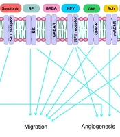
Tumor cells interact with nervous, stromal, and immune cells through specific receptors and produce soluble factors. These interactions may be driven throughout organs in distant places or inside the tumor that respond to tumor signals. Thus, understanding the interactions between cells may help to develop novel and more effective therapies against cancer. The neurotransmitter molecules such as catecholamine, dopamine, epinephrine, and NE have regulatory roles in breast cancer cell growth, migration, angiogenesis, and metastasis (Figure 1). Clarifying their functions in the biology of the tumor would highly enhance our comprehension of oncogenesis and open new windows for cancer treatment and diagnosis. Many neurotransmitters, including serotonin receptor antagonists, β-adrenergic receptor antagonists, dopamine receptor agonists, and ACh receptor antagonists, might have clinical applications in cancer therapy or combination drug therapies. Thus, blockage of some neurotransmitter actions may be a potent strategy for cancer therapy (Table 1). Additionally, targeting the possible correlations between the neurotrophic signaling and neo-neurogenesis inhibition and chemical or surgical denervation should be investigated more as therapeutic options for cancer treatment. Even so, more explorations are needed to stabilize the inclusion of these medicines in clinical cancer treatment strategies and to prevent side effects.

Figure 1.
Neurotransmitters involved in tumor growth, migration, angiogenesis and metastasis
Table 1.
Effective Neurotransmitters and the Neurotransmitter Inhibitors in Breast Cancer
| Neurotransmitters | Effects | Inhibitors |
|---|---|---|
| Dopamine | Tumor growth | Bromocriptine; miR‐4301 |
| Nitric oxide (NO) | Tumor growth; Angiogenesis | L-NMMA |
| Histamine | Tumor growth | OUP-186; Tamoxifen |
| ATP | Tumor growth; Metastasis | Aurovertin B; Citreoviridin, |
| Serotonin receptors (5-HT receptors or 5-hydroxytryptamine receptors) | Tumor growth | Tropisetron; Ketanserin, |
| Neurokinin-1 | Migration | Human hemokinin-1 (hHK-1) |
| GABA | Migration | - |
| Neuropeptide Y (NPY) | Migration; Metastasis; Angiogenesis | Y5R:L-152,804; Y2R:BIIE0246; Y1R:BIBP3226 |
| ACh | Angiogenesis | Muscarinic agonist (Carbachol) |
| Gastrin Releasing Peptide (GRP) | Metastasis | - |
| Norepinephrine | Metastasis | - |
| Tachykinins | Metastasis | - |
| Angiotensin 2 | Metastasis | DAMGO |
| Amino acids | Metastasis | SLC6A14 |