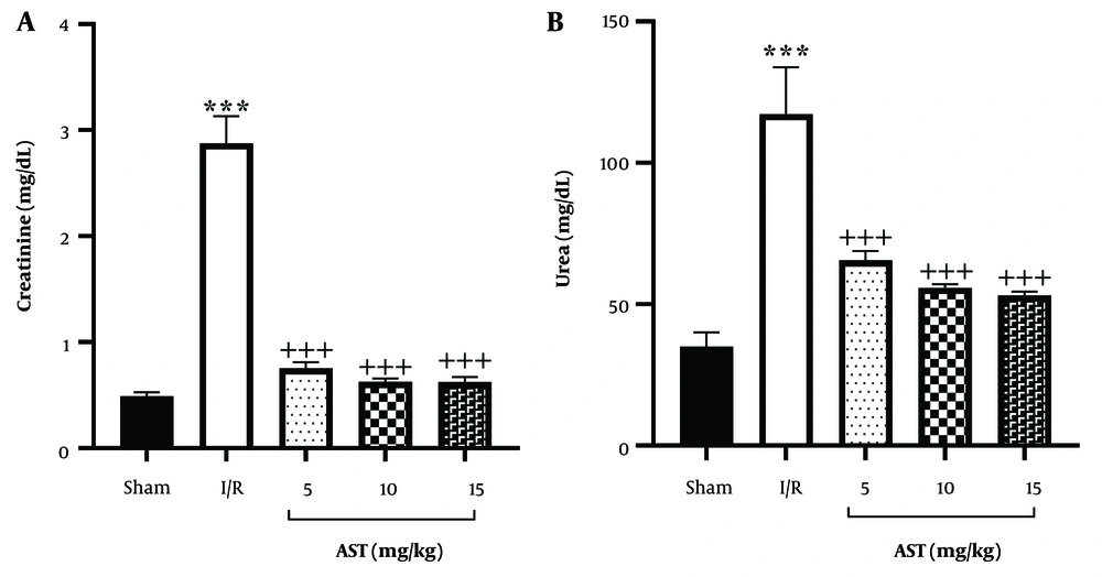
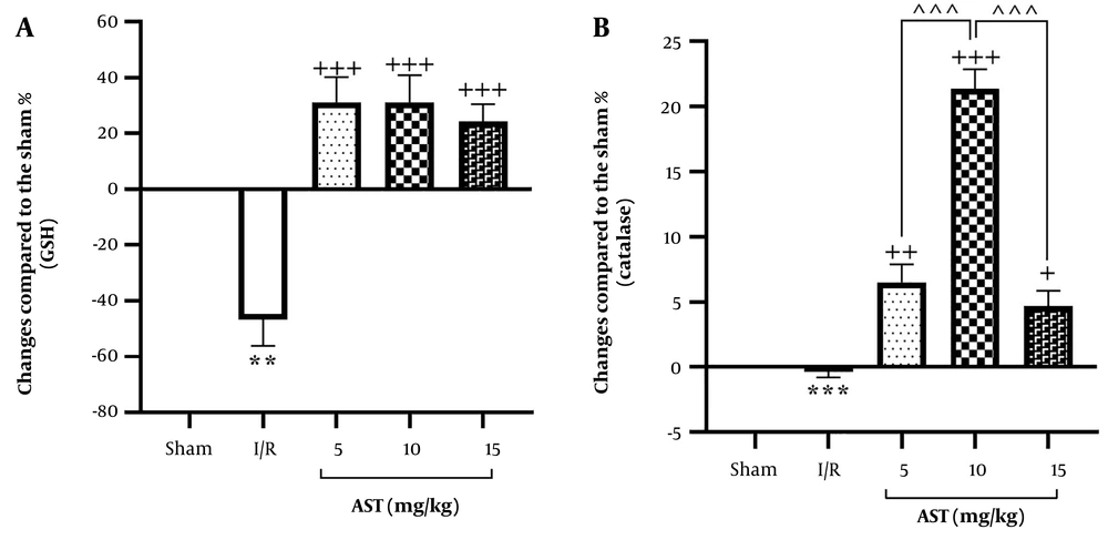
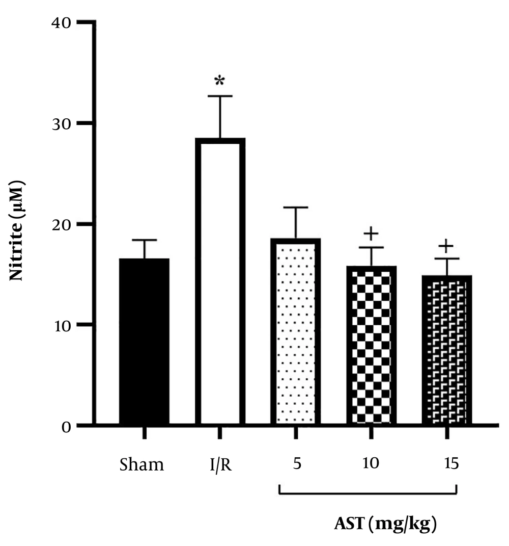
Astaxanthin showed considerable protective effects against I/R injury. Intraperitoneal administration of AST for seven consecutive days was associated with a reduction in oxidative stress markers such as nitrite and an increase in levels of key antioxidants such as GSH and catalase. This modulation helped improve kidney function and reduce tissue damage following I/R injury by regulating urea and creatinine levels (
Figure 5).
A schematic representation of study protocol and the effect of AST after renal I/R. AST, astaxanthin; GSH, glutathione; I/R, ischemia-reperfusion.
Acute kidney injury is characterized by a rapid decline in kidney function, often resulting from various insults, including I/R injury. I/R occurs when the blood supply to the kidney is temporarily interrupted and then restored, leading to a cascade of pathological changes that can result in significant renal damage (
16). Multiple dysregulated pathways contribute to the pathogenesis of I/R. Recent reports have introduced the canonical Wnt/β-catenin pathway as a protective mechanism, while the non-canonical Wnt pathway contributes to organ damage during I/R. The cross-talk between signaling pathways with Wnt signaling is also highlighted in controlling inflammation, oxidative stress, and apoptosis, forming a network that plays an important role in the modulation of I/R injury. Among the interconnected signaling mediators, the NF-κB, HIF-1α signaling, notch, PI3K/Akt, TGF-β, NMDA/HGF/c-Met, Rho/Rho-associated protein kinase, mitogen-activated protein kinase/extracellular signal-regulated kinase, Janus kinase/signal transducers and activators of transcription, and nuclear factor erythroid 2-related factor 2 signaling pathways deserve further investigation. Co-targeting Wnt signaling pathways and related interconnected signaling pathways offers a hopeful therapeutic candidate in I/R (
2).
As a critical interconnected dysregulated pathway handled by the Wnt/β-catenin pathway, oxidative stress mediators (i.e., Nrf2, catalase, GSH, nitrite) play important roles in I/R. During the ischemic phase, there is a reduction in renal blood flow, leading to inadequate oxygen delivery to kidney tissues. This results in cellular energy depletion, primarily through the depletion of ATP, and the accumulation of metabolic waste products (
17). The lack of oxygen triggers the production of ROS, which can damage cellular components, including lipids, proteins, and DNA (
18). Upon restoration of blood flow, a paradoxical increase in oxidative stress occurs due to the sudden influx of oxygen. This leads to further production of ROS, exacerbating cellular injury through mechanisms such as lipid peroxidation and apoptosis. The reperfusion phase also initiates an inflammatory response characterized by the release of pro-inflammatory cytokines (e.g., tumor necrosis factor-alpha and interleukins) and chemokines, which promote leukocyte adhesion and infiltration into renal tissues (
5,
19).
The main site of injury during I/R is the renal tubular epithelium. Ischemia causes tubular epithelial cell detachment, swelling, and necrosis (
20). The loss of tight junction integrity allows for back-leakage of glomerular filtrate, further reducing the glomerular filtration rate. Dying cells can form obstructive casts within tubules, contributing to oliguria and worsening acute kidney injury (
21,
22).
Given the complex interplay of oxidative stress and inflammatory responses in I/R injury, the protective effects of AST appear to be a promising therapeutic approach to reducing acute kidney injury. As a potent antioxidant, studies have shown that AST not only scavenges ROS but also enhances the body's endogenous antioxidant defenses (
23). In our study, increased levels of GSH and catalase, as well as decreased levels of nitrite, were evident after AST administration. This property helps restore the redox balance in kidney tissues during both ischemic and reperfusion phases. Previously, we demonstrated that pretreatment with AST (5 and 10 mg/kg) delivered via solid lipid nanoparticles significantly alleviates oxidative stress, as evidenced by reduced serum nitrite levels and enhanced concentrations of critical antioxidants such as catalase and GSH in the kidneys (
10). In a similar report, AST (5, 10, and 25 mg/kg) was orally used to play protective roles against renal I/R (
24). We also previously showed that AST in similar doses (e.g., 5 and 10 mg/kg) exhibited anti-neuropathic potentials against chronic constriction injury of the sciatic nerve (
9). In a similar range of dosages (5, 10, and 15 mg/kg), we confirmed the anti-nociceptive effects of AST through the l-arginine/nitric oxide (NO)/cyclic GMP (cGMP)/potassium channel (KATP) signaling pathway in mice (
25). Our current study is the first to provide the therapeutic role of naïve AST against renal I/R in rats.
Furthermore, a separate study indicated that AST at a concentration of 250 nM effectively mitigated the decline in viability of tubular epithelial cells exposed to H
2O
2, underscoring its protective role against oxidative stress (
26). Notably, Guo et al. reported that AST upregulates the heme oxygenase 1 axis and inhibits inflammatory pathways, such as the toll-like receptor 4 (TLR4)/MyD88/NF-κB axis, which are implicated in renal inflammation. This modulation further enhances its protective effects against acute kidney injury (
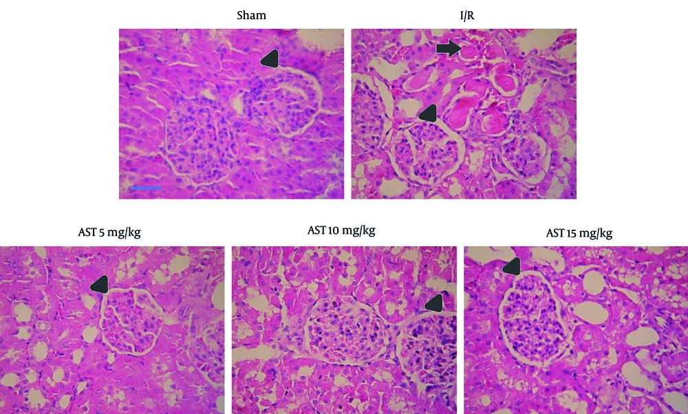
27). The cumulative effects of these pathological processes lead to acute tubular necrosis, characterized by significant impairment of renal function. As previously shown (
11,
28), in our study, renal I/R led to histological damage. Our results indicated that I/R caused tubular necrosis in the ascending thick portion of the Henle arc cortical areas of the proximal tubule. During I/R, we also observed an increased size of Bowman's space and numbers of red blood cells within the glomerular capillaries. In our study, AST decreased renal cortex tubule necrosis and reduced the numbers of red blood cells within the glomerular capillaries compared to the I/R group. If not promptly addressed, this can progress to chronic kidney disease or complete renal failure (
29).
Our findings suggest that reducing oxidative stress is closely associated with improved kidney function, as evidenced by lower levels of key biomarkers such as creatinine and urea. Effective management of oxidative stress may play a pivotal role in safeguarding renal health and facilitating recovery following episodes of diminished blood flow and subsequent restoration, thereby alleviating tissue damage and promoting overall kidney function. Moreover, the beneficial protective effects of AST on essential indicators of renal health, including serum urea and creatinine levels, were corroborated in a mouse model. The study revealed a significant decrease in pathohistological scores, the number of apoptotic cells, and the modulation of the level of superoxide dismutase and malondialdehyde, suggesting that AST effectively reduces cellular apoptosis and fibrosis (
26). We also previously showed the protective effects of a new formulation of AST (i.e., solid lipid nanoparticles) in combating kidney injury after I/R (
10).
In conclusion, AST demonstrates significant protective effects against renal I/R injury in rats, evidenced by reduced renal tissue damage and improved kidney function markers such as reduced plasma creatinine and urea levels. The AST enhances antioxidant levels, including GSH and catalase, while decreasing oxidative stress markers like serum nitrite. These findings highlight AST’s potential as a therapeutic agent for acute kidney injury. Further research is needed to explore its mechanisms and clinical applicability. Additionally, more studies are needed to develop strategies that critically modulate multiple interconnected signaling mediators in I/R damage.