
1. Background
Nanotechnology enables the development of tunable materials with enhanced physicochemical properties for pharmaceutical and textile applications. Nanoscale carriers improve compound retention, dispersibility, and interfacial interactions, making particle size and surface charge key determinants of adhesion and diffusion (1). Accordingly, agarwood oil (AO)–loaded chitosan nanoparticles (Ch-AO-NPs) may offer a more sustainable pathway towards functional textiles with reduced reliance on synthetic finishes. The use of natural actives and biodegradable biopolymers further supports interest in sustainable textile functionalisation. Chitosan (CS), a partially deacetylated biopolymer, is widely recognised for its biocompatibility, film-forming ability, and cationic nature. Through ionic gelation, protonated CS interacts with polyanions such as tripolyphosphate (TPP) to form nanoparticles in aqueous media without organic solvents (2). Careful consideration of these characteristics is essential to ensure optimal particle size, uniformity, and functional performance. Chitosan’s pH-dependent solubility, variable protonation, and tendency to aggregate at higher ionic strengths are critical parameters that directly influence the formulation process and long-term stability of CS nanoparticles (2). However, its positive charge, mucoadhesive behaviour, and affinity for negatively charged surfaces support its use as an eco-friendly nanocarrier for controlled release and textile functionalisation. For hydrophobic essential oils, emulsification before ionic crosslinking is often used to improve dispersion and facilitate nanoparticle formation (3). Aquilaria malaccensis is derived from Malaysia’s key agarwood-producing species, which is extensively utilised for fragrance and traditional medicine across Southeast Asia (4). Rich in sesquiterpenes and chromone derivatives, AO exhibits aromatic, antimicrobial, and antioxidant properties. Similar bioactive properties have also been reported in various A. malaccensis extracts (4). Despite its therapeutic relevance, AO remains underexplored in nanoparticle-based delivery systems compared with widely studied essential oils. Moreover, AO is associated with calming and stress-relief effects, making it ideal for medical fabrics. Its direct use is limited by high volatility and poor water compatibility, suggesting that CS encapsulation may be an effective strategy to improve stability and control release for bioactive textile applications (5). To understand their behaviour on fabrics, interactions between CS nanoparticles and textile fibres must be considered, especially since different fabric chemistries interact distinctly with nanoparticles, influencing adherence and durability. Chitosan nanoparticles adhere through electrostatic attraction, hydrogen bonding, and physical deposition, forming a coating capable of retaining and gradually releasing the encapsulated oil. Cotton’s (CO) hydrophilic cellulose promotes strong electrostatic and hydrogen-bond interactions with cationic CS, whereas polyester’s (PO) hydrophobic surface supports primarily physical adsorption (6). However, the presence of hydrophobic AO and citric-acid crosslinkers may enhance deposition onto PO by increasing hydrophobic affinity and providing additional binding sites. These interactions highlight the need to optimise nanoparticle properties for both fibre types. This study fabricated Ch-AO-NPs, optimised for size and dispersity, and evaluated their compatibility with cotton and polyester textiles.
2. Methods
Low-molecular-weight CS (75 - 85% deacetylation; Sigma-Aldrich, Iceland), sodium tripolyphosphate (TPP, ≥ 85%; Sigma-Aldrich, USA), and cellulose dialysis tubing (MWCO 14 kDa; Sigma-Aldrich, USA) were used as received. Tween 80, sodium hydroxide (NaOH), and glacial acetic acid were obtained from Chemiz (Malaysia). Aquilaria malaccensis (agarwood, AO) oil was supplied by Sheer Essence (India) via Benua Sains. Phosphate-buffered saline (PBS; Dulbecco A, pH 7.3; Oxoid, UK) was used for the release study. All reagents were analytical grade and used without further purification. Ultrapure water (18.2 MΩ·cm) was used throughout.
2.1. Methodology
2.1.1. Preparation of Ch-AO-NPs
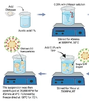
Agarwood oil–loaded chitosan nanoparticles (Ch-AO-NPs) were prepared using an emulsion-assisted ionic-gelation method adapted from Calvo et al. (7) and essential-oil nanoparticle formulations (3). A 0.25% (w/v) CS solution was prepared in 1% acetic acid and adjusted to pH 4.6 to ensure optimal protonation of CS (8), while a 0.1% (w/v) TPP solution was prepared separately. Tween 80 was added to the CS solution and mixed for 10 min before AO addition to form a pre-emulsion, followed by 1 h of stirring. TPP was added dropwise under constant stirring, and the mixture was stirred for an additional hour to complete nanoparticle formation. The pellet was frozen (-80 °C), lyophilised (-58°C, 72 h), and stored at -20°C until use (Figure 1). Emulsion-assisted ionic gelation was selected for its ability to efficiently encapsulate volatile essential oils within crosslinked CS matrices, yielding stable particles with the cationic character needed for textile adhesion, an advantage over less stable systems such as nanoemulsions.
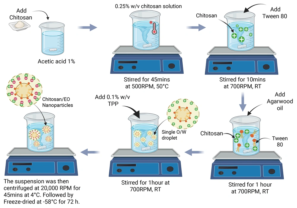
Figure 1.
Encapsulation process of Ch-AO-NPs
2.2. Experimental Design
To assess the effects of formulation variables on particle size, the CS:TPP ratio, CS:AO ratio, Tween 80 concentration, stirring speed, and stirring time were varied systematically while other conditions remained constant [Table D1 (Appendix D) in Supplementary File]. A one-factor-at-a-time (OFAT) approach was selected to isolate each parameter’s effect, providing a straightforward interpretation. This enabled the identification of an optimised formulation with the desired particle size and dispersity for subsequent encapsulation and stability analyses.
2.3. Particle Size, Polydispersity Index, and Zeta Potential Analysis
Particle size and Polydispersity Index (PDI) were measured by dynamic light scattering (DLS) at 25°C with a 172° backscatter angle, while zeta potential was determined by electrophoretic light scattering (Zetasizer Nano ZS, Malvern, UK). Short-term stability of the nanoparticle dispersion was evaluated by measuring particle size and PDI at days 0, 5, and 7 at 4°C, 25°C, and 40°C. Full results are presented in Appendix C. Nanoparticle morphology was observed via transmission electron microscopy (TEM; Talos L120C, FEI, USA).
2.4. Encapsulation Efficiency, Loading Capacity, and Yield
Encapsulation efficiency (EE) was determined indirectly using UV-Vis spectrophotometry (Thermo GENESYS 180, USA) at 248 nm by analysing AO content in the supernatant after centrifugation. Supernatants from unloaded CS nanoparticles served as blanks. A calibration curve was prepared using AO standards in ethanol. Encapsulation efficiency, loading capacity (LC), and yield were calculated using Equations 1 (3, 9).
Equation 1.
Equation 2.
Equation 3.
2.5. In vitro Drug Release Study
Ch-AO-NPs (10 mg) were dispersed in 2 mL of distilled water and loaded into pre-soaked 8 cm cellulose dialysis tubing. The dialysis bag was immersed in 100 mL PBS adjusted to pH 4.8 or 8.5 using 2 N NaOH or glacial acetic acid and maintained at 37°C in a shaking water bath (WNB-45; Memmert, Germany) to ensure uniform mixing. At predetermined intervals, 4 mL of the external medium was withdrawn for analysis and replaced with an equal volume of fresh, pre-warmed PBS of the same pH. Released AO was quantified using UV-Vis spectrophotometry at 248 nm, and cumulative release (%) was calculated, where represents the cumulative amount of AO released at time represents the total amount of AO initially present in the nanoparticles (Equation 4) (3).
Equation 4.
2.6. Textile Compatibility of Optimised Ch-AO-NPs
Cotton (CO) and polyester (PO) fabrics (50 × 20 mm) were treated with the optimised nanoparticle formulation using the bath-exhaustion method with slight modifications (10). Samples were immersed in a 12.5% (w/v) nanoparticle dispersion (1:20 bath ratio) containing 3% (w/v) citric acid and left to exhaust overnight, then pressed and dried at 40°C for 1 h. Wet pickup (measured from the wet weight after exhaustion) and over-the-weight-of-fibre (o.w.f., determined from the dry weight after treatment) quantified nanoparticle adherence, confirming the feasibility of Ch-AO-NP incorporation onto textile substrates. SEM micrographs were obtained using field-emission scanning electron microscopy (FESEM) to observe morphological differences between untreated and treated textile fibres.
2.7. Statistical Analysis
All experiments, including nanoparticle synthesis and textile treatments, were conducted in triplicate, with each batch measured in triplicate (biological and technical replicates). Data are presented as mean ± SD. Statistical significance was assessed using paired t-tests or one-way ANOVA with Tukey’s post hoc test (P < 0.05). Analyses were performed using IBM SPSS Statistics 29.0, and graphs were generated in GraphPad Prism 10.4.
3. Result and Discussion
3.1. Physicochemical Characteristics of Ch-AO-NPs
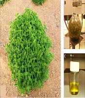
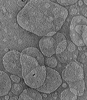
Ch-AO-NPs exhibited particle sizes ranging from 78.6 ± 5.2 nm to 2425 ± 402.3 nm across the formulations, with PDI values between 0.24 and 0.32, indicating narrow dispersity [Table D2 (Appendix D) in Supplementary File]. The optimised formulation (F10) produced the smallest and most uniform nanoparticles (90.75 ± 0.85 nm). Visual appearance correlated with DLS results: high oil loading (F8) produced milky, turbid suspensions, whereas lower oil loading (F3) yielded a more translucent dispersion. Insufficient stirring (F9) led to visible oil separation, while the optimised formulation displayed uniform opalescence with clear Tyndall scattering, characteristic of nanoscale systems (Figure 2). TEM imaging of the optimised formulation (Figure 3 and Appendix A, Figure A1 in Supplementary File) confirmed spherical, well-defined particles with dense cores, consistent with oil-loaded CS matrices. The morphology corroborates DLS measurements, validating the successful preparation of nanoscale, uniformly dispersed Ch-AO-NPs suitable for subsequent application testing.
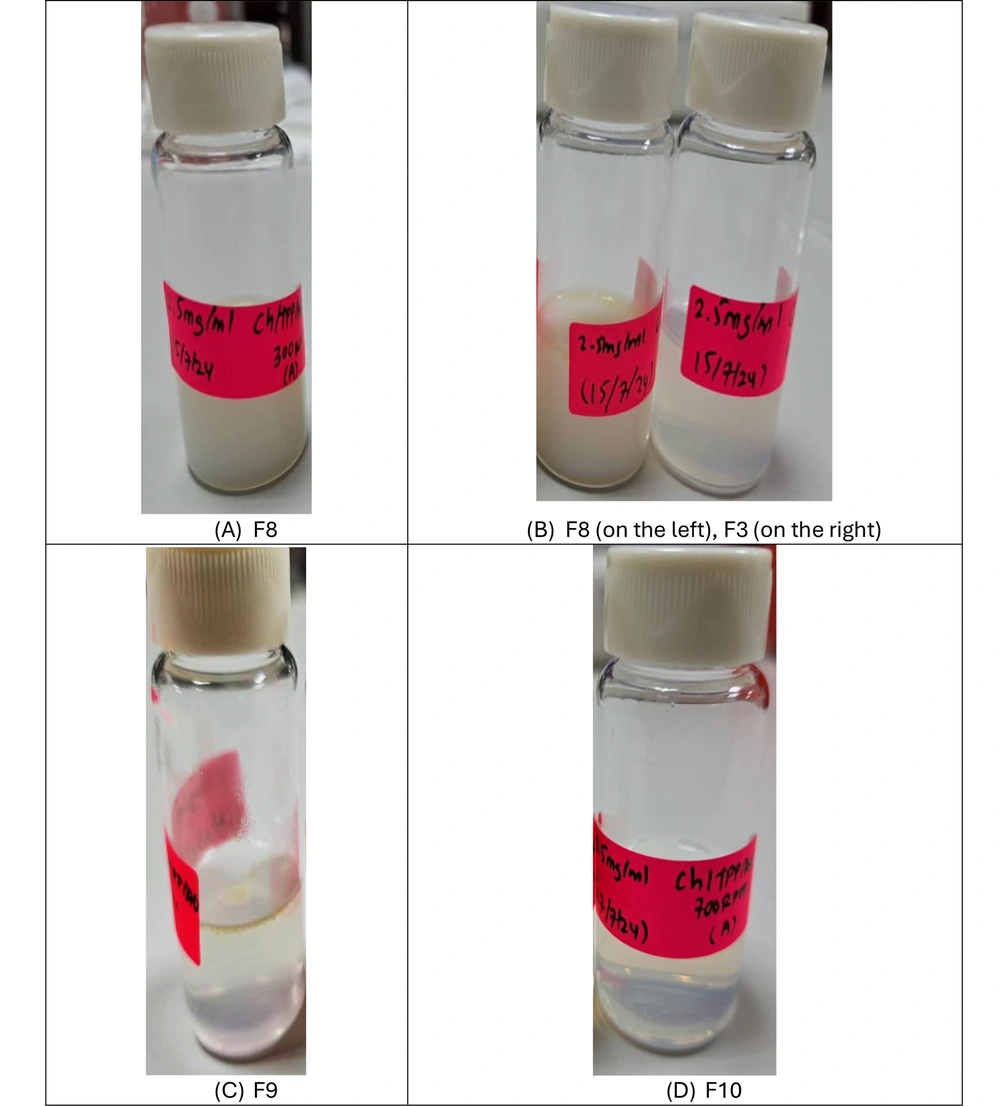
Figure 2.
Visual appearance of selected Ch-AO-NP formulations. (A) F8, (B) F3 (right) and F8 (left), (C) F9, and (D) F10
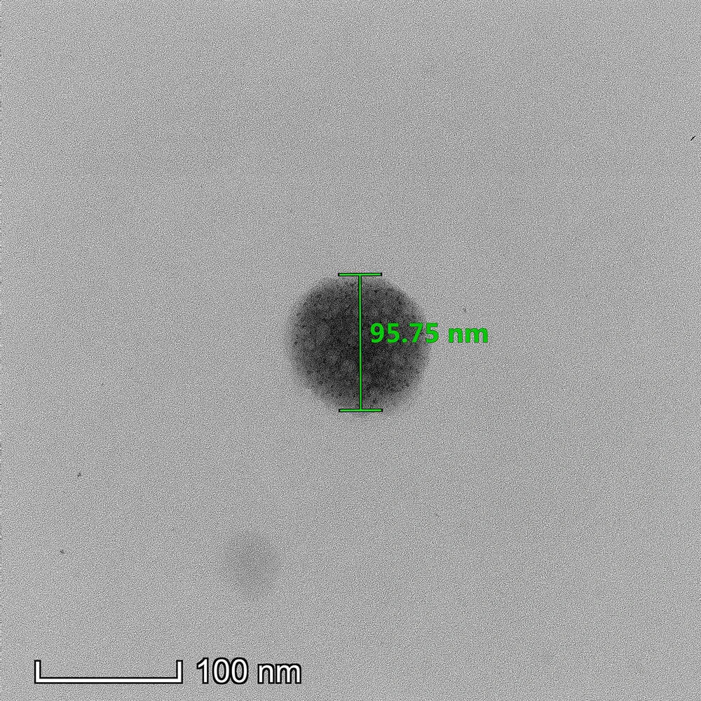
Figure 3.
TEM micrograph of optimised Ch-AO-NPs (F10/Z3). Scale bar = 100 nm.
3.2. Effect of Formulation Parameters on Particle Size
The particle size of Ch-AO-NPs was strongly influenced by formulation parameters, particularly the CS:TPP ratio, Tween 80 concentration, CS:AO ratio, and stirring speed. Formulations F1 - F4 (2:1 - 6:1) showed a biphasic trend, reaching a minimum size of 156.86 ± 6.76 nm at 5:1 (F3) before increasing at 6:1. Consistent with Liu and Ho (8), the 5:1 ratio produced the smallest particles, a behaviour attributed to a favourable charge balance that promotes efficient ionic bridging. Although stoichiometry was not determined experimentally, literature suggests that such ratios may facilitate compact crosslinking through multi-point or network-like interactions. At higher CS:TPP ratios, increased repulsion among protonated -NH₃⁺ groups reduces effective bridging, whereas excess TPP at lower ratios (e.g., 2:1) leads to over-crosslinking and aggregation (11). The 5:1 ratio, therefore, offers a favourable balance for forming stable, uniform nanoparticles, consistent with reports that charge ratio governs nanoparticle formation in related polyelectrolyte systems (12). Similarly, surfactant concentrations played a vital role in stabilising the system. Tween 80 concentrations (0.56 - 0.78% w/v; F3, F5, F6) were higher than the 0.001 - 0.1% typically used for polymeric nanoparticles (13), as essential oil systems require stronger emulsification due to their hydrophobicity and volatility. Similar use of 0.67% Tween 80 for peppermint oil encapsulation yielded ~500 nm particles (14), with size differences likely arising from formulation and volume effects. In this study, Tween 80 reduced interfacial tension and provided steric stabilisation, producing 150 - 250 nm particles and preventing aggregation. Its non-ionic nature ensures good biocompatibility, and concentrations up to 1% have shown no cytotoxicity or genotoxicity in human embryonic kidney (HEK-293) cells (15). Nonetheless, surfactant content must be balanced, as excessive levels may affect cell viability. In addition, the CS-to-oil ratio also influenced particle growth. Increasing AO content enlarged particle size from 156.86 ± 6.76 nm at 1:0.01 (F3) to 414.13 ± 99.36 nm at 1:0.03 (F7) and 1135.37 ± 236.69 nm at 1:0.05 (F8). Soltanzadeh et al. (16) attributed similar trends to extract migration and aggregation, while Froiio et al. (17) noted swelling of polymeric matrices by essential oils. Excess AO likely causes droplet coalescence and CS matrix swelling, producing larger particles. Nanoparticle size was strongly dependent on stirring speed, though no significant benefit was gained by extending the mixing time from one to two hours. Insufficient shear at low agitation (300 rpm, F9) created oversized particles (> 2 µm), whereas 700 rpm yielded significantly smaller and more uniform nanoparticles (F10 - F11). This size reduction is consistent with literature showing that increased agitation enhances TPP dispersion and accelerates ionic crosslinking. For instance, Al-nemrawi et al. (18) reported that efficient shear-mediated mixing improves CS-TPP interactions, leading to smaller, more narrowly distributed particles. Therefore, adequate mechanical energy is critical for preventing droplet coalescence and ensuring homogeneous network formation, though optimisation is essential to avoid using excessively high speeds that could induce aggregation by overcoming interparticle repulsion.
3.3. Optimised Formulation, Surface Charge, and Encapsulation Performance
As summarised in Table D3 (Appendix D in Supplementary File), the blank formulation (Z1) produced large particles (564.33 ± 90.99 nm) with a zeta potential of +27.37 ± 0.45 mV, reflecting strong electrostatic repulsion from protonated CS amines. Incorporation of Tween 80 (Z2) slightly reduced the surface charge (+24.87 ± 1.97 mV) while improving colloidal stability through steric hindrance (13). With AO loading, the optimised formulation (Z3/F10) yielded uniform nanosized particles (90.75 ± 0.85 nm) and a lower zeta potential (+17.59 ± 1.33 mV), consistent with partial shielding of surface amines by oil molecules (16). Although ±20 mV is a common stability benchmark, Tween 80 provided enough electrosteric support for short-term dispersion. Such reduced particle size is advantageous for textile-based antibacterial applications, as nanoscale CS carriers improve fibre adhesion and surface coverage, influence bioactive release behaviour, and enhance colloidal stability, as reported for encapsulated essential oil systems on textile substrates (19). In addition, essential-oil-loaded CS nanoparticles, including clove essential oil systems, have been shown to exhibit enhanced antibacterial activity, sustained release, and improved functional stability due to nanoscale encapsulation (20). The optimised nanoparticles also demonstrated high EE (80.23 ± 0.87%), LC (24.84 ± 1.85%), and acceptable yield (15.49 ± 0.66%), indicating effective retention of hydrophobic AO within the CS–TPP network while preserving nanoscale uniformity. Comparable efficiencies (> 70%) and LC values (2 - 23%) have been reported for other essential-oil nanocarriers, further confirming the suitability of this formulation strategy (3, 21). The observed decrease in surface charge following AO incorporation, together with the dense core regions identified in TEM images (Appendix A in Supplementary File), strongly suggests that AO is effectively associated with or embedded within the CS–TPP matrix rather than freely dispersed. This interpretation is further supported by similar electrostatic shielding and core-compaction effects reported in other essential-oil-loaded CS nanoparticle systems (3). These findings provide encouraging evidence of successful encapsulation, and future work can build on this by employing complementary techniques such as differential scanning calorimetry (DSC), solubility profiling, or microenvironment-sensitive spectroscopy to confirm AO localisation within the polymer network. Comparable CS-based encapsulation strategies have been reported for other essential oils, particularly in textile-related studies. For example, lime oil-loaded CS carriers prepared via emulsification followed by TPP-induced ionic interaction have been applied to CO fabrics, where stable oil-in-water emulsions were generated using high-speed homogenisation (≈13,000 rpm), yielding nanoscale particles typically in the 170–250 nm range (19). While the formulation sequence is broadly similar, the present Ch-AO-NPs were produced using a fully aqueous, low-energy ionic gelation approach, resulting in a smaller and more uniform particle size (~90 nm) and a compact internal morphology, as observed by TEM. In addition, unlike most previous studies that focus primarily on cellulosic substrates, the current work demonstrates nanoparticle deposition on both CO and PO fibres, as confirmed by SEM analysis.
3.4. Short-Term Stability of Ch-AO-NPs
Short-term stability results (Appendix C in Supplementary File) demonstrated minimal changes in particle size and PDI at both 4°C and 25°C over 7 days, confirming good colloidal stability under typical storage conditions. At ambient temperature (25°C), particle size increased by less than 15%, which aligns with expected short-term stability profiles for CS-TPP nanoparticle systems. This indicates that the textile formulation can maintain its integrity at room temperature, supporting its practical applicability in standard environments. In contrast, samples stored at 40 °C showed marked increases in size and PDI, highlighting sensitivity to elevated heat. Overall, these findings suggest that Ch-AO-NPs are well suited for use and storage at low and ambient temperatures, while heat stability remains an area for further optimisation. Full datasets are provided in Appendix C in Supplementary File.
3.5. In vitro Drug Release Profile
AO release was evaluated at pH 4.8 and 8.5, corresponding to the acidic pH of healthy skin and the alkaline shifts commonly reported in acute and chronic wound environments (22). Release profiles at both pH values exhibited a biphasic pattern, characterised by an initial burst release followed by a slower sustained phase. Faster release occurred at pH 4.8 due to increased protonation of CS, which promotes matrix swelling and accelerates diffusion (23), whereas reduced protonation at pH 8.5 limits swelling and slows the release of encapsulated oil. By 30 h, cumulative release reached approximately 85 - 90% at pH 4.8 compared with ~60% at pH 8.5, with statistically significant differences observed at multiple time points (4, 5, 6, and 30 h). This demonstrates a clear pH-dependent release behaviour, supporting the applicability of Ch-AO-NPs to wounds with variable physiological conditions. Complete release curves are provided in Appendix B in Supplementary File.
3.6. Textile Compatibility of Optimised Ch-AO-NPs
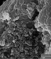
The optimised Ch-AO-NPs (F10/Z3) were evaluated using a mild bath-exhaustion method. Cotton and PO showed wet pickup values of 87.66 ± 4.78% and 81.06 ± 8.77%, and o.w.f. values of 5.10 ± 0.41% and 6.76 ± 0.57%, respectively. Cotton’s higher wet pickup reflects its hydrophilic cellulose structure, which contains free hydroxyl groups capable of forming electrostatic and hydrogen-bond interactions with CS (6). In contrast, polyester’s greater o.w.f. corresponds to its hydrophobic surface, which lacks reactive hydroxyl groups and therefore retains the AO-rich nanoparticles mainly through hydrophobic and van der Waals interactions, resulting in higher overall fibre loading (6). Citric acid further enhanced nanoparticle deposition, particularly on polyester, aligning with reports that citric acid improves CS attachment on both CO and PO by increasing reactive or adsorptive sites (6). These observations were further supported by SEM imaging (Figure 4), which showed visible surface deposition and morphological changes on treated CO and PO compared with their untreated counterparts, confirming successful nanoparticle adherence at the fibre level. These observations were further supported by SEM imaging (Figure 4), which showed visible surface deposition and morphological changes on treated CO and PO compared with their untreated counterparts, confirming successful nanoparticle adherence at the fibre level. Overall, these findings suggest that Ch-AO-NPs are able to deposit onto both textile types under gentle processing conditions.
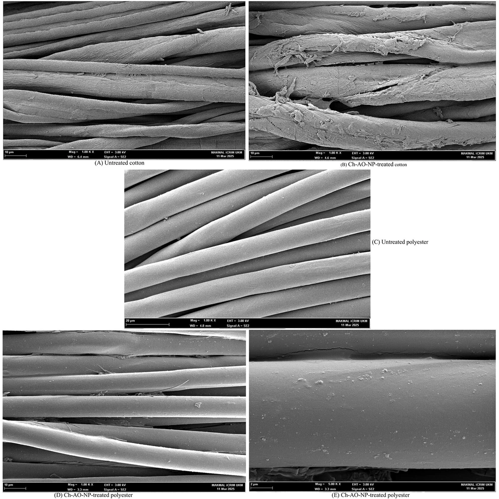
Figure 4.
SEM images of A, untreated cotton (CO); B, Ch-AO-NP-treated CO; C, untreated polyester (PO); and D-E, Ch-AO-NP-treated PO. Surface features observed on treated fibres suggest nanoparticle deposition, in agreement with o.w.f. measurements.
3.7. Limitations and Future Works
This study provides a strong foundation for future research by identifying key areas for enhancement. While functional performance was not confirmed due to the absence of antimicrobial, cytotoxicity, and anti-inflammatory assays, these tests present valuable opportunities for subsequent investigations. Textile durability was assessed at a preliminary level, suggesting that more comprehensive evaluations could further strengthen the findings. Stability was monitored for 7 days, offering initial insights; extending electrosteric stability studies to ≥ 30 days would provide a more robust understanding. The use of OFAT effectively clarified variable effects, and future optimisation can build on this by adopting a design of experiments (DoE) approach for greater efficiency. Finally, zeta potential and TEM offered indirect evidence of AO localisation, which can be complemented by advanced techniques such as DSC or solubility profiling to confirm these observations.
3.8. Conclusions
Ch-AO-NPs were successfully fabricated using an emulsion-assisted ionic gelation method, and formulation parameters enabled controlled tuning of nanoparticle size and structure. The optimised system demonstrated physicochemical traits consistent with effective AO encapsulation and stable nanoscale assembly. While biological assays and in situ textile evaluations were not performed in this study, the nanoparticles demonstrated physicochemical properties that suggest strong potential for dermal and textile applications. These applications represent promising directions for future research to validate and expand upon the current findings. The absence of long-term stability data and functional testing represents a key limitation that will guide subsequent investigations. Overall, the findings establish a promising platform for further work in dermal delivery and biofunctional textile research.