
1. Background
The efficient delivery of genetic material into cells is still a challenge in biotechnology, gene therapy, and nanomedicine, often determining the success of a therapeutic intervention (1, 2). While viral vectors represent an advanced solution, persistent concerns over their safety profile, potential for genomic integration, and immunogenicity have motivated the pursuit of sophisticated non-viral alternatives (3-5). Among these, cell-penetrating peptides (CPPs) have gained prominence as versatile and engineerable vehicles for transporting diverse cargoes including nucleic acids and proteins across cellular membranes (6, 7). Foundational CPPs like the TAT peptide from human immunodeficiency virus demonstrate powerful membrane-translocating ability (8). However, effective gene delivery is a multi-stage process requiring cellular entry; it also needs endosomal escape and nuclear import. Relying on a single functional motif is typically insufficient to overcome these sequential barriers and thereby necessitates a rational, multi-domain design strategy (9).
A significant limitation of many current CPP designs is their inability to efficiently navigate the entire delivery pathway from DNA complexation to nuclear delivery (10, 11). This reveals a critical research gap: The need for integrated, modular systems that combine specialized functions. The strategic combination of distinct functional domains such as those for DNA binding, membrane translocation, endosomal escape, and nuclear targeting connected by flexible linkers represents a promising approach to create cohesive and efficient delivery vehicles (12). In such chimeric peptides, each functional module serves a distinct purpose: DNA-binding domains (e.g., from H1 histone) ensure stable complex formation with genetic cargo (13); membrane-translocating sequences (e.g., TAT) mediate cellular entry (14); endosomal escape motifs (e.g., gp41FP or H5WYG) facilitate endosomal release (15); and nuclear localization signals (NLS) (e.g., SV40 NLS) enable nuclear entry (16). The overall activity of the peptide is governed by the selection of these motifs and their precise spatial arrangement, as the order of domains can profoundly influence the efficiency of each step, including nuclear accumulation (17).
2. Objectives
Building on the modular design concept, we engineered a multifunctional peptide capable of coordinating all key stages of gene delivery. The construct integrates a C-terminal H1 histone DNA-binding domain, membrane-translocating motifs, endosomal escape elements, linkers, and a NLS, each separated by flexible linkers to maintain functional independence. This architecture enables a viral-like delivery pathway in which the peptide condenses DNA, promotes cellular entry, supports endosomal escape, and directs nuclear targeting. The sequence was also computationally optimized for efficient bacterial expression, providing a practical platform for production and downstream functional evaluation.
3. Methods
3.1. Materials
Safe Stain, DNA ladder, and protein ladder (all from Sinaclon), as well as nickel-nitrilotriacetic acid (Ni-NTA) resin (ARG Biotech), were used. Additional chemicals including isopropyl-β-D-1-thiogalactopyranoside (IPTG), kanamycin, imidazole, and reagents for sodium dodecyl sulfate gel electrophoresis (SDS-PAGE), peptone, yeast extract, and agarose gel electrophoresis, along with various salts, were sourced from Merck (Germany), Sigma-Aldrich (USA), and reputable Iranian suppliers, and all of them were of analytical-grade chemicals.
3.2. Peptide Library Design
A rationally designed library of 30 chimeric peptides was generated to examine how different arrangements of functional motifs connected by flexible linkers influence gene delivery performance. Each construct combined key domains: A DNA-binding module consisting of two tandem 16-mer repeats from the C-terminal H1 histone, the classical TAT sequence for cellular entry, the endosomal escape motifs gp41FP and H5WYG, and an SV40-derived NLS. Flexible linkers (GGS, GGG, GG, G, WSQ, KPKP) were positioned to preserve independent folding and reduce steric interference (18). All peptides carried a C-terminal His₆-tag for purification and expression, and were designated as Peptide_X in Supplementary Table S1.
3.3. Sequence Assembly and Initial Quality Control
Peptide sequences were generated via manual design and automated Python 3.8 scripts within a combinatorial framework. Each sequence was checked for uniqueness to avoid duplicates and frameshift errors at the nucleotide level. Final peptide lengths and His₆-tag positions were validated to ensure compatibility with bacterial expression.
3.4. Computational Screening of the Peptide Library
The designed chimeric peptides underwent a comprehensive computational screening pipeline to evaluate key biophysical and physicochemical properties. Initial profiling was performed using the ExPASy ProtParam tool (19) to calculate fundamental parameters including GRAVY index, instability index, and charge distribution. Membrane interaction potential was assessed through hydrophobicity analysis using the Kyte & Doolittle scale via the Hydrophobicity Calculator, while solubility profiles and thermodynamic stability were evaluated using Protein-Sol (20) and ExPASy ProtScale. These analyses specifically examined peptide behavior under endosomal pH conditions. Three-dimensional structures were predicted using I-TASSER (21), with particular focus on secondary structure elements and solvent accessibility of DNA-binding residues. mRNA stability of coding sequences was analyzed through ViennaRNA Web Services. Finally, protein-DNA docking simulations were performed using HEX with parameters set to generate 100 docked structures from which the lowest-energy conformation was selected for analysis of binding interactions.
3.5. Recombinant Peptide Synthesis and Expression
The gene of Peptide_24 was synthesized by Shine Gene Co. (China). The synthesis included the insertion of NcoI and XhoI restriction sites prior to and following the gene sequence for digestion and ligation into the bacterial expression vector pET-28a(+) with a hexa His-tag sequence at the C-terminal. The recombinant plasmid was then introduced into competent Escherichia coli C41 (DE3) cells via heat shock and chemical transformation methods (22).
Following this, a single transformed colony was used to inoculate a 20 mL starter culture in Nutrient Broth supplemented with 50 µg/mL kanamycin, which was grown overnight at 37°C. This culture was then scaled up to 200 mL of fresh medium, and protein expression was induced with 0.1 mM IPTG at an OD₆₀₀ of ~0.8. Cells were harvested by centrifugation at 4°C, after three hours post-induction growth at 37°C, and the pellet was stored at -20°C for subsequent purification.
3.6. Peptide Purification and Preparation
Cell pellets were thawed and resuspended in denaturing lysis buffer (8 M urea, 50 mM Tris-HCl pH 8.0, 500 mM NaCl, 10 mM imidazole). The suspension was sonicated and centrifuged, and the resulting supernatant was loaded onto a Ni–NTA column pre-charged with nickel sulfate. The lysate was incubated with the resin for 60 minutes with gentle agitation to allow binding. The column was then subjected to an on-column refolding wash. This was performed with 10 mL of lysis buffer applied in 1 mL increments, with the urea concentration systematically reduced by 1 M in each subsequent wash step, from 8 M to 0 M. Following washing, the His₆-tagged peptides were eluted with elution buffer (50 mM Tris-HCl pH 8.0, 150 mM NaCl, 250 mM imidazole). Elution fractions were analyzed by SDS-PAGE to assess purity. Samples were denatured in buffer containing β-mercaptoethanol, heated to 95°C, and separated on 15% polyacrylamide gels, which were subsequently stained with Coomassie Brilliant Blue R-250. Fractions exhibiting a single predominant band at the expected molecular weight were pooled. The pooled sample was dialyzed three times for 3 hours each against dialysis buffer (50 mM Tris-HCl pH 8.0, 150 mM NaCl) at 4°C using a 3 kDa molecular weight cut-off membrane. After dialysis, the solution was centrifuged at 12,000 × g for 15 minutes at 4°C to pellet any insoluble aggregates. The concentration of the refolded peptide in the supernatant was determined by measuring the absorbance at 280 nm, using an extinction coefficient of 19,480 M⁻¹ cm⁻¹.
3.7. DNA-Binding Assay (Gel Retardation)
DNA-binding activity was evaluated by incubating 70 ng of pTZ57R/T plasmid with increasing amounts of peptide (5 – 50 µg). Complexes were resolved on a 0.8% agarose gel containing Safe-Stain and visualized under UV light to assess shifts in plasmid mobility indicative of peptide-DNA interaction.
4. Results
4.1. Physicochemical Profiling and Screening of Peptide Candidates
Physicochemical characterization using ProtParam, ProtScale, and solubility prediction tools confirmed that all 30 designed peptides exhibit favorable stability profiles (instability indices < 40), supporting their potential for biological applications (Table 1). Systematic screening identified distinct functional classes among the candidates. The peptides displayed a broad range of positive charges (21 – 43 residues) and predominantly hydrophilic character (GRAVY values: –0.368 to –1.328), essential for electrostatic interactions with nucleic acids. All candidates exceeded the minimum solubility threshold (> 0.45), with values ranging from 0.647 to 0.795, ensuring adequate solubility for delivery applications. Predicted half-lives were uniformly favorable across biological systems, indicating robust metabolic stability.
Table 1.
Physicochemical Properties and Multi-parameter Selection of 30 Peptide Candidates
| Peptide_# | Aliphatic Index | Instability Index | GRAVY | Half-Life (h) (Mammalian Reticulocytes, in vitro), (Yeast, in vivo), (E. Coli, in vivo) | Number of Positively Charged Residues | Number of Amino Acids | Molecular Weight (kDa) | pI | Predicted Scaled Solubility (> 0.45) | Hydrophobicity Scale |
|---|---|---|---|---|---|---|---|---|---|---|
| Peptide_1 | 51.04 | 21.57 | –0.783 | 30, > 20, > 10 | 21 | 96 | 10.33 | 10.96 | 0.718 | –0.74 |
| Peptide_2 | 55.78 | 19.04 | –0.498 | 30, > 20, > 10 | 21 | 116 | 12.25 | 10.96 | 0.694 | –0.42 |
| Peptide_3 | 66.72 | 22.54 | –0.368 | 30, > 20, > 10 | 21 | 116 | 12.39 | 10.96 | 0.647 | –0.29 |
| Peptide_4 | 51.28 | 30.08 | –1.083 | 30, > 20, > 10 | 35 | 109 | 12.13 | 12.17 | 0.733 | –1.10 |
| Peptide_5 | 42.61 | 32.11 | –1.277 | 30, > 20, > 10 | 35 | 115 | 13.04 | 11.96 | 0.721 | –1.28 |
| Peptide_6 | 42.61 | 33.18 | –1.277 | 30, > 20, > 10 | 35 | 115 | 13.04 | 11.96 | 0.721 | –1.27 |
| Peptide_7 | 45.66 | 25.17 | –0.897 | 30, > 20, > 10 | 35 | 129 | 13.92 | 12.17 | 0.731 | –0.89 |
| Peptide_8 | 53.24 | 36.6 | –0.840 | 30, > 20, > 10 | 29 | 105 | 11.24 | 12.16 | 0.738 | –0.84 |
| Peptide_9 | 59.47 | 11.99 | –0.713 | 30, > 20, > 10 | 25 | 94 | 10.14 | 11.29 | 0.736 | –0.70 |
| Peptide_10 | 53.24 | 36.6 | –0.840 | 30, > 20, > 10 | 29 | 105 | 11.24 | 12.16 | 0.694 | –0.84 |
| Peptide_11 | 51.28 | 30.08 | –1.083 | 30, > 20, > 10 | 35 | 109 | 12.13 | 12.17 | 0.695 | –1.10 |
| Peptide_12 | 45.66 | 25.17 | –0.897 | 30, > 20, > 10 | 35 | 129 | 13.92 | 12.17 | 0.738 | –0.89 |
| Peptide_13 | 55.50 | 26.48 | –0.780 | 30, > 20, > 10 | 35 | 129 | 14.06 | 12.17 | 0.738 | –0.76 |
| Peptide_14 | 55.50 | 28.32 | –0.780 | 30, > 20, > 10 | 35 | 129 | 14.06 | 12.17 | 0.738 | –0.76 |
| Peptide_15 | 50.52 | 19.46 | –0.782 | 30, > 20, > 10 | 21 | 97 | 10.43 | 10.96 | 0.721 | –0.74 |
| Peptide_16 | 49.00 | 15.41 | –0.959 | 30, > 20, > 10 | 25 | 100 | 11.05 | 11.04 | 0.731 | –0.93 |
| Peptide_17 | 61.70 | 19.48 | –0.808 | 30, > 20, > 10 | 25 | 100 | 11.18 | 11.04 | 0.730 | –0.77 |
| Peptide_18 | 42.61 | 32.11 | –1.277 | 30, > 20, > 10 | 35 | 115 | 13.04 | 11.96 | 0.730 | –1.28 |
| Peptide_19 | 45.29 | 36.36 | –1.328 | 30, > 20, > 10 | 35 | 121 | 14.08 | 11.75 | 0.695 | –1.29 |
| Peptide_20 | 50.00 | 32.18 | –1.016 | 30, > 20, > 10 | 35 | 141 | 16.01 | 11.75 | 0.695 | –0.96 |
| Peptide_21 | 47.93 | 29.29 | –0.959 | 30, > 20, > 10 | 35 | 135 | 14.96 | 11.96 | 0.795 | –0.93 |
| Peptide_22 | 42.61 | 33.18 | –1.277 | 30, > 20, > 10 | 35 | 115 | 13.04 | 11.96 | 0.795 | –1.27 |
| Peptide_23 | 44.14 | 39.46 | –1.055 | 30, > 20, > 10 | 29 | 111 | 12.14 | 11.94 | 0.795 | –1.03 |
| Peptide_24 | 43.75 | 39.7 | –1.049 | 30, > 20, > 10 | 29 | 112 | 12.20 | 11.94 | 0.730 | –1.02 |
| Peptide_25 | 43.75 | 37.41 | –1.060 | 30, > 20, > 10 | 29 | 112 | 12.29 | 11.94 | 0.795 | –1.03 |
| Peptide_26 | 43.75 | 38.67 | –1.060 | 30, > 20, > 10 | 29 | 112 | 12.29 | 11.94 | 0.795 | –1.03 |
| Peptide_27 | 39.85 | 16.9 | –1.098 | 30, > 20, > 10 | 37 | 133 | 14.25 | 11.21 | 0.795 | –1.08 |
| Peptide_28 | 39.85 | 17.96 | –1.098 | 30, > 20, > 10 | 37 | 133 | 14.25 | 11.21 | 0.795 | –1.08 |
| Peptide_29 | 38.97 | 15.19 | –1.282 | 30, > 20, > 10 | 43 | 136 | 15.00 | 11.28 | 0.721 | –1.27 |
| Peptide_30 | 38.97 | 17.65 | –1.282 | 30, > 20, > 10 | 43 | 136 | 15.00 | 11.28 | 0.721 | –1.27 |
Through multi-parameter optimization, seven peptides (Peptides 9, 15, 16, 21, 24, 27, and 28) were selected as lead candidates:
Peptide_9 exhibited the highest stability (Instability Index: 11.99), optimal hydrophilicity (GRAVY: -0.736), moderate positive charge (25 residues), and a compact structure (94 aa, 10.14 kDa), suggesting efficient membrane translocation and structural integrity.
Peptide_15 combined high stability (Instability Index: 19.46), moderate size (97 aa, 10.43 kDa), moderate positive charge (21 residues), and favorable physicochemical properties (GRAVY: –0.782), advantageous for crossing biological barriers.
Peptide_16 showed excellent stability (Instability Index: 15.41), balanced hydrophobicity (GRAVY: –0.959), moderate positive charge (25 residues), and moderate size (100 aa, 11.05 kDa), favoring cellular uptake without aggregation.
Peptide_21 (135 aa, 14.9 kDa) demonstrated the highest solubility (0.795), substantial positive charge (35 residues), and acceptable stability (Instability Index: 29.29), supporting strong DNA-binding in aqueous formulations.
Peptide_24, despite a higher instability index (39.70), was included due to its exceptional solubility (0.730), moderate charge (29 residues), balanced hydrophobicity (GRAVY: –1.049), and optimal size (112 aa, 12.20 kDa), making it suitable for formulations requiring high solubility.
Peptide_27 (133 aa, 14.3 kDa) was chosen for enhanced DNA-binding capacity, with 37 positively charged residues, good stability (Instability Index: 16.90), and balanced hydrophilicity (GRAVY: –1.098).
Peptide_28 (133 aa, 14.3 kDa) mirrored Peptide_27’s charge distribution (37 positive residues) and stability (Instability Index: 17.96), serving as a structural comparator.
Selection criteria emphasized balanced functional attributes: Positive charge density (25 – 37 residues), hydrophobicity within the CPP-preferred range (GRAVY: –0.7 to –1.1), molecular weight (10 – 15 kDa), structural stability (instability index < 30, with exceptions for compensatory traits), and high solubility (> 0.69). Peptides with extreme hydrophilicity (GRAVY < –1.25) were deprioritized due to potential membrane penetration limitations. The aliphatic indices (38.97 – 66.72) and pI values (10.96 – 12.17) of selected candidates further supported their thermostability and cationic nature, essential for DNA binding and cellular interactions. This multi-parameter strategy ensured the selection of candidates with the highest potential for success in downstream functional assays.
4.2. Structural and Biophysical Profiling of Lead Candidates
Hydrophobicity analysis using Kyte-Doolittle profiling revealed distinct amphipathic patterns across the seven selected peptides, all exhibiting the alternating hydrophobic-hydrophilic domains characteristic of effective CPPs (Table 1 and Supplementary Table S2). Three candidates emerged with particularly favorable profiles:
Peptide_9 demonstrated optimal structural characteristics with harmonized amphipathicity, featuring moderate hydrophobic peaks (+2.0 to +2.5) in terminal regions balanced by extensive hydrophilic segments. This pattern, combined with its exceptional stability (Instability Index: 11.99) and compact architecture (94 aa), suggests ideal properties for membrane translocation while maintaining structural integrity.
Peptide_27 displayed stronger hydrophobic character (+2.5 to +3.0 peaks) coupled with high positive charge density (37 aa), indicating enhanced DNA-binding capacity, though with potential aggregation concerns that would require formulation optimization.
Peptide_24 showed exceptionally balanced hydropathy with moderate hydrophobic domains (+1.5 to +2.0) distributed throughout its sequence, complemented by outstanding solubility (0.730) and optimal molecular size (112 aa). This profile suggests low aggregation propensity while retaining sufficient hydrophobicity for membrane interaction, making it particularly suitable for stable formulation development.
Comparative analysis identified specific limitations among the remaining candidates. Peptides_15 and _16 were characterized by predominant hydrophilicity, which may reduce their membrane penetration capability. Peptide_21 was found to exhibit a complex hydropathy profile with partially exposed hydrophilic surfaces. Despite its moderate-to-large size (135 aa, ~14.9 kDa), previous studies have demonstrated that CPPs of comparable dimensions can effectively traverse cellular membranes when amphipathicity and charge are optimally distributed (23, 24). Accordingly, peptide size was considered a minor, non-limiting factor for cellular uptake. Structural modeling using I-TASSER confirmed that Peptide_9 adopts a dual α-helical architecture conducive to both membrane interaction and DNA binding. In contrast, Peptide_24 was shown to exhibit balanced amphipathicity and favorable solvent accessibility, albeit with slightly reduced helicity.
Based on integrated analysis of hydrophobicity, structural stability, charge distribution, and molecular architecture, three candidates were prioritized for functional characterization:
Peptide_9 as the lead candidate for optimal balanced properties.
Peptide_27 for high-capacity DNA binding applications.
Peptide_24 for formulations requiring exceptional stability and solubility.
Further energy and charge profile analyses of Peptide_9 and Peptide_24 were performed using the Protein-Sol server. It was revealed that Peptide_24 exhibits a higher potential for effective endosomal escape and enhanced gene delivery (Supplementary Figure S1). Endosomal escape peptides are typically characterized by pH-sensitive behavior, whereby structural stability and limited membrane interaction are maintained under neutral pH conditions, such as those found in extracellular environments. In contrast, significant conformational changes are induced under acidic pH conditions representative of the endosomal compartment (approximately pH 5.0 - 6.5). Energy profile analysis, expressed as joules per amino acid, demonstrated that within the acidic pH range (pH 5.0 - 6.0) and across various ionic strengths, Peptide_24 undergoes a markedly greater increase in energy compared to Peptide_9. This increase was indicative of enhanced structural instability, a stronger propensity for conformational rearrangement, and increased membrane interaction. Such properties were shown to facilitate the transition of the peptide into an active amphipathic helical conformation, a critical feature for endosomal membrane disruption and efficient cargo release.
Additionally, charge profile analysis further supports Peptide_24's superior performance. At acidic pH, Peptide_24 maintains a slightly higher net positive charge compared to Peptide_9. This higher charge density is essential for forming stable complexes with negatively charged genetic cargo as well as for stronger electrostatic interactions with endosomal lipid membranes. Furthermore, this higher charge enhances the peptide’s ability to engage in mechanisms such as the "proton sponge" effect, which aids in endosomal escape (25, 26). The results from both energy and charge profiles strongly position Peptide_24 as the candidate for efficient endosomal escape and successful gene delivery.
4.3. Evaluation of mRNA Expression for Peptide_24
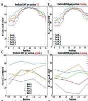
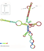
The mRNA for Peptide_24 [318 nt, 106 (+ 6 His-tag) aa] was analyzed for its thermodynamic and translational characteristics (Figures 1 and 2). Its composition is 102 A, 59 C, 91 G, and 68 U, yielding a 46.5% G + C content, providing a good balance for stability and efficient expression. The secondary structure shows a stable monomeric fold (MFE = –75.9 kcal/mol) to protect the mRNA from degradation and extend its lifespan. Analysis of dimer formation (ΔGmonomer ≈ –76 vs. ΔGdimer ≈ –30 kcal/mol) confirmed that the single-stranded form is thermodynamically favored, reducing the risk of molecule aggregation. A localized constraint was identified near the start codon: The AUG codon is partially base-paired within a stem-loop, which slightly hinders ribosome access. However, analysis of structural ensembles showed the molecule is flexible (MFE frequency = 0.01%, diversity = 80.04), meaning it constantly shifts between different shapes. This dynamic behavior allows the AUG codon to be temporarily exposed for translation initiation as seen in the expression profile (Figure 4A).

Figure 1.
Structural and functional dynamics of the Peptide-24 mRNA; the minimum free energy illustrates a stable monomeric fold (MFE = -75.9 kcal/mol), contributing to mRNA longevity. A key structural constraint is observed near the start codon (AUG) (highlighted in red), where partial base-pairing within a stem-loop moderately limits ribosome accessibility.

Figure 2.
Structural and functional analysis of the Peptide-24 mRNA reveals secondary-structure features that contribute to its stability. The centroid secondary structure shows that a notable structural constraint forms near the start codon (AUG) (highlighted in red), where partial base-pairing within a stem–loop moderately reduces ribosomal accessibility and may influence translation initiation.

Figure 3.
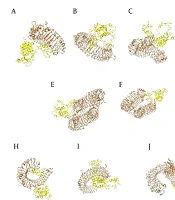
HEX docking predicted poses 1 and 2 of Peptide_24 with a double-stranded DNA fragment (extracted from PDB: 1AIO) and detailed view of the Peptide_24 sequence and motifs.
This mild, inherent limitation on translation initiation aligns with biosafety objectives. While Peptide_24's cationic density is crucial for interacting with its cargo, it also carries a risk of cytotoxicity through non-specific binding to cell membranes. Therefore, a moderated initiation rate helps prevent excessive protein production and subsequent cellular stress, acting as a built-in safety mechanism. This controlled expression level is intentionally maintained in our design to provide a safety margin during initial validation tests.
4.4. Docking Analysis of Peptide-DNA Interactions
To assess the DNA-binding potential of the designed peptides, molecular docking was performed using HEX. HEX is a fast rigid-body docking program that employs spherical polar Fourier correlations to evaluate both shape complementarity and electrostatic interactions between macromolecules. This algorithm allows efficient sampling of rotational and translational degrees of freedom, making it suitable for modeling peptide–nucleic acid interactions. Prior to docking, a representative double-strand DNA fragment was extracted from PDB entry 1AIO, and its structure was energy-minimized to remove steric clashes and optimize backbone geometry.
Docking of Peptide_24 against the optimized DNA fragment revealed two energetically favorable binding modes, with total docking energies (E_total) of −574.3 (Pose 1) and −545.0 (Pose 2), respectively (Figure 3 and Supplementary Figure S2). These two docking poses correspond to distinct DNA-interaction regions, both localized in segments of the peptide enriched in positively charged residues (Lys and Arg). Structural inspection of the complexes indicated that these cationic residues are oriented toward the negatively charged phosphate backbone of DNA, resulting in strong hydrogen and electrostatic attractions and good spatial complementarity.
![Biochemical characterization of purified peptide and its DNA-binding activity; A, sodium dodecyl sulfate gel electrophoresis (SDS-PAGE) analysis of purified peptide; samples were heat-denatured and separated on a 15% polyacrylamide gel. Lanes (left to right): Elution 1-3, protein; B, gel retardation assay evaluating peptide-DNA complex formation with increasing peptide concentrations [Lane 1: Plasmid (P; 70 ng); MWM: 1Kb DNA molecular weight marker; 2: P+ 5 μg peptide; 3: P+ 10 μg peptide; 4: P+ 15 μg peptide; 5: P+ 20 μg peptide; 6: P+ 30 μg peptide; 7: P+ 40 μg peptide; 8: P+ 50 μg peptide]; progressive retardation of plasmid migration indicates effective peptide-DNA complex formation, confirming the DNA-binding capacity of the purified peptide.](https://brieflands.com/journals/jrps/articles/168266/figures/jrps-14-1-168266-g001-preview.webp "Biochemical characterization of purified peptide and its DNA-binding activity; A, sodium dodecyl sulfate gel electrophoresis (SDS-PAGE) analysis of purified peptide; samples were heat-denatured and separated on a 15% polyacrylamide gel. Lanes (left to right): Elution 1-3, protein; B, gel retardation assay evaluating peptide-DNA complex formation with increasing peptide concentrations [Lane 1: Plasmid (P; 70 ng); MWM: 1Kb DNA molecular weight marker; 2: P+ 5 μg peptide; 3: P+ 10 μg peptide; 4: P+ 15 μg peptide; 5: P+ 20 μg peptide; 6: P+ 30 μg peptide; 7: P+ 40 μg peptide; 8: P+ 50 μg peptide]; progressive retardation of plasmid migration indicates effective peptide-DNA complex formation, confirming the DNA-binding capacity of the purified peptide.")
Figure 4.
Biochemical characterization of purified peptide and its DNA-binding activity; A, sodium dodecyl sulfate gel electrophoresis (SDS-PAGE) analysis of purified peptide; samples were heat-denatured and separated on a 15% polyacrylamide gel. Lanes (left to right): Elution 1-3, protein; B, gel retardation assay evaluating peptide-DNA complex formation with increasing peptide concentrations [Lane 1: Plasmid (P; 70 ng); MWM: 1Kb DNA molecular weight marker; 2: P+ 5 μg peptide; 3: P+ 10 μg peptide; 4: P+ 15 μg peptide; 5: P+ 20 μg peptide; 6: P+ 30 μg peptide; 7: P+ 40 μg peptide; 8: P+ 50 μg peptide]; progressive retardation of plasmid migration indicates effective peptide-DNA complex formation, confirming the DNA-binding capacity of the purified peptide.
The pose with the lower energy score (−574.3) exhibited tighter packing and more extensive electrostatic interfaces, suggesting it represents the primary binding mode, whereas the second pose (−545.0) may reflect an alternative register of interaction. In both cases, the interaction pattern is consistent with non-sequence-specific DNA binding mediated by hydrogen and electrostatic interactions. This mechanism is commonly observed in cell-penetrating and DNA-condensing peptides. Thus, docking results support the ability of Peptide_24 to form stable peptide–DNA complexes through multiple positively charged regions, which is essential for efficient gene delivery.
4.5. Expression and Purification of the Peptide
The recombinant His₆-tagged peptide was successfully expressed and purified under denaturing conditions using Ni–NTA affinity chromatography. SDS–PAGE analysis revealed a distinct and predominant band at the expected molecular weight (12.2 kDa) in the elution fractions, indicating successful expression and purification of the target peptide (Figure 4A).
4.6. Assessment of DNA-Binding Activity
The gel retardation assay demonstrated the DNA-binding ability of the purified peptide. A progressive decrease in plasmid migration was observed with increasing peptide concentrations (5 − 50 µg). Indeed, this phenomenon supports effective complex formation between the cationic purified peptide and plasmid DNA. At higher peptide ratios, complete retardation of plasmid movement was evident, indicating the strong DNA-binding affinity and functional bioactivity of the purified peptide (Figure 4B).
5. Discussion
Our study demonstrates the power of rational and modular design in surmounting the persistent challenge of efficient gene delivery. Unlike conventional carriers that often falter at critical intracellular hurdles, our lead candidate Peptide_24 functions as a versatile molecular guide, seamlessly coordinating the entire journey (27). A cornerstone of its design is a pH-sensitive 'conditional activation' mechanism for endosomal escape. As suggested by the energy and net charge profiles obtained from Protein-Sol predictions (Supplementary Figure S1), Peptide_24 is predicted to remain structurally stable at physiological pH, while potentially undergoing a conformational shift under acidic endosomal conditions (pH 5.0 - 6.0). This pH-dependent behavior may enhance membrane interaction and could facilitate endosomal cargo release; however, these observations are based on in silico analyses and require experimental validation. This targeted activation directly circumvents a primary bottleneck in non-viral gene delivery — endosomal entrapment — that represents a significant leap beyond previous designs (27).
The principal novelty of our work lies not in designing new motifs, but in their strategic integration into a unified and synergistic architecture. We demonstrate that through careful spatial organization and flexible linkers, distinct functional modules — DNA-binding, cellular uptake, endosomal escape, and nuclear targeting — can be combined to operate in concert. The concept mimics a streamlined and viral-like delivery pathway. This multi-domain strategy, validated by our comprehensive physicochemical profiling and docking studies (Table 1, Figure 4 and Supplementary Figure S2), ensures efficient progression through each intracellular stage. A comparison of Peptide_24 with reference peptides like TAT and MPG-NLS reveals advantages in multiple aspects (18). Unlike TAT, which has weak endosomal escape efficiency, Peptide_24 incorporates a pH-sensitive motif that triggers stronger conformational changes under acidic endosomal conditions (pH 5.0–6.5), more effectively aiding cargo release. Moreover, while TAT lacks a specific NLS, Peptide_24 includes the SV40 NLS, ensuring active nuclear import. Similar to the MPG-based peptides reported by Majidi et al., Peptide_24 utilizes H1 histone motifs for DNA binding. Additionally, Peptide_24 boasts an optimized solubility profile (0.730) and a negative GRAVY index (–1.049), ensuring that it remains monomeric and active in physiological buffers, unlike the more hydrophobic MPG-based peptides that tend to aggregate. While MPG peptides typically rely on static membrane-interaction domains, Peptide_24 utilizes a pH-responsive mechanism where, as the pH drops from 7.4 to 5.0, the peptide’s energy increases (from 0.730 to 0.762 kcal/mol). This rise in energy, accompanied by a shift in the instability index from 42.06 to 43.14, represents a 'conditional instability' strategically engineered into the peptide. This programmed flexibility allows the molecular chassis to transition from a stable state during transport to an active, membrane-disrupting conformation within the acidic environment of the endosome, thereby promoting efficient cargo escape (18).
When contextualized within the landscape of recent advances, the distinct advantages of our platform become clear. Previous strategies have often focused on combining limited functions. For instance, the fusion of cell-penetrating and membrane-disruptive motifs in CM(18)-TAT(11) enhanced cytoplasmic delivery but operated through constitutive membrane disruption rather than a triggered mechanism (28). Similarly, while pioneering work on multidomain peptides (MDPs) has leveraged supramolecular self-assembly to boost uptake and endosomal escape (29, 30), these systems typically lack integrated nuclear targeting. Our Peptide_24 builds upon these concepts by incorporating four distinct functions into a single chimeric polypeptide chain with a pH-triggered escape mechanism to enable a more coordinated and potentially safer intracellular journey.
Further comparisons highlight our design's versatility. The CHAT peptide, though rationally designed for DNA delivery, incorporates a limited set of functional modules and notably lacks a NLS (31). In tissue engineering, the GET peptide achieved efficient transfection within scaffolds but was not designed as a standalone and adaptable platform for diverse cargoes (32). Moreover, while high-throughput screens of CPPs for small interfering RNA delivery have identified promising candidates, many remain single-function entities without modular pH-responsive architectures (33). In contrast, Peptide_24 embodies a tunable "molecular chassis," integrating DNA condensation, cellular uptake, pH-triggered endosomal escape, and nuclear import into one system, thereby offering a broader applicability from plasmid DNA to larger ribonucleoprotein complexes.
The rational design of advanced CPPs, such as the modular Peptide_24 developed in this study, is driven by the goal of clinical translation for nucleic acid and drug delivery. Although more than three decades of research have demonstrated the strong preclinical potential of CPPs for intracellular delivery across diverse therapeutic areas (34, 35), no standalone CPP-based therapeutic has yet received FDA or EMA approval (35), underscoring persistent translational challenges related to pharmacokinetics, specificity, immunogenicity, and therapeutic index in humans (35). Nevertheless, CPPs continue to be actively explored as enabling components in advanced delivery strategies, particularly for biologics requiring intracellular access, including cancer vaccines, adoptive cell therapies, and transport across biological barriers such as the blood-brain barrier (ClinicalTrials.gov ID NCT01493596) (34, 35).
In this context, the modular peptide platform described here should be viewed not as a final therapeutic entity, but as a foundational framework for the development of intelligent delivery systems. Its adaptable architecture allows future engineering, including the incorporation of tissue-specific targeting ligands or optimization for emerging cargoes such as clustered regularly interspaced short palindromic repeats–Cas9 ribonucleoprotein complexes. The encouraging results obtained from binding and functional assays (Figure 3), together with in silico and in vitro validation of DNA-binding and pH-responsive properties, support these prospective applications and position this platform as a gateway to next-generation CPP-based therapeutics.
It is acknowledged that the present findings are primarily based on computational predictions and in vitro validation. Therefore, comprehensive in vivo investigations will be required to rigorously assess peptide stability, biodistribution, immunogenicity, scalability of production, long-term safety, and therapeutic efficacy within complex physiological environments. Addressing these factors will be essential for bridging rational peptide design with clinical feasibility and for advancing CPP-based delivery systems from promising experimental tools toward realistic clinical translation. Future work will focus on rigorous molecular and cellular biology evaluation, including confocal microscopy to track intracellular trafficking, endosomal escape assays, and reporter gene expression studies to fully quantify the delivery performance of Peptide_24 and its optimized variants.
5.1. Conclusions
This study successfully demonstrates the power of rational and modular design in creating an efficient multi-functional peptide for gene delivery. Through systematic integration of specialized domains connected by flexible linkers, we have developed Peptide_24 as an optimized vector that coordinates the critical stages of gene delivery: DNA complexation, cellular internalization, pH-responsive endosomal escape, and nuclear targeting. The principal novelty of our approach lies in the strategic unification of known functional motifs into a synergistic, viral-inspired architecture that operates in concert, rather than in the discovery of new motifs. Our integrated approach combining computational design with experimental validation yielded a peptide with superior biophysical properties, confirmed DNA-binding capability, and a unique pH-triggered activation mechanism for enhanced endosomal escape. The modular architecture presented here represents more than just an effective gene delivery vehicle; it establishes a versatile "molecular chassis" that can be adapted for diverse therapeutic applications. While further investigation is needed to validate its performance in vivo, our findings provide a suitable foundation for the development of next-generation non-viral delivery systems. This work advances the field of synthetic gene delivery by demonstrating how rational integration of functional motifs can create coordinated, viral-inspired systems with enhanced efficacy and specificity.