
1. Background
Cutaneous wound repair is a coordinated process involving hemostasis, inflammation, cell proliferation and migration, extracellular matrix deposition, angiogenesis, re-epithelialization, and matrix remodeling (1, 2). Fibroblasts are particularly important during the proliferative and remodeling phases because they populate the wound bed and synthesize structural extracellular matrix proteins, including type I and type III collagen (1, 2). However, matrix deposition and degradation must remain balanced, as dysregulated remodeling can contribute to fibrosis and abnormal scar formation (2).
Cell-free regenerative approaches have received increasing attention because a substantial proportion of the effects attributed to therapeutic cells are mediated by secreted paracrine factors rather than durable engraftment (3-6). Conditioned medium (CM) is a practical representation of the cellular secretome and may contain growth factors, cytokines, extracellular vesicles, lipids, nucleic acids, and other soluble mediators (3-6). Studies of stem-cell- and fibroblast-derived conditioned media have reported effects on dermal fibroblast proliferation, migration, and extracellular-matrix-related gene expression, although CM composition and potency depend on cell source, donor characteristics, passage, culture conditions, conditioning duration, and downstream processing (3-6).
Free conditioned medium has practical limitations for local delivery because soluble factors may rapidly diffuse from, or be cleared from, the application site. Hydrogels are attractive delivery matrices because their hydrated polymer networks can retain water, conform to irregular tissue surfaces, resemble selected features of the extracellular matrix, and enable controlled release of incorporated bioactive molecules (7-10). Recent reviews also emphasize the growing use of hydrogel systems in three-dimensional cell culture and regenerative biomaterial design (7, 8, 10).
Conditioned-medium-loaded hydrogels have therefore been investigated as a strategy to combine secretome-associated biological activity with the physical advantages of a biomaterial carrier (9, 11). Related in vitro studies have shown that hydrogel-based constructs and secretome-rich preparations can influence fibroblast proliferation and migration (11, 12).
2. Objectives
The present study evaluated a gelatin–alginate hydrogel loaded with human dermal fibroblast-conditioned medium (HDF-CM). The objective was to characterize the material and determine whether delivery of HDF-CM via the hydrogel altered in vitro fibroblast metabolic activity, migration, and extracellular-matrix-related gene expression compared with the untreated control, hydrogel alone, or free HDF-CM. We hypothesized that the HDF-CM-loaded hydrogel would provide sustained protein release and elicit stronger fibroblast responses relevant to skin repair than free HDF-CM or hydrogel alone.
3. Methods
3.1. Study Design
This completed, controlled in vitro experimental study compared four treatment conditions: untreated human dermal fibroblasts (HDF) control, hydrogel alone, free HDF-CM, and HDF-CM-loaded hydrogel. Hydrogel physicochemical characterization and fibroblast functional and gene-expression outcomes were evaluated using independent experimental observations. Technical replicate measurements were averaged within each independent experiment and were not treated as independent biological observations. The overall experimental workflow is shown in Figure 1.
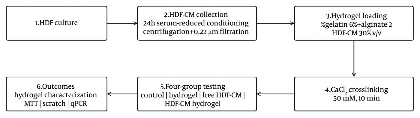
Figure 1.
Experimental workflow for preparation and evaluation of the HDF-CM-loaded gelatin–alginate hydrogel. The workflow used HDF-CM, a 30% v/v loading ratio, release sampling through day 7, and MTT as the metabolic activity assay. HDF, human dermal fibroblast; HDF-CM, human dermal fibroblast-conditioned medium; CM, conditioned medium; qPCR, quantitative polymerase chain reaction.
3.2. Human Dermal Fibroblast Source, Culture, and Ethics
Primary HDFs were expanded in Dulbecco modified Eagle medium supplemented with 10% fetal bovine serum and 1% penicillin–streptomycin at 37°C in a humidified 5% CO2 atmosphere.
3.3. Preparation and Quality Control of HDF-Conditioned Medium
At approximately 70 - 80% confluence, HDF cultures were washed twice with phosphate-buffered saline (PBS) and incubated in serum-reduced medium for 24 h. Conditioned medium was collected, centrifuged to remove cells and debris, passed through a 0.22-µm filter, aliquoted, and stored at −80°C until use. Each aliquot underwent a single freeze–thaw cycle. Total protein was quantified using the bicinchoninic acid assay.
3.4. Hydrogel Formulation and HDF-CM Loading
Gelatin (6% w/v) and sodium alginate (2% w/v) were dissolved separately in sterile aqueous buffer and combined under gentle stirring. After cooling to near-physiological temperature, HDF-CM was incorporated at 30% v/v. The mixture was cast into sterile molds and ionically crosslinked with 50 mM calcium chloride for 10 min. The hydrogels were then washed briefly with sterile buffer to remove unbound calcium and equilibrated before testing. Blank hydrogels were prepared identically without HDF-CM.
3.5. Hydrogel Characterization
Hydrogel morphology was assessed by scanning electron microscopy after freeze-drying. Swelling was calculated from wet and dry masses and expressed as [(Wwet − Wdry)/Wdry] × 100. In vitro mass loss was assessed by incubating preweighed hydrogel samples in PBS containing lysozyme at 37°C and determining the residual dry mass. HDF-CM protein release was evaluated by incubating loaded hydrogels in PBS at 37°C and measuring released protein by bicinchoninic acid assay at 24 h, day 3, and day 7. Cumulative release was expressed as a percentage of the initially loaded protein. No day-14 value was included in the analysis or figures.
3.6. Experimental Groups and Treatment Exposure
The four biological assay groups were untreated HDF control, blank gelatin–alginate hydrogel/hydrogel extract, free HDF-CM, and HDF-CM-loaded gelatin–alginate hydrogel. Free HDF-CM and hydrogel-delivered HDF-CM were compared under matched exposure conditions. Technical replicate wells were averaged within each independent experiment before statistical analysis (Table 1).
Table 1.
Experimental Groups Used for Biological Assays a
| Group | Treatment | Purpose |
|---|---|---|
| G1 | Untreated HDF control | Baseline cellular response |
| G2 | Blank gelatin–alginate hydrogel/hydrogel extract | Biomaterial control |
| G3 | Free HDF-CM | Effect of conditioned medium without carrier |
| G4 | HDF-CM-loaded gelatin–alginate hydrogel | Combined delivery system |
a Abbreviations: HDF, human dermal fibroblast; HDF-CM, human dermal fibroblast-conditioned medium; CM, conditioned medium.
3.7. Cell Metabolic Activity Assay
HDFs were seeded in 96-well plates and exposed to the four treatment conditions for 72 h. Metabolic activity was measured using the MTT assay. After incubation with MTT reagent, formazan was solubilized, and absorbance was recorded. Values were normalized to the untreated control.
3.8. Scratch-Wound Migration Assay
HDFs were grown to near confluence in six-well plates. A linear scratch was generated using a sterile pipette tip, detached cells were removed by gentle washing, and the assigned treatments were applied. Images were acquired at 0 and 24 h at predefined locations. Scratch closure was calculated as [(A0 − A24)/A0] × 100, where A0 and A24 represent the cell-free scratch area at 0 and 24 h, respectively (12). Image analysis was performed using the same measurement approach across all treatment groups.
3.9. Gene-Expression Analysis
Total RNA was extracted from treated HDFs and reverse-transcribed to cDNA. Quantitative polymerase chain reaction was performed for COL1A1, COL3A1, and MMP1, with GAPDH as the reference gene. Relative expression was calculated using the 2−ΔΔCt method after confirming acceptable amplification efficiency. Technical qPCR measurements were averaged for each independent observation before statistical analysis (Table 2).
Table 2.
qPCR Analysis Targets a
| Targets | Analysis Role | Reference Gene | Relative-expression Method |
|---|---|---|---|
| COL1A1 | Target gene | GAPDH | 2−ΔΔCt |
| COL3A1 | Target gene | GAPDH | 2−ΔΔCt |
| MMP1 | Target gene | GAPDH | 2−ΔΔCt |
| GAPDH | Reference gene | — | — |
a COL1A1, collagen type I alpha 1 chain; COL3A1, collagen type III alpha 1 chain; MMP1, matrix metalloproteinase 1; GAPDH, glyceraldehyde-3-phosphate dehydrogenase.
3.10. Statistical Analysis
Data are reported as mean ± standard deviation (SD) for independent observations (n = 3 per group for the biological endpoints reported in Appendix A in the Supplementary File). Technical replicate measurements were averaged before inferential testing. For each biological endpoint, residual normality and homogeneity of variance were explored using the Shapiro–Wilk and Levene tests, respectively. Because n = 3 per group provides limited power for assumption testing, these checks were interpreted cautiously, and raw observations are provided in Appendix A in the Supplementary File. Four-group comparisons were performed using one-way ANOVA, followed by Tukey’s honestly significant difference testing for multiplicity-adjusted pairwise comparisons. Effect size was summarized using eta-squared (η2), and Tukey 95% confidence intervals (CIs) were reported for prespecified comparisons of G4 versus G1, G2, and G3. A two-sided P < 0.05 was considered statistically significant.
4. Results
4.1. Hydrogel Physical Characteristics and HDF-CM Protein Release
The gelatin–alginate formulation formed a stable, hydrated hydrogel suitable for HDF-CM incorporation. The mean swelling ratio was 615 ± 42%, and the residual dry mass at day 7 was 71 ± 5%. Cumulative protein release reached 34 ± 3% at 24 h, 53 ± 4% at day 3, and 69 ± 4% at day 7, indicating an initial release phase followed by slower release through day 7 (Table 3).
Table 3.
Hydrogel Characterization and Cumulative HDF-CM Protein Release a
| Parameter | Result | Interpretation |
|---|---|---|
| Swelling ratio | 615 ± 42% | High water uptake |
| Residual mass, day 7 | 71 ± 5% | Moderate in vitro mass loss |
| Protein released, 24 h | 34 ± 3% | Initial release phase |
| Protein released, day 3 | 53 ± 4% | Intermediate release |
| Protein released, day 7 | 69 ± 4% | Sustained release through day 7 |
a Values are expressed as mean ± SD. Abbreviations: SD, standard deviation; HDF-CM, human dermal fibroblast-conditioned medium.
4.2. Fibroblast Metabolic Activity
Metabolic activity differed among groups (one-way ANOVA, F(3, 8) = 123.21, P < 0.001, η2 = 0.979). The HDF-CM-loaded hydrogel group showed the highest mean activity (118.7 ± 2.5% of control). Tukey-adjusted comparisons showed that G4 exceeded G1 by 23.7 percentage points (95% CI, 18.7 - 28.7; adjusted P < 0.001), G2 by 26.7 points (95% CI, 21.7 - 31.7; adjusted P < 0.001), and G3 by 11.0 points (95% CI, 6.0 - 16.0; adjusted P = 0.0005). The G1–G2 comparison was not significant (adjusted P = 0.293) (Table 4).
Table 4.
Fibroblast METABOLIC ACTIVITY at 72 H a
| Groups | Metabolic Activity (% of Control) |
|---|---|
| G1 Control | 95.0 ± 1.0 |
| G2 Hydrogel | 92.0 ± 1.0 |
| G3 Free HDF-CM | 107.7 ± 2.5 |
| G4 HDF-CM hydrogel | 118.7 ± 2.5 |
a Values are expressed as mean ± SD. Abbreviations: HDF-CM, human dermal fibroblast-conditioned medium; SD, standard deviation.
4.3. Fibroblast Migration
Scratch closure at 24 h differed among groups (F(3, 8) = 436.44, P < 0.001, η2 = 0.994). Mean closure was 77.3 ± 1.5% in G4, compared with 32.7 ± 1.5% in G1, 40.3 ± 1.5% in G2, and 59.0 ± 2.0% in G3. Tukey-adjusted mean differences for G4 were 44.7 percentage points versus G1 (95% CI, 40.3 - 49.0), 37.0 versus G2 (95% CI, 32.7 - 41.3), and 18.3 versus G3 (95% CI, 14.0 - 22.7); all adjusted P < 0.001 (Table 5).
Table 5.
Fibroblast Scratch-Wound Closure at 24 H a
| Groups | Scratch Closure at 24 h (%) |
|---|---|
| G1 Control | 32.7 ± 1.5 |
| G2 Hydrogel | 40.3 ± 1.5 |
| G3 Free HDF-CM | 59.0 ± 2.0 |
| G4 HDF-CM hydrogel | 77.3 ± 1.5 |
a Values are expressed as mean ± SD. Abbreviations: HDF-CM, human dermal fibroblast-conditioned medium; SD, standard deviation.
4.4. Extracellular-Matrix-Related Gene Expression
COL1A1 expression differed among groups (F(3, 8) = 713.46, P < 0.001, η2 = 0.996). Relative to G4, Tukey-adjusted mean differences were 1.22-fold versus G1 (95% CI, 1.12 - 1.31), 1.12-fold versus G2 (95% CI, 1.02 - 1.21), and 0.53-fold versus G3 (95% CI, 0.44 - 0.63); all adjusted P < 0.001. COL3A1 expression also differed among groups (F(3, 8) = 803.54, P < 0.001, η2 = 0.997), with G4 exceeding G1 by 1.08-fold (95% CI, 1.00 - 1.16), G2 by 1.01-fold (95% CI, 0.93 - 1.09), and G3 by 0.50-fold (95% CI, 0.42 - 0.58); all adjusted P < 0.001. MMP1 differed among groups (F(3, 8) = 210.71, P < 0.001, η2 = 0.988); G4 was lower than G1 by 0.40-fold (95% CI, −0.47 to −0.33), G2 by 0.50-fold (95% CI, −0.57 to −0.43), and G3 by 0.20-fold (95% CI, −0.27 to −0.13), with adjusted P ≤ 0.0001 for these comparisons (Table 6).
Table 6.
Relative Gene Expression After Treatment a
| Group | COL1A1 Fold Change | Col3a1 Fold Change | MMP1 Fold Change |
|---|---|---|---|
| G1 Control | 1.00 ± 0.05 | 1.00 ± 0.03 | 1.00 ± 0.04 |
| G2 Hydrogel | 1.10 ± 0.02 | 1.07 ± 0.02 | 1.10 ± 0.02 |
| G3 Free HDF-CM | 1.68 ± 0.04 | 1.59 ± 0.04 | 0.80 ± 0.02 |
| G4 HDF-CM hydrogel | 2.22 ± 0.04 | 2.08 ± 0.04 | 0.60 ± 0.02 |
a Values are expressed as mean ± SD. Abbreviations: COL1A1, collagen type I alpha 1 chain; COL3A1, collagen type III alpha 1 chain; MMP1, matrix metalloproteinase 1; HDF-CM, human dermal fibroblast-conditioned medium.
4.5. Assumption Checks and Adjusted Pairwise Comparisons
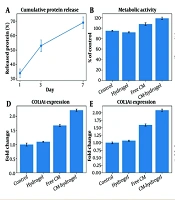
Shapiro–Wilk tests of ANOVA residuals did not indicate marked departures from normality (P = 0.281 - 0.844 across endpoints), and Levene tests did not indicate unequal variances (P = 0.552 - 0.971). Given only three independent observations per group, these tests have low power and were therefore considered supportive rather than definitive. Raw independent observations are provided in Appendix A of the Supplementary File. Prespecified pairwise comparisons are summarized in Table 7, and a graphical summary of hydrogel release and fibroblast outcomes is shown in Figure 2.
Table 7.
Prespecified Tukey-Adjusted Comparisons of HDF-CM-Loaded Hydrogel Versus Comparator Groups a
| Endpoints and G4 vs Comparator | Mean Difference | 95% CI | Tukey-adjusted P |
|---|---|---|---|
| Metabolic activity | |||
| G1 | +23.67 | 18.66 to 28.67 | < 0.001 |
| G2 | +26.67 | 21.66 to 31.67 | < 0.001 |
| G3 | +11.00 | 5.99 to 16.01 | 0.0005 |
| Scratch closure | |||
| G1 | +44.67 | 40.33 to 49.00 | < 0.001 |
| G2 | +37.00 | 32.66 to 41.34 | < 0.001 |
| G3 | +18.33 | 14.00 to 22.67 | < 0.001 |
| COL1A1 | |||
| G1 | +1.22 | 1.12 to 1.31 | < 0.001 |
| G2 | +1.12 | 1.02 to 1.21 | < 0.001 |
| G3 | +0.53 | 0.44 to 0.63 | < 0.001 |
| COL3A1 | |||
| G1 | +1.08 | 1.00 to 1.16 | < 0.001 |
| G2 | +1.01 | 0.93 to 1.09 | < 0.001 |
| G3 | +0.50 | 0.42 to 0.58 | < 0.001 |
| MMP1 | |||
| G1 | −0.40 | −0.47 to −0.33 | < 0.001 |
| G2 | −0.50 | −0.57 to −0.43 | < 0.001 |
| G3 | −0.20 | −0.27 to −0.13 | 0.0001 |
a Abbreviations: G1, untreated control; G2, blank hydrogel; G3, free HDF-CM; G4, HDF-CM-loaded hydrogel; CI, confidence interval; HDF-CM, human dermal fibroblast-conditioned medium. Positive differences indicate higher G4 values; negative differences indicate lower G4 values. Mean differences and 95% CIs are in percentage points for metabolic activity and scratch closure and in fold for COL1A1, COL3A1, and MMP1.
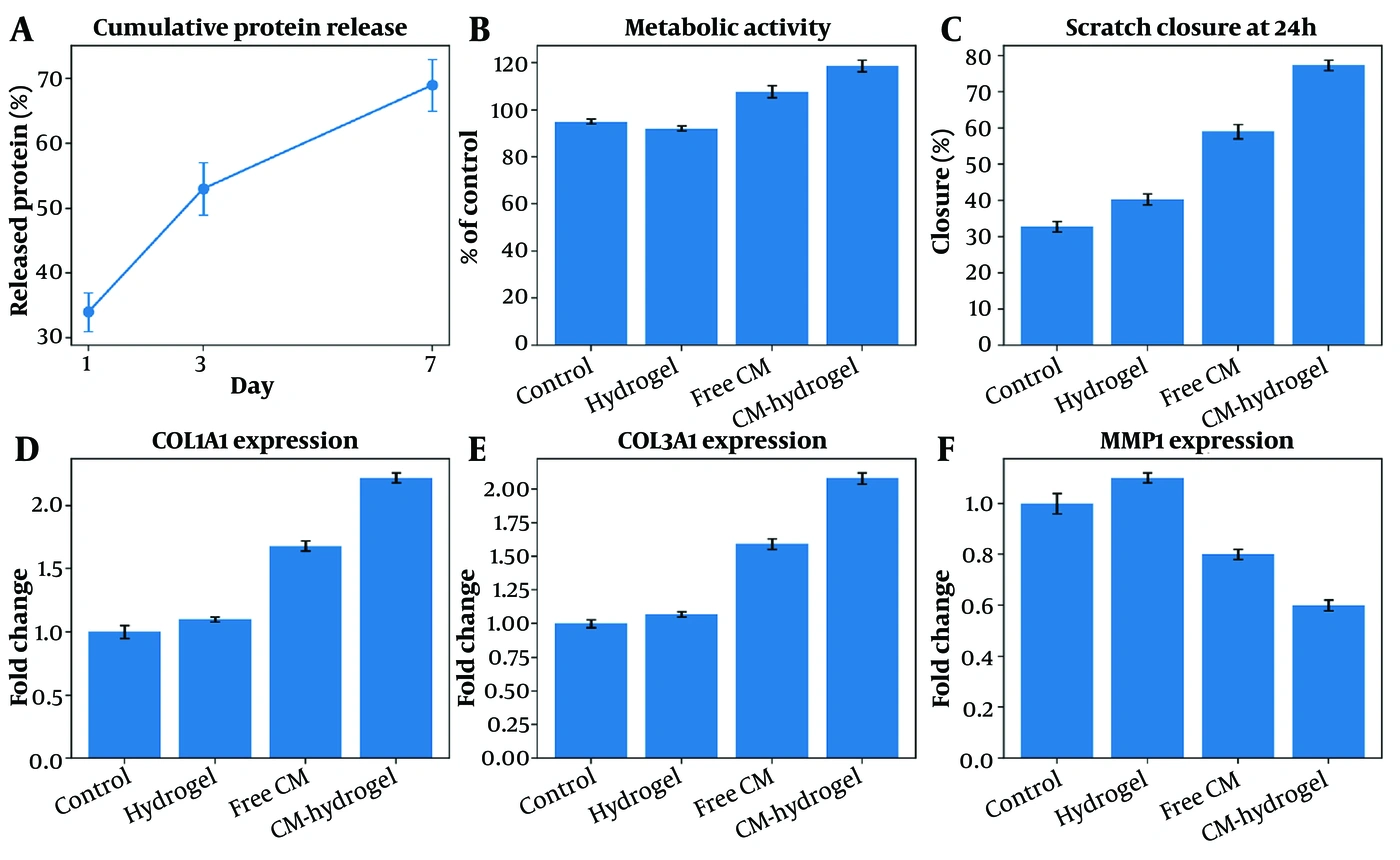
Figure 2.
Graphical summary of hydrogel release and fibroblast outcomes. (A) Cumulative protein release through day 7. (B) Metabolic activity measured by MTT. (C) Scratch closure at 24 h. (D-F) Relative COL1A1, COL3A1, and MMP1 expression. All panels use HDF-CM terminology. HDF-CM, human dermal fibroblast-conditioned medium; MTT, 3-(4,5-dimethylthiazol-2-yl)-2,5-diphenyltetrazolium bromide.
5. Discussion
This study combined a gelatin–alginate delivery matrix with HDF-CM and evaluated both material behavior and fibroblast responses relevant to skin repair. The HDF-CM-loaded hydrogel exhibited high water uptake and released protein over seven days, while the biological assays showed increased fibroblast metabolic activity and migration, as well as a shift in COL1A1, COL3A1, and MMP1 expression, compared with free HDF-CM or blank hydrogel. These observations support a carrier-associated difference in vitro; however, they should not be interpreted as evidence of complete skin regeneration.
The material findings are consistent with the broader hydrogel literature, in which hydrated polymer networks are used to maintain a moist environment and enable controlled local delivery of bioactive agents (7-11). The observed initial release followed by slower release is compatible with diffusion and network–cargo interactions in hydrated matrices. However, total protein release alone does not establish preservation of individual growth factors or extracellular-vesicle bioactivity; future studies should therefore quantify selected HDF-CM components before loading and after release.
Conditioned media contain heterogeneous paracrine factors that can influence fibroblast survival, proliferation, migration, and matrix regulation (3-6). The increased metabolic activity and scratch closure observed with HDF-CM exposure are therefore biologically plausible and align with prior reports of secretome-mediated fibroblast responses (3, 4, 11, 12). The stronger response in the hydrogel-delivered group may reflect prolonged local presentation; however, the current design cannot distinguish release kinetics from concentration effects unless free and hydrogel-delivered HDF-CM are rigorously matched for protein dose and batch.
The increase in COL1A1 and COL3A1 together with lower MMP1 expression indicates a shift in extracellular-matrix-related transcription. This pattern may be relevant to matrix formation, but it is not equivalent to increased collagen protein deposition or improved tissue repair. Collagen accumulation and matrix degradation are temporally regulated during healing, and excessive or prolonged collagen synthesis can contribute to fibrosis (1, 2, 13). Accordingly, future experiments should pair transcript measurements with collagen protein assays and broader remodeling markers.
The use of a cell-free HDF-CM formulation may offer practical advantages over direct administration of living cells, including simplified storage and delivery. Nevertheless, conditioned medium is intrinsically heterogeneous, and donor source, passage, serum exposure, conditioning time, storage, and batch processing can alter its composition (5, 6, 9). Reproducible translation will require predefined manufacturing controls, potency assays, and reporting of batch-to-batch variability.
The scope of the present study is limited to in vitro fibroblast responses relevant to skin repair. The study did not directly assess keratinocyte re-epithelialization, endothelial angiogenesis, inflammatory responses, collagen deposition, or in vivo wound closure. Therefore, the findings should not be generalized to whole-skin regeneration or clinical efficacy. Future work should extend the system to keratinocyte, endothelial, and immune-cell models and, if supported by those results, to an appropriately designed in vivo wound model.
5.1. Conclusions
A gelatin–alginate hydrogel incorporating HDF-CM showed sustained protein release through day 7 and was associated with increased fibroblast metabolic activity and scratch closure, higher COL1A1/COL3A1 expression, and lower MMP1 expression in this in vitro model. These findings are restricted to fibroblast responses relevant to skin repair and do not demonstrate whole-skin regeneration or in vivo therapeutic efficacy. Verification in independent biological replicates, standardized HDF-CM batches, multicellular assays, and in vivo studies is required before translational conclusions can be drawn.
5.2. Limitations and Future Work
• The biological sample size was small; independent replication should be expanded to improve precision and the robustness of assumption checks.
• HDF-CM composition is heterogeneous and should be characterized using representative soluble factors and, where relevant, extracellular-vesicle-associated markers.
• Free HDF-CM and hydrogel-delivered HDF-CM should be compared at rigorously matched protein doses and across dose-response and time-course conditions.
• Fibroblast metabolic activity, migration, and mRNA expression do not establish whole-skin regeneration. Future studies should assess collagen protein deposition, keratinocyte re-epithelialization, endothelial angiogenesis, inflammatory-cell responses, and in vivo wound closure.
• Protein stability and biological potency after hydrogel encapsulation and release require direct measurement.