Journal of Semnan University of Medical Sciences
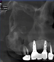
Image Credit:Koomesh
Outline
Tooth Suspended in the Maxillary Sinus (Case Report)
Authors

Abstract
Background: The presence of an ectopic tooth in a non-tooth-bearing area is a rare occurrence, attributable to various factors such as developmental anomalies, pathological processes, or iatrogenic activities. This phenomenon can manifest unilaterally or bilaterally. In certain cases, patients may experience symptoms including pain, swelling, and chronic or recurrent sinusitis, although it may sometimes be discovered incidentally.
Case report: In the present study, we examine a case of an ectopic third molar in the maxillary sinus. A 29-year-old male patient visited the Shahid Beheshti Dental School for dental implant treatment. Cone beam computed tomography (CBCT) images revealed the presence of an ectopic third molar in the left maxillary sinus. The patient reported no history of pain or swelling in the affected area and had no significant systemic medical history. Due to the patient's non-cooperation, surgical treatment and follow-up sessions were not possible for this case.
Conclusion: The importance of this article lies in its contribution to recording rare instances of ectopic teeth. These reports increase understanding and aid in the better management of such anomalies.
Highlights
References
- 1.Ramanojam S, Halli R, Hebbale M, Bhardwaj S. Ectopic tooth in maxillary sinus: Case series. Ann Maxillofac Surg. 2013;3(1):89-92. [PubMed ID:23662268]. [PubMed Central ID:PMC3645620]. https://doi.org/10.4103/2231-0746.110075.
- 2.Elango S, Palaniappan SP. Ectopic tooth in the roof of the maxillary sinus. Ear Nose Throat J. 1991;70(6):365-6. [PubMed ID:1893885].
- 3.Bodner L, Tovi F, Bar-Ziv J. Teeth in the maxillary sinus--imaging and management. J Laryngol Otol. 1997;111(9):820-4. [PubMed ID:9373546]. https://doi.org/10.1017/s0022215100138721.
- 4.Hashemipour MA, Tahmasbi-Arashlow M, Fahimi-Hanzaei F. Incidence of impacted mandibular and maxillary third molars: a radiographic study in a Southeast Iran population. Med Oral Patol Oral Cir Bucal. 2013;18(1):e140-5. [PubMed ID:23229243]. [PubMed Central ID:PMC3548634]. https://doi.org/10.4317/medoral.18028.
- 5.Beriat GK, Beriat N, Yalcinkaya E. Ectopic molar tooth in the maxillary sinus: a case report. Clin Dent Res. 2011;35(2):35-40.
- 6.Rathee M, Jain P. Embryology, Teeth. Treasure Island (FL): StatPearls Publishing; 2025.
- 7.Al-Ani RM, Aldelaimi TN, Khalil AA. Ectopic Upper Third Molar in Maxillary Sinus: A Case Report and Literature Review. Indian J Otolaryngol Head Neck Surg. 2022;74(Suppl 3):4718-21. [PubMed ID:36742931]. [PubMed Central ID:PMC9895750]. https://doi.org/10.1007/s12070-021-03039-0.
- 8.Jain P, Rathee M. Anatomy, Head and Neck, Tooth Eruption. Treasure Island (FL): StatPearls Publishing; 2025.
- 9.Shetty P, Shetty S, Agarwal N, Srivastava P. Third molar in the orbital floor: A precarious presentation of odontogenic keratocyst-A case report and review of literature. Clin Case Rep. 2018;6(9):1854-61. [PubMed ID:30214778]. [PubMed Central ID:PMC6132149]. https://doi.org/10.1002/ccr3.1714.
- 10.Anand R, Kieu A, Arulraj E, Ilangovan G, D.A N, AshwathNarayana G, et al. A Rare Case of an Intra-nasal Ectopic Tooth in a Young Woman. Cureus. 2021. https://doi.org/10.7759/cureus.19370.
- 11.El-Sayed Y. Sinonasal teeth. J Otolaryngol. 1995;24(3).
- 12.Pracy JP, Williams HO, Montgomery PQ. Nasal teeth. J Laryngol Otol. 1992;106(4):366-7. [PubMed ID:1613354]. https://doi.org/10.1017/s0022215100119516.
- 13.Yusuf H, Quayle AA. Intracondylar tooth. Int J Oral Maxillofac Surg. 1989;18(6):323. [PubMed ID:2516098]. https://doi.org/10.1016/s0901-5027(89)80027-x.
- 14.Toranzo Fernandez M, Terrones Meraz MA. Infected cyst in the coronoid process. Oral Surg Oral Med Oral Pathol. 1992;73(6):768. [PubMed ID:1437053]. https://doi.org/10.1016/0030-4220(92)90028-o.
- 15.Sharma S, Chauhan JS. Bilateral ectopic third molars in maxillary sinus associated with dentigerous cyst-A rare case report. Int J Surg Case Rep. 2019;61:298-301. [PubMed ID:31401439]. [PubMed Central ID:PMC6699555]. https://doi.org/10.1016/j.ijscr.2019.07.072.
- 16.Masalha M, Schneider S, Kassem F, Koren I, Eliashar R, Margulis A, et al. Endoscopic treatment of ectopic teeth in the maxillary sinus. J Clin Exp Dent. 2021;13(3):e227-e33. [PubMed ID:33680324]. [PubMed Central ID:PMC7920564]. https://doi.org/10.4317/jced.57905.
- 17.Lai YT, Luk YS, Fung KH. Anomalous morphology of an ectopic tooth in the maxillary sinus on three-dimensional computed tomography images. J Radiol Case Rep. 2013;7(2):11-6. [PubMed ID:23705035]. [PubMed Central ID:PMC3661307]. https://doi.org/10.3941/jrcr.v7i2.1227.
- 18.Safi Y, Mortazavi H, Iranparvar Alamdari M. Ectopic Deciduous Molar in Maxillary Sinus: An Extremely Rare Case Report. Journal of Iranian Medical Council. 2022. https://doi.org/10.18502/jimc.v5i1.9591.
- 19.Almomen A, Alkhudair B, Alkhatib A, Alazzah G, Ali Z, Al Yaeesh I, et al. Ectopic maxillary tooth as a cause of recurrent maxillary sinusitis: a case report and review of the literature. J Surg Case Rep. 2020;2020(9):rjaa334. [PubMed ID:33024531]. [PubMed Central ID:PMC7524603]. https://doi.org/10.1093/jscr/rjaa334.
- 20.Topal Ö, Dayısoylu EH. Ectopic Tooth in the Maxillary Sinus. Turk Arch Otorhinolaryngol. 2017;55(3):151-2. [PubMed ID:29392075]. [PubMed Central ID:PMC5782996]. https://doi.org/10.5152/tao.2017.2308.
Copyright
© 2025, Author(s). This open-access article is available under the Creative Commons Attribution 4.0 (CC BY 4.0) International License (https://creativecommons.org/licenses/by/4.0/), which allows for unrestricted use, distribution, and reproduction in any medium, provided that the original work is properly cited.
Similar Articles
Root Canal Treatment of a Maxillary Second Molar with Two Palatal Roots: A Case Report
Dehghan R, Ghahramani Y. Root Canal Treatment of a Maxillary Second Molar with Two Palatal Roots: A Case Report. Thrita J Neu. 2018;7(1):e69575. doi: https://doi.org/10.5812/thrita.69575
Radicular cyst associated with a second primary molar: Case Report
Seyedmajidi M, Foroughi R, Gholami A. Radicular cyst associated with a second primary molar: Case Report. J Inflamm Dis. 2024;18(4):e155853. doi:
Diagnosis and Treatment of an Odontogenic Maxillary Sinusitis: A Case Report Study
Soghli N, Johari M, Panjnoush M. Diagnosis and Treatment of an Odontogenic Maxillary Sinusitis: A Case Report Study. Thrita J Neu. 2020;9(2):e108248. doi: https://doi.org/10.5812/thrita.108248
Evaluation of Panoramic Radiography at Showing Width of the Dental Follicle
Seyedmajidi SK, Haghanifar S, Mehdizadeh M, Seyedmajidi M, Bijani A, et al. Evaluation of Panoramic Radiography at Showing Width of the Dental Follicle. Zahedan J Res Med Sci. 2014;16(7):. doi:
The Prevalence of Three Rooted Primary Mandibular Molars in Zahedan
Amiri tehrani N, Heydari A. The Prevalence of Three Rooted Primary Mandibular Molars in Zahedan. Zahedan J Res Med Sci. 2008;10(3):e94521. doi:
- Scopus by DOI: 0
Last Update: 2 weeks ago
- Scopus by Title: 0
Last Update: 2 weeks ago
- Scopus by Title (Ref): 0
Last Update: 2 weeks ago
- CrossRef: 0
Last Update: 1 week ago
- OpenAlex: 0
Last Update: 1 day ago
- OpenCitations: 0
Last Update: 4 days ago
