


1. Background
2. Objectives
3. Methods
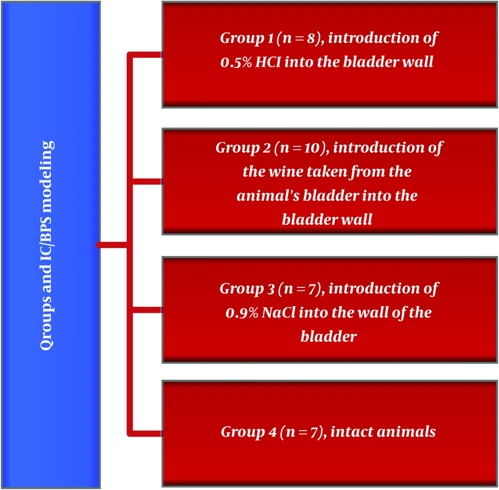
Groups of animals with stimulated IC/BPS
4. Results
| Groups | Cell Types | |||
|---|---|---|---|---|
| Neutrophils | Lymphocytes | Eosinophils | Mast Cells | |
| 1 (n = 8) | 89.375 ± 14.927a [68; 112] | 48.375 ± 7.614a. b [36; 59] | 2.125 ± 3.226a [0; 8] | 1.125 ± 1.642 [0; 4] |
| 2 (n = 10) | 0.866 ± 1.884 [0; 6] | 29.866 ± 10.183a. b [12; 49] | 0.333 ± 0.899 [0; 3] | 14.200 ± 5.796a [3; 26] |
| 3 (n = 7) | 1.428 ± 2.699 [0; 7] | 2.285 ± 3.728b [0; 10] | 0.142 ± 0.377b [0; 1] | 0 |
| 4 (n = 7) | 0 | 0.375 ± 1.060 [0; 3] | 0.375 ± 0.744 [0; 2] | 0 |
aStatistical significance between groups (P < 0.05 - 0.001)
bStatistical significance with group 4 (control group)
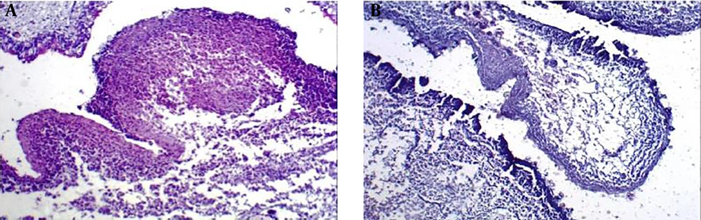
The histological preparation of the bladder after creating a model of IC/BPS by injection of 0.5% HCl solution into the bladder of experimental animals. Mixed lymphocyte and neutrophil infiltration, extensive necrosis (×400 magnification, hematoxylin-eosin staining).
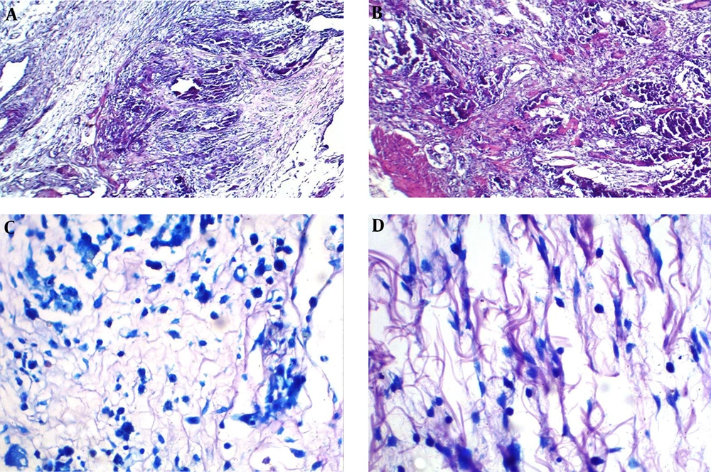
The histological preparation of the bladder after creating a model of IC/BPS by introducing urine taken from the animal’s bladder into the wall of the bladder. (A) Developing necrosis and mixed inflammatory infiltration (×100 magnification, hematoxylin-eosin staining). (B) Developing necrosis, giant multinucleated histiocytes and mixed inflammatory infiltration (×100 magnification, hematoxylin-eosin staining). (C) Mast cell infiltration in the bladder wall (×400 magnification, May-Grünwald-Giemsa staining). (D) Mast cells infiltration in the bladder wall of experimental animal (×400 magnification, May-Grünwald-Giemsa staining).
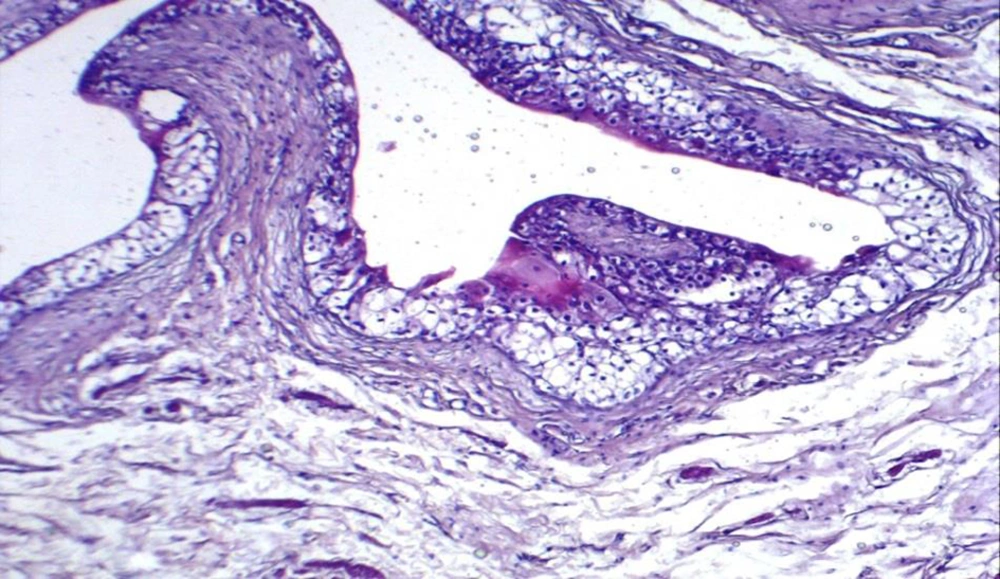
The histological preparation of the bladder after creating a model of IC/BPS by injection of 0.9% NaCl into the wall of the bladder. Single lymphocytes against the background of edema, (×40 magnification, hematoxylin-eosin staining).
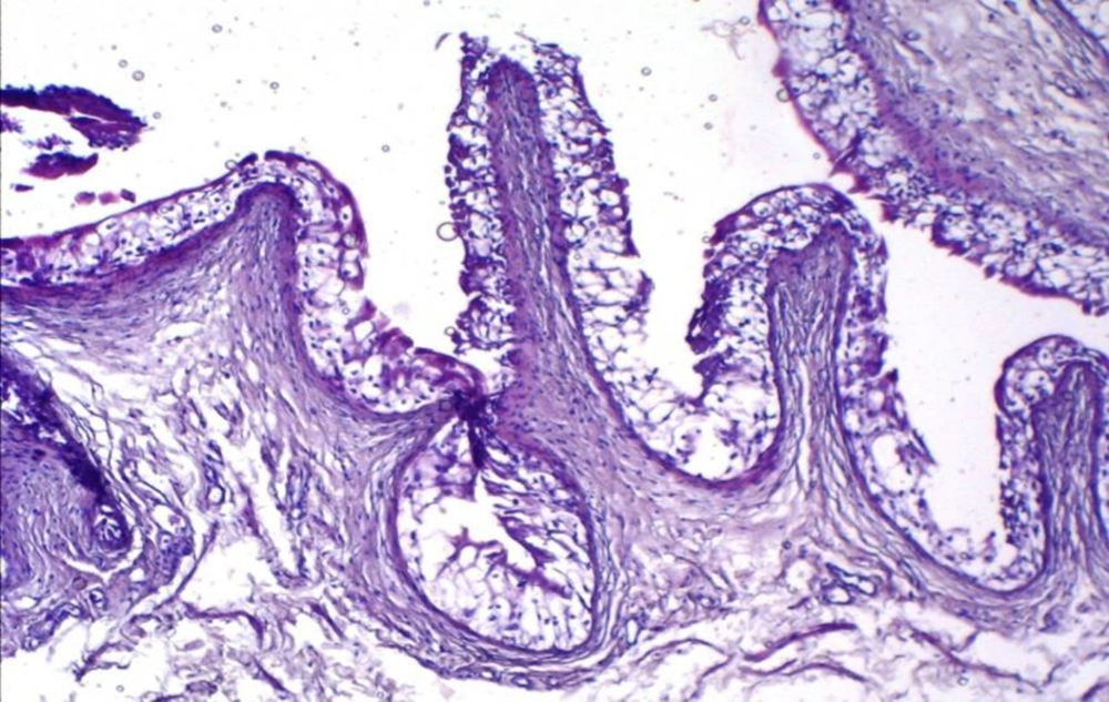
The histological preparation of an intact animal. The wall of the animal bladder of the intact group (control) without any features (×40 magnification, hematoxylin-eosin staining).

