A 67-year-old woman presented with lower urinary tract symptoms (LUTSs), intermittent right flank, and suprapubic pain that had been ongoing for 1.5 years prior to presentation. She was a known hypertensive but controlled with medications. No positive surgical history. There was one episode of microscopic hematuria, but other laboratory investigations (including creatinine, complete blood count, and liver function tests) were normal. Urine cytology was negative for malignancy.
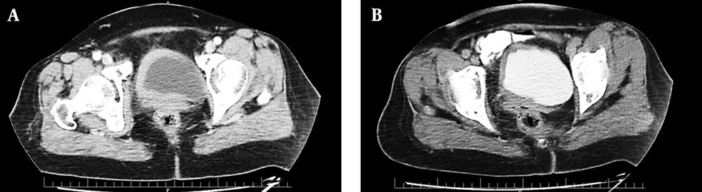
Abdominopelvic ultrasonography revealed an asymmetric diffused bladder wall thickening with a maximum diameter of 14 mm, involving the right lateral and right half of the anterior and posterior wall of the bladder with extension to the right ureteropelvic junction (UVJ), causing mild to moderate hydroureteronephrosis in the right side (pelvic anterior-posterior diameter [APD]: 9 mm). This was later confirmed by a computed tomography (CT) urogram, which also revealed mild right hydronephrosis and mild hydronephrosis of the right kidney and asymmetric bladder wall thickening in the right side of the urinary bladder (
Figure 1), in addition to multiple osteoblastic lesions in the bones, suggestive of malignancy. Therefore, a whole-body bone scan was performed, revealing multiple bone lesions in the left sacroiliac (SI) joint, thoracic, and upper lumbar spine.
Computed tomography (CT) scan with intravenous (IV) contrast of the pelvis in the arterial phase (A) and delayed excretory phase (B). An axial section showing asymmetric hyperdense segmental urinary bladder wall thickening involving the right side. The contrast enhancement is 77 Hounsfield units (HU) (from 48 HU before IV contrast injection). There are no signs of involvement of adjacent structures (uterus and adnexa).
In the next step, she had transurethral resection of the bladder tumor (TURBT), revealing a neoplastic mass mainly around the right ureteral site that was deviated from the trigon to the left. A compressive effect had deformed the right ureter to the point that it was impossible to cross a 6 Fr. ureteroscope. The rest of the bladder mucosa was severely pale. Diffuse lesions without a distinct base were resected up to the muscular layer as much as possible.
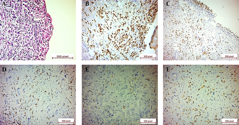
Microscopic examination of the specimen revealed a neoplastic tissue composed of sheets of discohesive single tumoral cells and cords of cells with enlarged, occasionally laterally placed nuclei and pale eosinophilic cytoplasms. A few tumoral cells with signet ring features were also present. The urothelium was ulcerated, and the neoplastic cells diffusely infiltrated the lamina propria and muscularis propria (
Figure 2). Immunohistochemical (IHC) staining showed positive immunoreactions for CK7, GATA3, ER, and CD138. Also, GCDFP-15 and CEA were positive in the scattered tumoral cells (
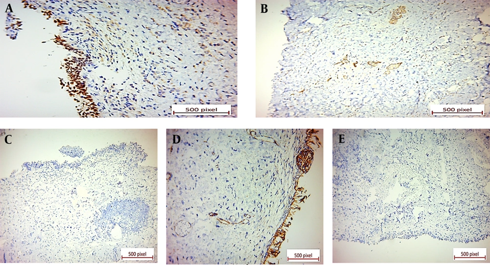
Figure 2). P63, E-cadherin, CDX2, B-catenin, and CK20 revealed negative immunoreactions (
Figure 3).
(A) Ulcerated urothelium underlined by discohesive tumoral cells with occasional signet ring features (×20). (B) Positive CK7 cytoplasmic immunostaining of tumoral cells in bladder tissue (×40). (C) Positive GATA3 nuclear immunostaining of tumoral cells in bladder tissue (×20). (D) Positive ER nuclear immunostaining of tumoral cells in bladder tissue (×40). (E) Positive GCDFP-15 cytoplasmic immunostaining in a few scattered tumoral cells in bladder tissue (×40). (F) Positive CEA cytoplasmic immunostaining in scattered tumoral cells in bladder tissue (×40).
Negative immunoreactions: (A) Negative P63 immunostaining of tumoral cells in bladder tissue. The benign surface urothelium shows positive nuclear staining for P63 (×40). (B) Negative E-cadherin immunostaining of tumoral cells in breast tissue. Some benign breast acini show positive staining (×20). (C) Negative CDX2 immunostaining of tumoral cells in bladder tissue (×20). (D) Negative B-catenin immunostaining of tumoral cells in bladder tissue (×40). (E) Negative CK20 immunostaining of tumoral cells in bladder tissue (×20).
The pathologist reported a carcinoma with plasmacytoid/signet ring features and added a note saying that this tumor would be considered a primary tumor of the bladder if metastasis of lobular carcinoma of the breast could be clinically ruled out.
Physical examination of the breasts and lymph nodes was then performed with no pathological findings. The vaginal and cervical exams and the Pap smear test were also normal. Tumor markers, such as CA 19-9, CA 125, CEA, and CA 15-3, were detected in the normal range. Transvaginal ultrasonography was performed without any additional findings. Both axillary and breast ultrasonography and bilateral mammography were normal, and no enlarged axillary lymph nodes were present (Breast Imaging Reporting and Data System 1 (BI-RADS 1)).
A fluorodeoxyglucose positron-emission tomography/computed tomography (FDG PET/CT) scan from vertex to mid-thigh was requested. In the chest, an asymmetrical soft tissue density nodule in the superolateral quadrant of the right breast with mild metabolic activity was detected. Ipsilateral right axillary adenopathy was also observed, along with right hilar FDG-avid adenopathy. Bilateral subpleural hypermetabolic ground glass nodules were noted in the lung field, suggestive of ground glass metastasis. Moreover, extensive mixed lytic sclerotic bone lesions in the axial and appendicular skeleton were reported.
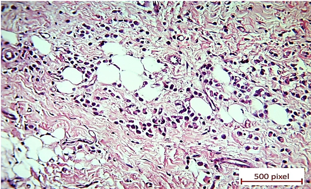
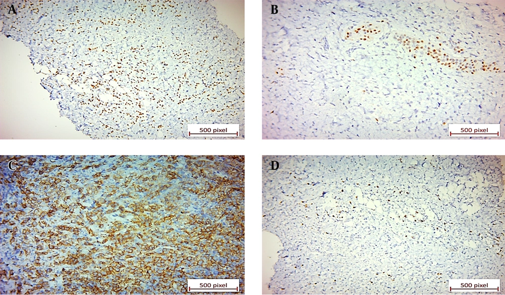
Eventually, an ultrasound sonography-guided core needle biopsy of the right breast nodule and right axillary lymph node was performed. Microscopic evaluation displayed invasive lobular carcinoma of the breast (
Figure 4) in the presence of lobular intraepithelial neoplasm (LIN) and lymphovascular invasion (LVI). The right axillary lymph node was also involved, along with the extranodal extension. The tumoral cells were positive for ER, PR, and Her2/neu in the IHC study, and Ki-67 was positive in about 5% - 10% of these cells (
Figure 5).
Diffuse infiltration of lobular carcinoma in the breast as single tumoral cells (×20)
(A) Positive ER nuclear immunostaining of tumoral cells in breast tissue (×20). (B) Positive PR nuclear immunostaining in 1% of tumoral cells in breast tissue. Some benign breast acini are positive for PR (×20). (C) Complete strong membranous HER2 immunostaining in more than 10% of tumoral cells in breast tissue (score 3; ×20). (D) Ki-67 shows nuclear immunostaining of 7% of tumoral cells in breast tissue (×20).
Considering the patient’s preference to preserve her breasts, her age, and the presence of distant metastases, it was decided to start palliative chemotherapy. Today and after 8 courses of palliative chemotherapy with adriamycin, endoxan, and paclitaxel regimens, 12 courses of bone metastasis treatment with Zometa, and daily hormone therapy with letrozole (2.5 mg) in 24-month follow-up, the patient is well, and the urinary symptoms have fully disappeared.