


1. Introduction
2. Case Presentation
2.1. Case 1
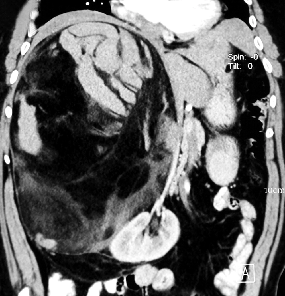
CECT Abdomen Showing a 35 x 19 cm Suprarenal Mass Hounsfield Density (HU-19)
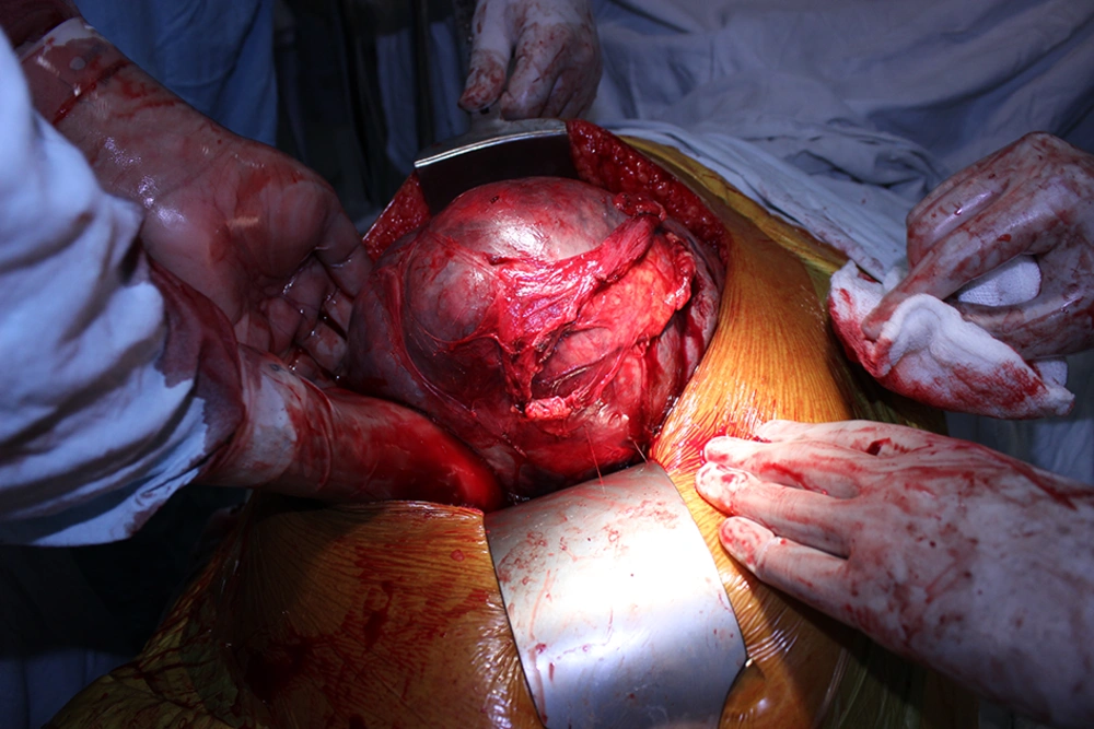
Intraoperative Picture Showing a Large Suprarenal Mass
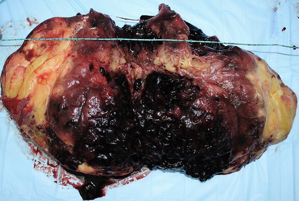
Resected Specimen With a Size of 38 x 20 x 16 cm and Weight of 5200 Grams With Well Circumscribed Tumor Showing Multiple Areas of Hemorrhage
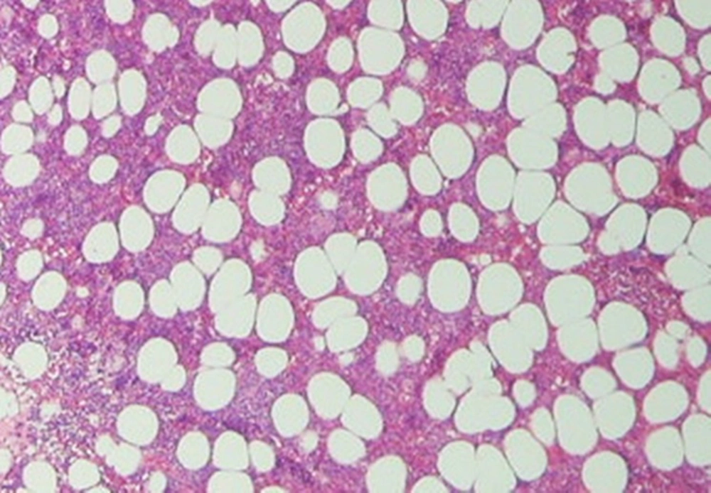
Histopathology Showing Adiposities Admixed With Hematopoitic Cells Suggestive of Myelolipoma of Adrenal Gland
2.2. Case 2
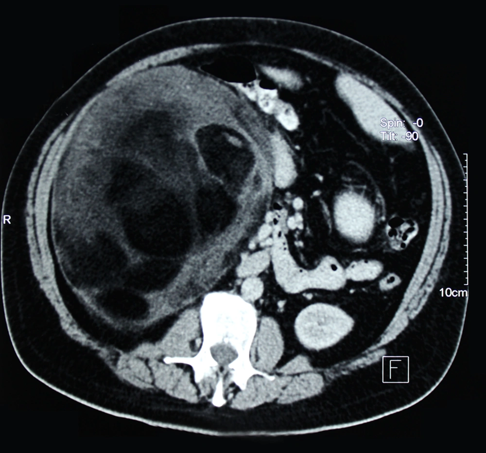
CECT Abdomen Showing a 23 × 21 cm Suprarenal Mass With Fat and Fluid Density Within it Pushing the Kidney Downwards
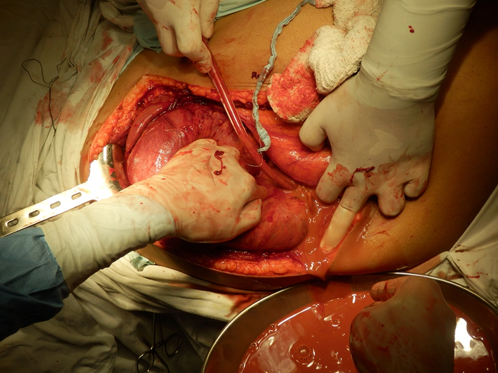
Intraoperative Picture Showing a Large Suprarenal Mass With Pus Filled Within it
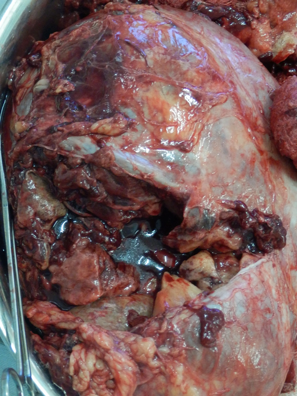
Resected Specimen Showing Tumor With Residual Cavity After Aspiration of Pus
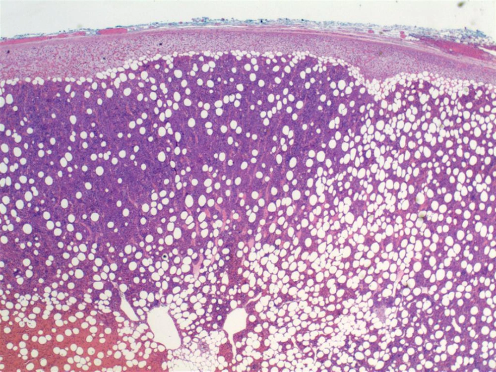
Histopathology Showing a Mixture of Adipose and Myeloid Elements Suggestive of Myelolipoma of the Adrenal Gland
