


1. Background
Trauma is one of the most important causes of death in patients under the age of 40 years and the third most common cause of death overall (1). Chest injuries comprise one of the most important causes of death in the first minutes after trauma. Blunt chest wall trauma is responsible for more than 10% of all traumas in patients referred to emergency departments worldwide. Rib fractures due to blunt traumas are not common in children due to their chest flexibility but are frequent in the elderly (2).
Following trauma, management and evaluation of patients begin based on advanced trauma life support (ATLS). Diagnostic procedures in patients with stable vital signs usually begin with simple chest radiography or, in those with clinical suspicion of injury, a computed tomography (CT) scan. Portable ultrasound (US) in trauma patients is a non-invasive, accessible, and rapid diagnostic method for detecting the accumulation of blood and other intra-abdominal fluids in the pleural space and pericardium (3, 4). On the other hand, studies have shown that 33 - 50% of rib fractures are not detected on chest radiography, rendering the prognosis of patients more complex and difficult to determine using simple radiographic images (5). Although chest CT scan is a highly sensitive and specific method for detecting even minor rib fractures and their complications while patients are lying down, patient management is still complicated with concerns about the high radiation dose, costs, and the impossibility of transferring critically ill patients to the radiology center (6-8).
Point-of-care ultrasound (POCUS), on the other hand, is a cheap and fast method applicable in patients with unstable conditions and can accelerate reaching an immediate diagnosis, especially in the emergency department (ED) (6-8) and where radiography and CT scans are not possible, such as rural or war zones, or in space voyages. However, extensive research is required to evaluate this method’s power of gear fracture detection and its complications. Studies have been performed around the world on the sensitivity and specificity of ultrasound for diagnosing rib fractures and their complications following impenetrable injury. In one study, researchers concluded that high-resolution US can detect rib lesions better than plain radiography (9). Other studies found that ultrasound was more sensitive than chest radiography in detecting rib fractures (10). Another study noted that ultrasound with a negative result in areas with the most tenderness significantly reduced the likelihood of rib fractures (11). The sensitivity of ultrasound in the diagnosis of fractures, hemothorax, pneumothorax, and contusion has been reported at 37.2%, 60.7%, 93.1%, and 16.7%, respectively, with a specificity of 100% for all conditions (12). Another study showed that ultrasound had a higher diagnostic accuracy than radiography in detecting fractures (13). However, the above-mentioned studies’ sample sizes were mostly inadequate. Therefore, more research is required to obtain more credible evidence (1).
2. Objectives
The present study aimed to determine and compare the diagnostic value of POCUS and CT scans in discovering rib fractures and related complications in patients with blunt chest wall trauma.
3. Methods
3.1. Study Design and Setting
The present prospective cross-sectional study (October 2017-April 2018) was conducted on all adult (> 18 years) patients with blunt chest wall trauma who were referred to the Shahid Rajaei Hospital of Shiraz, the main trauma center in the south of Iran, which is affiliated to the Shiraz University of Medical Sciences.
3.2. Study Population
The current study was conducted on all eligible patients referred to the ED. Inclusion criteria were being adult, the diagnosis of blunt chest trauma, having stable vital signs and clinical condition, and suffering from no active and life-threatening bleeding. Exclusion criteria were being overweight, interfering with performing CT scans, undergoing previous pulmonary resuscitation, having an interval of > 3 hours between ultrasound and CT scans, and having a chest tube or undergoing any other intervention before the ultrasound. Moreover, the patients who required emergency actions, including airway management, and those whose parents did not sign the written informed consent form were excluded from the study.
3.3. Sample Size and Sampling Method
A sample size of n = 65 was observed to deliver a 95% confidence interval (CI) [standard deviation (SD) of 5%] with an estimated sensitivity of 98.1% (α = 5%, β = 20%), designating a frequency of 30% for pneumothorax (14). The sample size was determined using the method of Arkin and Wachtel (15) and Medcalc software for Windows. In order to increase the power of the study, 113 patients who met the inclusion criteria were enrolled. We used convenience sampling to enroll the participants.
3.4. Study Protocol and Interventions
In this study, the protocol was completely explained to the patients, and all individuals signed a written informed consent form. Following clinical assessments, eligible patients underwent chest POCUS using a portable ultrasonography machine (Fujifilm SonoSite, Inc., USA) equipped with a 7.5 MHz linear probe and a low-frequency (3.5 MHz) probe by trained third-year emergency medicine (EM) residents supervised by an EM-attending physician (who was an Iranian board-certified doctor, a faculty member, and an expert in performing POCUS). It is noteworthy that POCUS is one of the topics covered in the EM curriculum in Iran, so EM residents in Iran become fully experts in this regard during their residency periods. In addition, the residents who participated in this study attended a 2-day workshop on lung ultrasound. Each lung field was scanned in the mid-alveolar anterior intercostal space (2-4) and the medial axial intercostal space (6-8). The presence of any discontinuity in the periosteum of the rib bone indicated a fracture (Figure 1). Due to the limitations of POCUS in the examination of subscapular ribs and the fact that in patients with trauma, many different positions cannot be instructed to the patient, the examination of the posterior rib was excluded from the study’s protocol (10). After recording the results, the patients were referred to the Radiology Department for performing lung CT scans within less than three hours from POCUS. The lung CT scan was performed without contrast agent injection, and the results were reported under the supervision of radiology attending physicians, who were also Iranian radiology board-certified faculty members. Then, using the PACS system, all CT scan images were examined with a standard method by blinded examiners (i.e., identification was based on patients’ national IDs). All the data obtained were documented in a data collection form. Finally, the results of POCUS were compared with the findings of lung CT scans as the gold standard.
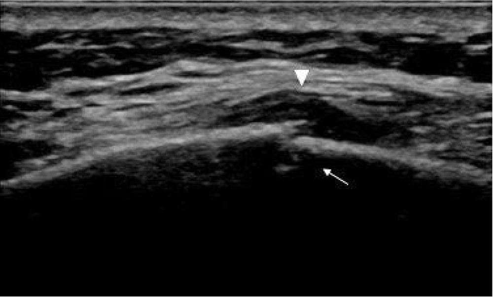
Figure 1.
Ultrasound view in patients with rib fractures. Any discontinuity in the periosteum of the bone indicated a rib fracture (the triangle). Hypodense areas indicated extensive hematoma (the arrow).
3.5. Statistical Analysis
All data were entered into statistical package for social sciences (SPSS) software version 19.0 (SPSS Inc., Chicago, IL, USA) and MedCalc Statistical Software version 13.3.3 for Windows (MedCalc software bvba, Ostend, Belgium; 2014). Sensitivity, specificity, positive predictive value (PPV), negative predictive value (NPP), positive likelihood ratio (PLR), negative likelihood ratio (NLR), and accuracy were calculated. In order to determine the best sensitivity and specificity and to obtain the area under the curve (AUC), the receiver operating characteristic (ROC) curve was plotted. Kappa was used to assess the level of agreement between POCUS results and radiology reports. The data were presented as mean ± SD for continual variables and as number (percentage) for categorical ones. A two-sided P-value less than 0.05 and a CI of 95% were utilized to identify statistically significant differences.
4. Results
Among the 1432 patients referred to the Shahid Rajaei Hospital of Shiraz with blunt and penetrated chest traumas during the study period, 894 (62.43%) had blunt chest wall traumas, 113 of whom were finally enrolled in this study (Figure 2).
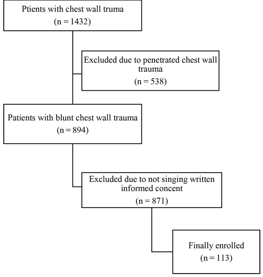
Figure 2.
The flowchart showing the process of patient enrollment
As shown in Table 1, the mean ± SD of age was 44.07 ± 22.07 years, and 90 (79.56%) patients were male. The mean ± SD of the international space station (ISS) was 17.07 ± 11.92. The most common sites of associated injuries were the extremities (51.3%) and the head (41.6%), respectively.
Table 1.Demographic and Clinical Characteristics of the Participants a
| Variables | Values |
|---|---|
| Age (y) | 44.07 ± 22.07 |
| Gender | |
| Female | 23 (20.35) |
| Male | 90 (79.65) |
| Damage reasons | |
| Pedestrian | 47 (41.59) |
| Road accidents | 14 (12.39) |
| Same-level or same-surface falls | 23 (20.35) |
| Falling from height | 29 (25.67) |
| ISS | 17.07 ± 11.92 |
| Systolic blood pressure (mmHg) | 129.64 ± 25.36 |
| Diastolic blood pressure (mmHg) | 81.88 ± 16.71 |
| Heart rate per minute | 100.82 ± 22.57 |
| Respiratory rate per minute | 18.25 ± 3.78 |
| Sites of associated injuries | |
| Extremities | 58 (51.3) |
| Head | 47 (41.6) |
| Abdomen | 31 (27.4) |
| Face | 15 (13.3) |
Abbreviations: ISS, injury severity score; mmHg, millimeters of mercury; SD, standard deviation.
a Values are presented as No. (%) or mean ± SD.
Figure 3 shows the distribution of the severity of the abbreviated injury scale (AIS) based on associated injuries in patients with blunt chest walls. In total, 91 patients (80.5%) had at least one associated injury; 58 patients (51.3%) had an injury to one of their organs; 47 patients (41.6%) had a head injury; 31 patients (27.4%) had an abdominal injury, and 15 people (13.3%) had facial injuries. The total incidence of these co-injuries was more than 100% due to the fact that some patients had more than one co-injuries.
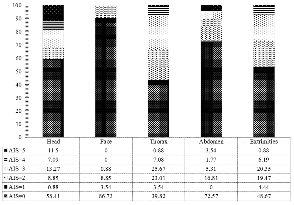
Figure 3.
Distribution of abbreviated injury scale severity based on associated injuries
Overall, 75 (66.37%) patients had at least one rib fracture based on CT scan imaging, while 62 (54.9%) patients were identified with at least one fracture based on POCUS findings. Figure 4 depicts the distribution of patients based on the number of broken ribs on CT scan and POCUS.
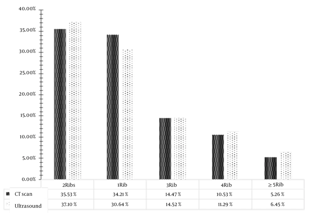
Figure 4.
Distribution of patients based on the number of broken ribs
Table 2 shows the diagnostic value of POCUS to identify patients with one, two, three, four, and five rib fractures. The chi-square test showed that there was a significant association between the findings of the CT scan and POCUS (all P values < 0.001). Compared with the CT scan, POCUS delivered a specificity of beyond 97% and an accuracy of > 84% in identifying all types of fractures and complications of fractures. Moreover, the results showed that the greater the number of broken ribs, the greater the sensitivity of POCUS in correctly detecting fractured ribs.
Table 2.The Diagnostic Value of Ultrasound in Comparison with CT Scan in Identifying Rib Fractures in Patients with Blunt Chest Injury
| Ultrasound Findings | CT Scan | Detection Power (95% CI) | ||||||||
|---|---|---|---|---|---|---|---|---|---|---|
| Positive | Negative | Kappa | P-Value | Sensitivity | Specificity | PPV | NPV | Positive LR | Negative LR | |
| One rib fracture | ||||||||||
| Positive | 19 (73.1) | 0 (0) | 0.8 | < 0.001a | 73.08 (52.21-88.43) | 100 (95.85-100) | 100 | 92.55 (86.84-95.9) | - | 0.27 (0.14-0.51) |
| Negative | 7 (26.9) | 87 (100) | ||||||||
| Two rib fractures | ||||||||||
| Positive | 21 (77.8) | 2 (2.3) | 0.8 | < 0.001 a | 77.78 (57.74-91.38) | 97.67 (91.85-99.72) | 91.30 (72.45-97.67) | 93.33 (87.35-96.60) | 33.44 (8.38-133.54) | 0.23 (0.11-0.46) |
| Negative | 6 (22.2) | 84 (97.7) | ||||||||
| Three rib fractures | ||||||||||
| Positive | 9 (81.8) | 0 (0) | 0.9 | < 0.001 a | 81.82 (48.22-97.72) | 100 (96.45-100) | 100 | 98.08 (93.57-99.44) | - | 0.18 (0.05-0.64) |
| Negative | 2 (18.2) | 102 (100) | ||||||||
| Four rib fractures | ||||||||||
| Positive | 7 (87.5) | 0 (0) | 0.9 | < 0.001 a | 87.50 (47.35-99.68) | 100 (96.55-100) | 100 | 99.06 (94.38-99.85) | - | 0.12 (0.02-0.78) |
| Negative | 1 (12.5) | 105 (100) | ||||||||
| Five rib fractures | ||||||||||
| Positive | 4 (100) | 0 (0) | 1 | < 0.001 a | 100 (39.76-100) | 100 (96.67-100) | 100 | 100 | - | 0 |
| Negative | 0 (0) | 109 (100) | ||||||||
| Hemothorax | ||||||||||
| Positive | 12 (85.7) | 0 (0) | 0.9 | < 0.001 a | 85.71 (57.19-98.22) | 100 (96.34-100) | 100 | 98.02 (93.21-99.44) | - | 0.14 (0.09-0.27) |
| Negative | 2 (14.3) | 99 (100) | ||||||||
| Subperiosteal Hematoma | ||||||||||
| Positive | 69 (80.2) | 0 (0) | 0.7 | < 0.001 a | 80.23 (70.25-88.04) | 100 (87.23-100) | 100 | 61.36 (50.92-70.86) | - | 0.20 (0.13-0.30) |
| Negative | 17 (19.8) | 27 (100) | ||||||||
Abbreviations: LR, likelihood ratio; NPV, negative predictive value; PPV, positive predictive.
a Statistically significant.
In addition, POCUS attained a sensitivity of 80.23% (95%CI: 70.25 - 88.04) and specificity of 100 (95%CI: 87.23 - 100) in the diagnosis of hemothorax, as well as a sensitivity of 80.23% (95%CI: 70.25 - 88.04) and specificity of 100% (95%CI: 87.23 - 100) in the detection of subperiosteal hematoma. It should be noted that no cases of pneumothorax were observed on POCUS, while 13 (11.5%) patients had pneumothorax on CT scans.
5. Discussion
The use of POCUS in patients with chest injuries (whether blunt or penetrating) has been increasing in recent years. Studies have shown that compared to anterior-posterior (AP) and lateral radiographs, ultrasound has a higher diagnostic value in the diagnosis of pneumothorax, hemothorax, pulmonary contusion, pneumonia, pleural effusion, and alveolar diseases, as well as other injuries (16, 17).
The present study aimed to determine and compare the diagnostic value of POCUS and CT scans in identifying rib fractures and other complications in patients with blunt chest wall traumas. We found that the specificity of POCUS compared with CT scan in identifying all types of rib fractures was more than 97%, with accuracy beyond 84%. Moreover, our results showed that the greater the number of broken ribs, the greater the sensitivity of POCUS in correctly diagnosing fractured ribs. Also, it was found that POCUS had a sensitivity of 80.23% and a specificity of 100% in detecting hemothorax, and the sensitivity and specificity were 80.23% and 100% for the diagnosis of subperiosteal hematomas.
Many studies have been performed to compare the diagnostic value of ultrasound with radiographic images, and two of these studies used CT scans as the gold standard (11, 18). In one study, the accuracy, sensitivity, and specificity were obtained as 80%, 91.2%, and 72.7%, respectively, where both POCUS and CT scans were performed and evaluated by an EM physician (11). In our study, CT scan images were reviewed by radiologists and interpreted in non-emergency situations, and POCUS was performed by 3rd-year EM residents under the supervision of an attending EM physician. In our study, only two cases with rib fractures on POCUS did not match the findings of CT scans, indicating higher specificity and sensitivity compared to the values mentioned in the recent study. In another similar experiment, 93 patients who had no rib fractures based on radiographs and CT scans examined by two thoracic surgeons and two radiologists were identified to have no rib fractures in reality; on the other hand, 64 patients (68.8%) had rib fractures based on ultrasound (18).
Considering that we found no report comparing the sensitivity and specificity of ultrasound vs. CT scan, it was not possible to compare our results with others. In a recent study, the sensitivity of ultrasound compared to radiography was estimated to be 98.31% (13). The sensitivity of ultrasound in the diagnosis of rib fractures was obtained about 92% in another report (19). In our study, the sensitivity of POCUS compared to CT scan was investigated, which was lower than the above values. This seems logical regarding that the above studies used radiographic images for comparisons.
In terms of the side effects associated with rib fractures, in one study, ultrasound had a sensitivity of 93.3% and specificity of 99.6% in diagnosing pneumothorax in traumatic patients (20). In another study conducted by radiology residents on 169 suspected cases of pneumothorax, the sensitivity, specificity, and positive and negative predictive values of ultrasound were 47%, 99%, 87%, and 93%, respectively, compared to CT scan (21). Moreover, in a study on 176 patients, ultrasound sensitivity was reported to be 98%, while supine sensitivity was 75.5% (22). No pneumothorax was identified in our study. This may be related to our inclusion and exclusion criteria and due to differences in the study design and patient selection, justifying the different sensitivity and specificity of POCUS in our study compared to other studies. Other possible factors explaining these differences may include the method of performing ultrasound (supine, sitting, lateral decubitus), complete dependence of POCUS on the operator, time and place conditions (emergency and non-emergency), and limitations such as obesity, large breasts, restrictions of ultrasound in examining subscapular and infraclavicular ribs, the presence of life-threatening injuries, and the need for immediate diagnostic actions.
5.1. Limitations and Strengths
Our study had several limitations, some of which were technical due to the limitations of POCUS itself in examining fractures in subscapular ribs, in obese patients, and in those with large breasts. In order to prevent any bias, posterior rib fractures were not examined in this study. In order to examine posterior ribs (except for subscapular ribs, which were not fully assessable by ultrasound), the patient needs to be taken out of the supine position and placed in an appropriate position. This is not normally possible in those with traumas due to the risk and suspicion of spinal cord injury. The patient’s position can only be changed if a CT scan confirms that the spine is healthy. On the other hand, since POCUS is completely operator-dependent, it is better to perform multicenter studies to better comment on the diagnostic value of POCUS compared to CT scans. Despite the limitations mentioned, our study is one of the few studies that have compared POCUS with CT scan as the gold standard. In addition, our relatively suitable sample size has increased the power of this study.
5.2. Conclusions
The results of this study showed that POCUS, conducted using a portable US machine and by emergency physicians in the ED, could precisely recognize rib fractures, hemothorax, and subperiosteal hematomas in patients with blunt chest wall traumas. On the other hand, with increasing the number of damaged ribs, the accuracy and other diagnostic parameters of POCUS increased dramatically. Hence, this modality can be considered an appropriate diagnostic tool for the management of traumatic patients in emergency departments.

