


1. Background
The orbits can be described as a pair of cone-shaped bony housings surrounding and thus protecting the organs of vision (1). They have a complex anatomy, containing extraocular muscles, adipose tissue, and many important neurovascular structures (2). There are canals, fissures, and foramina in the apex region that serve as passageways for nerves and vessels (3). The optic nerve passes through the optic foramen (OTF) to enter the orbit (4). Because of these connecting pathways, infections and diseases in the orbit can spread to the sinus and surrounding areas (5). One of the important anatomical structures adjacent to the orbital apex is the sphenoid sinus, which has a complex structure with difficult accessibility (6). It is known as the most variable cavity in the human body (7). It is surrounded by many vital structures, and its relationship is subject to several anatomic variations. Due to its adjacency to important anatomical structures within a limited space, thorough knowledge of the detailed anatomy of the orbital apex and the surrounding structures is crucial in understanding the spreading pattern of different diseases and also in planning and performing safe surgeries with minimum complications in this region (8, 9).
Orbital decompression is a surgery that reduces intra-orbital pressure by removing tissues around the orbit, such as fat and bone. In this surgical procedure, the volume of the orbit is increased, thus minimizing the pressure on the optic nerve (10). Indications for orbital decompression include selected cases of thyroid eye disease, ethmoiditis with orbital complications, exophthalmos, optic neuropathy, diplopia, orbital hemorrhage, progressive proptosis, and different orbital tumors, including benign, malignant, and metastatic (11-13). Among the different techniques for orbital decompression, the trans-nasal technique is being used the most as a result of the development of endoscopic surgical techniques and the improvement of endoscopic instruments (14, 15). When maximum decompression is needed, it may become necessary to expand the orbital opening in the posterior direction to the OTF and the annulus of Zinn (orbital apex) (4). When the annulus of Zinn is positioned posterior to the anterior wall of the sphenoid sinus, in order to achieve complete decompression, it might become inevitable to involve the sphenoid sinus and remove some part of it. Involving the sphenoid sinus raises the risk of serious complications (4).
Preventing these complications cannot be achieved without comprehensive and detailed knowledge of anatomy, the ability to predict possible difficulties, expertise, and adequate experience (16, 17). Several variants of the orbital apex anatomy and its surrounding structures have been discussed in the literature (4, 5, 9, 18). However, due to the extensive range of anatomical variations in this complex region, there is still a need for additional research in this area.
2. Objectives
The aim of this study is to determine radiologically the relative position of the OTF to the anterior face of the sphenoid sinus (AFS) (Figure 1). Other important bony landmarks that are used and measured in this study are the carotid prominence (CP) in the posterior sphenoid sinus and the medial orbital wall length (MOWL). Our findings might be a useful aid for surgeons in their planning for surgeries such as orbital decompression.
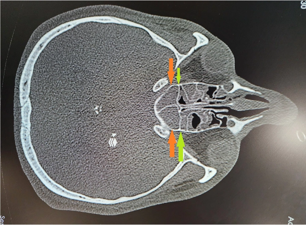
Figure 1.
Relationship between the anterior face of sphenoid sinus (green arrows) and optical foramen (orange arrows)
3. Methods
This cross-sectional study was carried out at Imam Khomeini Medical Faculty Hospital (Ahvaz, Iran) between March 21 and September 22, 2022. The study was approved by the institutional review board/ethics committee of Ahvaz Jundishapur University of Medical Sciences (IR.AJUMS.HGOLESTAN.REC.1402.096). All research stages were in accordance with the principles of the Declaration of Helsinki. Eighty computed tomography (CT) scans of the orbits and sinuses (160 orbits) were included from the Picture Archiving and Communication System (PACS) server at the Radiology Department between March and September 2022. A 64 multi-slice CT scanner (Siemens Medical Solutions, Erlangen, Germany) was used to take cranial CT images. Obtained images were multi-planar (axial, coronal, and sagittal) bone window slices taken from the paranasal sinuses, with thin (1.5 mm) axial slices. The orbitomeatal line was used for standardizing the imaging planes. Patients aged 17 years or older who had normal CT scans of orbits and sinuses, regardless of the reasons for the scans, were included in the study. Subjects with any major sinus or orbital abnormality as seen on CT scans, or cases with a past history of any kind of paranasal, sinus, or orbital surgery, trauma, or any abnormal findings such as benign and malignant lesions in the orbital/sinus area were excluded from the study. Other exclusion criteria included massive polyps of the sphenoid sinus, any orbital or cranial deformation involving the sphenoid sinus, or any other marked facial deformity.
3.1. Sample Size Calculation
The required sample size was estimated based on the results of a previous study. In the study conducted by Cinaroglu et al. (5), the proportion of individuals with symmetrical positioning of the OTF between the right and left orbits was reported as P = 0.609. Considering a significance level of α = 0.05 and a margin of error d = 0.25, the minimum required sample size was calculated to be 63. This estimation was performed using PASS software, based on the Score with Continuity Correction method. However, a total of 80 participants were ultimately included in the study due to the availability of sufficient data and to enhance the precision of the data analysis.
A single radiologist was exclusively trained in identifying the specific landmarks in order to review the CT scans and undertake measurements. Anatomical landmarks were identified by multiplanar evaluation of images. The first image on the axial plane, which identified the structures to be measured, was used for the tomographic analysis in the anterior to posterior directions. If two landmarks were not identified in the same slice, tilting was done so both landmarks were identifiable. The following measurements were performed.
By examination of the obtained CT slices, the locations of the medial part of the OTF, the superolateral area of the AFS where it joins the medial orbital wall (MOW), and the CP in the posterior area of the sphenoid sinus were determined. The CP is caused by the protrusion of a segment of the internal carotid artery in the posterior wall of the sphenoid sinus; the point of maximum convexity was chosen as the landmark for measurements. The relative position of the OTF and the AFS was reported as anterior, posterior, and equal to each other. The OTF’s position was reported as symmetrical if it was the same in both the right and left orbit, and it was considered asymmetrical if the location was different between the two orbits of the same patient.
A digital caliper was used to measure the front-to-back distance (mm) between the OTF and the AFS, as well as the distances from both the OTF and AFS to the CP in thin axial slices (Figure 1). The acquired data were rechecked to ensure accuracy and consistency prior to analysis. We used Stata version 14 (StataCorp.) software to perform the statistical analysis. An independent t-test was performed to compare the mean values based on sex and laterality (right/left) (Table 1). A Pearson correlation test was performed to investigate the correlation between all measurements and variables (age, sex, and laterality) included in the study. A P-value < 0.05 was considered to be statistically significant. The results of the descriptive statistics for the categorical variables are reported as number (%) and for continuous variables are reported as mean ± SD.
Table 1.The Mean Values of Parameters Based on Sex
| Orbit; Parameters | N | Mean ± SD | P-Value |
|---|---|---|---|
| Right | |||
| OTF-AFS | 0.365 | ||
| Male | 40 | 3.528 ± 2.4943 | |
| Female | 40 | 3.040 ± 2.2862 | |
| MOWL | < 0.001 | ||
| Male | 40 | 38.935 ± 2.4707 | |
| Female | 40 | 37.010 ± 2.2027 | |
| AFS-CP | 0.398 | ||
| Male | 40 | 7.817 ± 3.2130 | |
| Female | 40 | 7.205 ± 3.2333 | |
| OTF-CP | 0.229 | ||
| Male | 40 | 6.960 ± 2.3403 | |
| Female | 40 | 6.325 ± 2.3420 | |
| Left | |||
| OTF-AFS | 0.548 | ||
| Male | 40 | 3.715 ± 2.4795 | |
| Female | 40 | 3.393 ± 2.4795 | |
| MOWL | ≤ 0.001 | ||
| Male | 40 | 38.563 ± 2.3192 | |
| Female | 40 | 36.678 ± 2.3946 | |
| AFS-CP | 0.407 | ||
| Male | 40 | 8.533 ± 2.9325 | |
| Female | 40 | 7.972 ± 3.0800 | |
| OTF-CP | 0.808 | ||
| Male | 40 | 6.780 ± 2.5764 | |
| Female | 40 | 6.910 ± 2.1801 |
Abbreviations: SD, standard deviation; OTF, optic foramen; AFS, anterior face of sphenoid; MOWL, medial orbital wall length; CP, carotid prominence.
4. Results
One hundred and sixty orbits of 80 subjects (males = 40 and females = 40) were included. The subjects were aged between 17 and 75 years, with an average age of 46.81 ± 16.74 years. The CT scans that were entered in the study were all reported to be normal.
Findings of the relative position of the OTF to the AFS (regardless of sex and orbit side): Among the 160 evaluated orbits, the OTF was positioned anterior to the AFS in 84 (52.5%), it was in an equal position to the AFS in 37 (23.1%), while it was posterior to the AFS in the remaining 39 (24.3%) (Table 2). The finding was symmetrical in 50% of patients (n = 40).
Table 2.Position of the Optic Foramen Relative to the Anterior Aspect of the Sphenoid Sinus Based on Sex a
| Sex | Right | Left | Total | |||||||||
|---|---|---|---|---|---|---|---|---|---|---|---|---|
| Equal | Anterior | Posterior | Total | Equal | Anterior | Posterior | Total | Equal | Anterior | Posterior | Total | |
| Male | 6 (15) | 25 (62.5) | 9 (22.5) | 40 (100) | 11 (27.5) | 22 (55) | 7 (17.5) | 40 (100) | 17 (23.1) | 47 (58.8) | 16 (20) | 80 (100) |
| Female | 8 (20) | 20 (50) | 12 (30) | 40 (100) | 12 (30) | 17 (42.5) | 11 (27.5) | 40 (100) | 20 (25) | 37 (46.3) | 23 (28.7) | 80 (100) |
| P-value | 0.635 | 0.391 | 0.45 | |||||||||
a Values are expressed as No. (%).
In the right orbit, the mean distance when the OTF was positioned anterior to the AFS was 4.778 ± 1.56 mm, and when the OTF was posterior to the AFS, the mean distance was 4.195 ± 1.62 mm. In the left orbit, the corresponding rates were 3.86 ± 2.168 mm and 5.29 ± 2.03 mm, respectively (Table 3). The mean distance of the OTF to the AFS was 4.35 ± 1.378 mm when the OTF was anterior to the AFS and 4.7 ± 1.80 mm when the OTF was located posterior to the AFS.
Table 3.Mean Values of Parameters Based on the Position of the Optic Foramen Relative to the Anterior Surface of the Sphenoid Sinus (mm)
| Parameters | N | Mean ± SD | Min-Max | 95% CI for Mean (Lower-Upper Bound) |
|---|---|---|---|---|
| OTF-AFS (right); mm | ||||
| Equal | 14 | 0.00 ± 0.00 | 0 - 0 | 0 - 0 |
| Anterior | 45 | 4.778 ± 1.56 | 2 - 8.7 | 4.284 - 5.259 |
| Posterior | 21 | 4.195 ± 1.62 | 1.5 - 8 | 3.434 - 4.956 |
| OTF-AFS (left); mm | ||||
| Equal | 23 | 0.00 ± 0.00 | 0 - 0 | 0 - 0 |
| Anterior | 39 | 3.86 ± 2.168 | 1.1 - 6.8 | 3.422 - 4.297 |
| Posterior | 18 | 5.29 ± 2.03 | 2.7 - 9 | 4.313 - 6.276 |
Abbreviations: SD, standard deviation; OTF, optic foramen; AFS, anterior face of the sphenoid sinus.
The outcomes of other measurements, including the average MOWL and the average distance between the OTF and AFS to the CP, were 37.79 ± 2.29 mm, 6.74 ± 2.25 mm, and 7.87 ± 2.76 mm, respectively (Table 1). The localization of the OTF relative to the AFS is shown in Table 2. In the right orbit, the AFS and the OTF were in the same positions (anteroposteriorly) in 15% of men (n = 6) and 20% of women (n = 8), while in the left orbit, the corresponding rates were 27.5% (n = 11) and 30% (n = 12), respectively. The OTF was anterior to the AFS in 62.5% (n = 25) of men and 50% (n = 20) of women in the right orbit, whereas in the left orbit, the corresponding frequencies were 55% (n = 22) and 42.5% (n = 17), respectively. In the right orbit, the OTF was posterior to the AFS in 22.5% of men (n = 9) and 30% of women (n = 12), while in the left orbit, the corresponding rates were 17.5% (n = 7) and 27.5% (n = 11), respectively.
By evaluating both women and men, in the right orbit, the OTF was in front of the AFS in 56.2% of cases (n = 45), in an equal position to it in 17.5% (n = 14), and posterior to it in 26.2% of subjects (n = 21). In the left orbit, the corresponding rates were 48.7% (n = 39), 28.7% (n = 23), and 22.5% (n = 18), respectively. Using the chi-square test, the position of the OTF relative to the AFS (anterior/posterior/equal) based on sex and laterality (right/left) was evaluated (Table 2). No statistically significant correlations were found between any of the measurements and variables, sex, and the side of the orbit (P > 0.05). The mean distance and the relative position between the OTF and the AFS are presented in Table 3.
The mean values of measurements such as MOWL, the distance between the OTF and the AFS, the distance between the OTF and the CP, and the distance between the AFS and the CP, stratified by sex, are presented in Table 1. An independent t-test was performed to compare the mean values based on sex and laterality (right/left). According to the results, MOWL was the only parameter that was significantly different between the two sexes; in both right and left orbits, the mean values of MOWL were significantly higher in men compared to women (P-value in right orbit = 0.00 and left orbit = 0.001). Regarding other parameters, there was no statistically significant difference between men and women in both right and left orbits (Table 1). The Pearson correlation test showed a negative correlation between AFS-CP and MOWL and a positive correlation between OTF-CP and AFS-CP in both men and women.
5. Discussion
In the current research, the relative relationship and correlation between the two landmarks (OTF and the anterior face of the sphenoid sinus) were evaluated in CT images of subjects in Ahvaz city, Iran. According to our findings, the OTF was positioned anterior to the AFS in 52.5% of the cases, it was in an equal position to the AFS in 23.1%, while it was posterior to the AFS in the remaining 24.3%. The finding was symmetrical in 50% of patients (n = 40).
According to our findings, the OTF was mainly anterior (52.5%) to the AFS in both males and females; this was more often seen in males than females (62.5% of males versus 50% of females in the right orbit and 55% of males versus 42.5% of females in the left orbit) (Table 2). In the right orbit, the mean distance when the OTF was positioned anterior to the AFS was 4.778 ± 1.56 mm, and when the OTF was posterior to the AFS, the mean distance was 4.195 ± 1.62 mm. In the left orbit, the corresponding rates were 3.86 ± 2.168 mm and 5.29 ± 2.03 mm, respectively (Table 3). The mean distance of the OTF to the AFS was 4.35 ± 1.378 mm when the OTF was anterior to the AFS and 4.7 ± 1.80 mm when the OTF was located posterior to the AFS. The results of other measurements, including the average MOWL and the average distance between the OTF and AFS to the CP, were 37.79 ± 2.29 mm, 6.74 ± 2.25 mm, and 7.87 ± 2.76 mm, respectively (Table 1).
The superolateral part of the anterior sphenoid sinus wall (where it meets the MOW) was the chosen landmark for measurements in this study. The reason for selecting this specific point of the anterior sinus wall (as an alternative to the ostium or other areas of the wall) was to assess the accessibility of this area from a surgical perspective, especially in cases where there is a need for maximal posterior orbital decompression to the level of the orbital apex. In addition, although in a few instances the anterior sinus wall may be well located vertically in the coronal plane, in the majority of cases, due to high structural variability, the anterior wall has variable inclinations in different planes (4, 19). Therefore, from a surgical standpoint, for performing surgery in the orbital apex region, this superolateral insertion point may have the highest level of practical clinical significance. In cases where the optical foramen was located posterior to the anterior sinus wall, it might be necessary to involve the sphenoid sinus by removing some part of it (sphenoidectomy) to reach the orbital apex in maximal orbital decompression surgeries (9).
This involvement by itself potentially increases the risk of surgery because of the proximity of the sphenoid sinus to a number of vital anatomical structures such as the optic canal, carotid artery, and other structures in the skull base (9). Injury to the carotid artery could result in profuse hemorrhage, which is an emergency situation. Therefore, the importance of planning prior to the surgery and evaluating the necessity of involving the sphenoid sinus cannot be overstated. In our study, it was inevitable to involve the sphenoid sinus during this intervention in 24.3% of the cases.
In cases where the optical foramen was located anterior to the anterior sinus wall, it indicates that part of the optical canal was also located anterior to the sinus wall. Therefore, surgeries in the posterior ethmoidal sinus area or the anterior part of the sphenoid sinus could increase the risk of optic canal and subsequently optic nerve injury (9). This was the case in 52.5% of the subjects. The high standard deviation obtained from different measurements indicates the high level of variation in the orbital apex area in different individuals and even between the right and left sides in one person. No statistically significant correlations were found between the obtained results and age and sex. Higher study populations in the future might reveal possible correlations among different variables.
In recent years, three similar studies have been conducted by Aujla et al. (4), Cinaroglu et al. (5), and Nguyen et al. (9), evaluating the position of the optical foramen relative to the anterior wall of the sphenoid sinus in CT scans and measuring the distance from the optical foramen and anterior wall of the sphenoid sinus to the CP. We will compare our results to these previous studies in the following section.
Our results showed that the optical foramen was positioned anterior to the sphenoid sinus wall in 52.5% of orbits, while it was located posterior in 24.3% of the orbits, and in the remaining 23.1%, it was at the same level as the sinus wall. These results are well in line with previous findings reported by Nguyen et al. and Cinaroglu et al., revealing that the optical foramen was positioned anterior to the anterior wall of the sphenoid sinus in 50% and 51.5% of the orbits, respectively. A study by Aujla et al. (4) revealed a 40.6% anterior position of the OTF relative to the sinus wall, which shows a mostly posterior position of the OTF contrary to our study and reports by Cinaroglu et al. and Nguyen et al. (5, 9). Slight discrepancies between the findings could be attributed to race and sex variables.
In the study by Aujla et al. (4) and Nguyen et al. (9), the relative position of the OTF to the anterior face of the sphenoid sinus was reported as being either anterior or posterior to the sphenoid sinus (3). Nevertheless, in our study, similar to the article by Cinaroglu et al. (5), the relative position of the OTF was categorized as being anterior, posterior, or in an equal position to the sphenoid sinus wall. It seems that more precise and detailed information can be obtained by reporting the relative position of the optical foramen to the anterior sinus wall as anterior, posterior, and equal. Furthermore, the sex factor was taken into account in our study.
In the current study, the relative position of the OTF to the anterior sinus wall was symmetrical in 50% (n = 40) of the cases. In the previous studies by Nguyen et al., Cinaroglu et al., and Aujla et al. (4, 5, 9), symmetry was reported as 65%, 60.9%, and 80%, respectively, which were all higher than the 50% symmetry reported in our study. In the study by Cinaroglu et al. (5), where the sex factor was also evaluated, symmetry was higher in females (75%) compared to males (46.9%). However, contrary to these findings, our results suggest a higher rate of symmetry in males (55%) relative to females (45%).
Among the symmetric cases, regardless of sex, in 70.7% the OTF was anterior to the AFS, in 22% it was located posterior to the AFS, and it was positioned at the same level in the remaining 7.3%. These findings could be useful in cases that need revision surgery after previous surgeries that may have altered the anatomical landmarks and in cases with unilateral involvement of lesions or diseases. The mean distance of the OTF to the AFS was 4.35 ± 1.378 mm when the OTF was anterior to the AFS and 4.7 ± 1.80 mm when the OTF was located posterior to the AFS, which is similar to findings reported by other studies (1, 9).
The average distance of the OTF and the AFS to the CP, located in the posterior sinus wall, was 6.74 ± 2.25 mm and 7.87 ± 2.76 mm, respectively, which falls well within the range of variations reported previously by other authors (4, 9). There is, of course, a need for surgeons to be familiar with the relationship between these important anatomical landmarks in order to minimize the risk of complications such as injuring the carotid artery and other vital structures. The average length of the MOWL was reported as 37.79 ± 2.29 mm, which is in line with previous studies (4, 20-23).
Regarding the sex factor, all the measurements obtained in this study were slightly higher in males compared to females, which is in agreement with the findings reported by Enatsu et al. (8).
Finally, a number of potential limitations need to be considered. First, key landmarks used for measurements are not usually identifiable on a single axial CT slice. In order to obtain a proper view for measurements on a patient, CT planes may require reconstruction by tilting the plane and thickening the slices. It might be useful to integrate such a reconstruction in the preoperative protocols so the need for manual reconstruction of the images will be eliminated. Another limitation was that all measurements were taken by a single radiologist. In order to increase accuracy and reduce errors, it is recommended that measurements be taken by at least two individuals. Artificial intelligence could also be applied in future studies to better help address challenges in certain areas.
5.1. Conclusions
Based on the results of CT scans, the OTF lies anterior to the anterior sphenoid sinus wall in nearly half of the orbits (52.5%), is positioned posterior in a quarter of cases (24.3%), and is at the same level in the remaining quarter of orbits (23.1%). Additionally, the OTF was in a symmetrical position in the two orbits in 50% of subjects. The data collected in the current study provides detailed measurements in the area of the sphenoid sinus, carotid canal, and optic canal, and our findings could be a useful aid for surgeons to avoid complications while performing surgeries such as orbital decompression. It should be noted that there may be differences in terms of race and sex.



