


1. Background
Chronic kidney disease (CKD) affects more than 500 million individuals worldwide, representing approximately 10% of the adult population. More than 1.5 million people rely on renal replacement therapies, such as dialysis or transplantation, to survive (1). Kidney transplantation remains the preferred treatment for patients with end-stage renal disease (ESRD), offering superior quality of life and long-term survival compared with dialysis (2). Global estimates suggest that more than 1.4 million patients with CKD have undergone kidney transplantation, with an annual growth rate of 8% (3). In addition to recent advances in surgical techniques and pharmacotherapeutic protocols for renal transplantation, perioperative fluid and acid-base management plays a critical role in ensuring adequate graft perfusion and optimizing clinical outcomes. Furthermore, educational interventions have significantly improved all domains of health-related quality of life and reduced the incidence of acute post-transplant complications, including infection, acute rejection, and hospital readmission, among patients undergoing kidney transplantation (4, 5). Despite these therapeutic advances and improvements in graft survival over recent decades, acute rejection (AR) and DGF remain major obstacles to successful transplantation outcomes (6). Acute rejection, an immune-mediated response against the transplanted kidney, has decreased in prevalence but continues to compromise graft longevity (7). Delayed graft function, which is typically observed after deceased-donor transplantation, is defined by the requirement for dialysis within the first postoperative week and is frequently associated with ischemia-reperfusion injury leading to ATN (8). This condition not only increases the risk of AR but also negatively affects graft and patient survival (9-11).
The incidence of DGF varies according to donor type and immunosuppression regimen, ranging from less than 5% in living-donor transplants to more than 80% in non-heart-beating donors (12, 13). Despite medical advances, DGF rates have remained relatively unchanged, partly because of the increasing use of marginal donors, including expanded-criteria donors and donors after cardiac death (14). Delayed graft function presents both diagnostic and therapeutic challenges because its causes are diverse and include immunologic rejection, surgical complications, drug toxicity, recurrent disease, and ATN (8).
2. Objectives
Given the clinical significance and multifactorial etiology of DGF, timely differentiation among its causes is essential for effective management. This study aimed to determine the prevalence of ATN and acute rejection in kidney transplant recipients with DGF and to identify associated immune and nonimmune risk factors.
3. Methods
3.1. Study Design and Population
This retrospective observational study included adult patients who underwent kidney transplantation at our center between 2010 and 2016. Using a census sampling method, we reviewed the medical records of kidney donors and recipients. The inclusion criteria were as follows: 1) recipient age ≥ 18 years; 2) clinically diagnosed DGF, in accordance with the aforementioned definition, followed by allograft biopsy; and 3) availability of complete medical records for donor and recipient variables. Patients were excluded if they had 1) a primary nonfunctioning graft, defined as a graft that never achieved sufficient function to allow discontinuation of dialysis; 2) death due to non-transplant-related causes, such as accidental death or unrelated malignancy, occurring before graft biopsy or within the first postoperative week; or 3) an inadequate biopsy specimen, defined as a biopsy containing fewer than 7 glomeruli or no arterial cross-section.
3.2. Definitions and Data Collection
All cases of DGF were confirmed by histopathological evaluation of allograft biopsy specimens. Biopsy findings were categorized as ATN, acute rejection according to the diagnostic criteria of the Banff 2017 Kidney Meeting Report, both ATN and rejection, or the absence of both conditions. The following variables were extracted from patient records: recipient and donor age, sex, body mass index (BMI), blood group and Rh status, underlying kidney disease, induction immunosuppressive regimen, donor cause of death, and serum creatinine levels.
3.3. Statistical Analysis
Data were analyzed using SPSS software version 17.0 (SPSS Inc., Chicago, IL, USA). Normality of distribution was assessed using the Kolmogorov-Smirnov test and visual inspection of histograms. Descriptive statistics were reported as mean ± standard deviation (SD) for continuous variables and frequency (percentage) for categorical variables. Group comparisons were performed using the Pearson chi-square test or Fisher exact test, as appropriate, for categorical variables and the independent t-test for continuous variables. A P value of < 0.05 was considered statistically significant.
4. Results
A total of 203 kidney transplant recipients who underwent allograft biopsy due to DGF were initially included. However, 8 patients were excluded because of inadequate biopsy specimens, resulting in a final study population of 195 patients. The mean recipient age was 42.46 ± 14.21 years (range, 19 - 75 years), including 105 males (53.8%) and 90 females (46.2%). Recipient baseline characteristics are summarized in Table 1.
Table 1.Baseline Characteristics of Recipients
| Variables | Recipients (n = 195) |
|---|---|
| Recipient age (y) | 42.46 ± 14.21 |
| Recipient sex | |
| Male | 105 (53.8) |
| Female | 90 (46.2) |
| Recipient BMI (kg/m2) | 21.74 ± 4.81 |
| Recipient blood group | |
| A | 66 (33.8) |
| B | 49 (25.1) |
| O | 67 (34.4) |
| AB | 13 (6.7) |
| Recipient Rh status | |
| Positive | 183 (93.8) |
| Negative | 12 (6.2) |
| Underlying kidney disease | |
| Hypertension | 41 (21.0) |
| Diabetes mellitus | 29 (14.9) |
| Polycystic kidney disease | 9 (4.6) |
| Renal stone | 11 (5.6) |
| Glomerulonephritis | 18 (9.2) |
| Unknown | 85 (29.7) |
| Other | 29 (14.9) |
| Induction regimen | |
| None | 45 (23.1) |
| Thymoglobulin | 126 (64.6) |
| Alemtuzumab | 8 (4.1) |
| Basiliximab | 3 (1.5) |
| Daclizumab | 13 (6.7) |
Donors had a mean age of 38.67 ± 13.84 years (range, 18 - 80 years), including 110 males (56.4%) and 85 females (43.6%). The primary causes of donor death were trauma (43.1%), intracranial hemorrhage (19.5%), cardiac events (12.3%), and other causes (25.1%). The mean serum creatinine levels on the first and third post-transplant days were 3.71 ± 3.72 mg/dL and 6.26 ± 3.27 mg/dL, respectively. Donor baseline characteristics are presented in Table 2.
Table 2.Baseline Characteristics of Donors
| Variables | Donors (n = 195) |
|---|---|
| Donor age (y) | 38.67 ± 13.84 |
| Donor sex | |
| Male | 110 (56.4) |
| Female | 85 (43.6) |
| Donor blood group | |
| A | 65 (33.3) |
| B | 52 (26.7) |
| O | 66 (33.8) |
| AB | 12 (6.2) |
| Donor Rh status | |
| Positive | 184 (94.4) |
| Negative | 11 (5.6) |
| First creatinine level (mg/dL) | 3.71 ± 3.72 |
| Third creatinine level (mg/dL) | 6.26 ± 3.27 |
| Cause of death | |
| Trauma | 84 (43.1) |
| Intracranial hemorrhage | 38 (19.5) |
| Cardiac events | 24 (12.3) |
| Other | 49 (25.1) |
Histopathological evaluation of kidney biopsies revealed ATN in 155 patients (79.5%) and rejection in 86 patients (44.1%). Among these patients, 55 (28.2%) showed evidence of both ATN and rejection, 100 (51.3%) had isolated ATN, and 31 (15.9%) had isolated rejection. Notably, 9 patients (4.6%) had neither ATN nor rejection. The distribution of biopsy findings is illustrated in Figure 1.
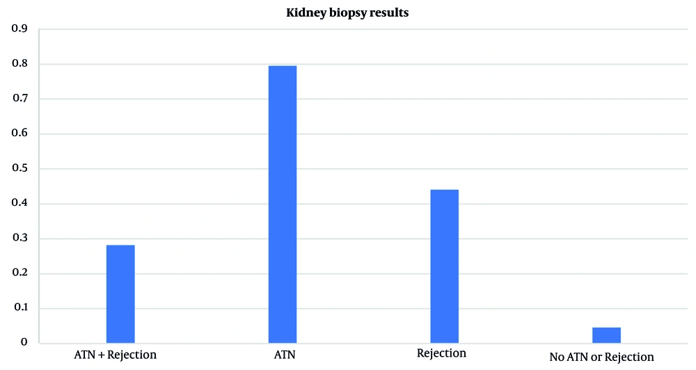
Figure 1.
Osteoporosis prevalence by treatment group analysis. The difference was statistically significant (P = 0.01)
A total of 186 kidney transplant recipients were categorized into 3 groups: patients with ATN (n = 100), patients with graft rejection (n = 31), and patients with both conditions (ATN plus rejection; n = 55). Further analyses compared clinical and donor-related variables among these categories. Donor age was comparable across all groups (P > 0.05). However, donor sex distribution differed significantly, with the ATN plus rejection group having a higher proportion of female donors (60%) than the ATN group (41%; P = 0.02) and rejection group (29.1%; P = 0.006). The cause of donor death also differed significantly between the ATN and rejection groups (P = 0.01); trauma was most frequent in the ATN group (45%), whereas intracranial hemorrhage was most common in the rejection group (41.9%).
Recipient age and BMI were similar across groups. In contrast, recipient sex distribution varied substantially, with P values of 0.03 for the ATN versus rejection comparison, 0.01 for the ATN versus ATN plus rejection comparison, and 0.001 for the rejection versus ATN plus rejection comparison. The rejection group was predominantly male (77.4%), whereas the ATN plus rejection group was predominantly female (65.5%). The distribution of recipients' underlying kidney diseases did not differ significantly between groups (P > 0.05). Clinically, the first recorded creatinine level was significantly higher in the ATN plus rejection group (5.03 ± 3.74 mg/dL) than in the rejection group (1.76 ± 1.55 mg/dL; P = 0.001). The level in the ATN group (3.89 ± 4.10 mg/dL) was also significantly higher than that in the rejection group (P = 0.006). No significant difference was found in third creatinine levels. Significant differences were also observed in the use of induction immunosuppressive regimens (P = 0.03 for ATN vs rejection, P = 0.002 for ATN vs ATN plus rejection, and P = 0.009 for rejection vs ATN plus rejection). Thymoglobulin was used most frequently in the rejection (96.7%) and ATN (68%) groups, whereas nearly half of the ATN plus rejection group (49.1%) received no induction therapy (Table 3).
Table 3.Comparison of Immune and Nonimmune Characteristics Between Patients with ATN and Rejection in Biopsy Results
| Variables | ATN group (n = 100) | Rejection group (n = 31) | ATN + rejection group (n = 55) | ATN vs rejection P value | ATN vs ATN + rejection P value | Rejection vs ATN + rejection P value |
|---|---|---|---|---|---|---|
| Donor age (y) | 38 ± 13.88 | 41.64 ± 14.35 | 38.38 ± 13.94 | 0.21 | 0.87 | 0.31 |
| Donor sex | 0.29 | 0.02 | 0.006 | |||
| Male | 59 (57.3) | 22 (21.4) | 22 (21.4) | |||
| Female | 41 (49.4) | 9 (10.8) | 33 (39.8) | |||
| First creatinine level (mg/dL) | 3.89 ± 4.10 | 1.76 ± 1.55 | 5.03 ± 3.74 | 0.006 | 0.09 | 0.001 |
| Third creatinine level (mg/dL) | 6.78 ± 3.77 | 6.05 ± 2.95 | 5.64 ± 2.56 | 0.71 | 0.17 | 0.77 |
| Cause of death | 0.01 | 0.87 | 0.92 | |||
| Trauma | 45 (57.0) | 10 (12.7) | 24 (30.4) | |||
| Intracranial hemorrhage | 17 (47.2) | 13 (36.1) | 6 (16.7) | |||
| Cardiac events | 11 (45.8) | 0 | 13 (54.2) | |||
| Other | 27 (57.4) | 8 (17.0) | 12 (25.6) | |||
| Recipient age (y) | 41.81 ± 13.52 | 46.32 ± 15.38 | 41.61 ± 14.78 | 0.11 | 0.93 | 0.16 |
| Recipient sex | 0.03 | 0.01 | 0.001 | |||
| Male | 56 (56.6) | 24 (24.2) | 19 (19.2) | |||
| Female | 44 (50.6) | 7 (8.0) | 36 (41.4) | |||
| Recipient BMI (kg/m2) | 21.21 ± 5.62 | 23 ± 6.23 | 22.03 ± 4.36 | 0.76 | 0.59 | 0.83 |
| Underlying kidney disease | 0.06 | 0.47 | 0.94 | |||
| Hypertension | 20 (48.8) | 4 (9.8) | 17 (41.4) | |||
| Diabetes mellitus | 13 (46.4) | 8 (28.6) | 7 (25.0) | |||
| Polycystic kidney disease | 3 (37.5) | 5 (62.5) | 0 | |||
| Renal stone | 5 (50.0) | 1 (10.0) | 4 (40.0) | |||
| Glomerulonephritis | 13 (72.2) | 2 (11.1) | 3 (16.7) | |||
| Unknown | 32 (58.2) | 9 (16.4) | 14 (25.4) | |||
| Other | 14 (53.8) | 2 (7.7) | 10 (38.5) | |||
| Induction regimen | 0.03 | 0.002 | 0.009 | |||
| None | 17 (37.8) | 1 (2.2) | 27 (60.0) | |||
| Thymoglobulin | 68 (58.1) | 30 (25.6) | 19 (16.2) | |||
| Alemtuzumab | 4 (50.0) | 0 | 4 (50.0) | |||
| Basiliximab | 2 (66.7) | 0 | 1 (33.3) | |||
| Daclizumab | 9 (69.2) | 0 | 4 (30.8) |
5. Discussion
Kidney transplantation remains the optimal treatment for patients with ESRD, providing substantial improvements in quality of life and survival compared with maintenance dialysis (15). However, despite advances in immunosuppressive regimens and postoperative care, acute renal allograft rejection remains a major clinical challenge that threatens long-term graft survival. This issue underscores the complexity of managing transplant recipients, who are inherently at risk because of both their underlying condition and the immunosuppressive therapies required to maintain graft viability (15).
This study highlights that, although the incidence of acute rejection has decreased substantially over the past few decades, particularly with the use of potent agents such as calcineurin inhibitors and mycophenolate mofetil, its presence, even when subclinical, can have lasting implications for graft function (15, 16). Notably, subclinical rejection, often identified through protocol biopsies, may progress silently and result in interstitial fibrosis and tubular atrophy, ultimately contributing to chronic allograft nephropathy and death-censored graft loss (16).
One of the most pressing concerns is the apparent disconnect between decreased rates of acute rejection and the plateau in long-term graft survival. This paradox suggests that other factors, including overimmunosuppression, calcineurin inhibitor toxicity, and undetected or inadequately treated subclinical antibody-mediated rejection, may offset the benefits achieved through improved early rejection control (17). The distribution of induction immunosuppressive regimens differed among our study groups. The high use of thymoglobulin in the isolated rejection group reflects our center protocol for patients perceived to be at high immunological risk. Conversely, the finding that nearly half of the ATN plus rejection group received no induction therapy is striking. This observation raises the question of whether inadequate initial immunosuppression contributed to the development of rejection in patients who were also experiencing ischemic injury (ATN).
This study also emphasizes the importance of identifying and mitigating risk factors such as presensitization, human leukocyte antigen mismatches, donor quality, particularly extended-criteria donors, and DGF (18). Similarly, the findings of this manuscript suggest that donor sex was significantly associated with biopsy outcomes, with female donors overrepresented in the ATN plus rejection group. This aligns with some prior studies suggesting that donor-recipient sex mismatch, particularly female-to-male donation, may be associated with differential immunological risks, potentially mediated by minor histocompatibility antigens. Additionally, the difference in donor cause of death between the ATN and rejection groups, with more trauma in the ATN group and more intracranial hemorrhage in the rejection group, may relate to differences in donor management, inflammatory milieu, or organ quality, which could predispose grafts to different types of injury.
The complex pathophysiology of DGF, involving ischemia-reperfusion injury, inflammation, and immune activation, represents a multifactorial process that adversely affects post-transplant outcomes (19). Innovations in donor management and organ preservation, such as machine perfusion and hypothermic strategies, may help reduce its incidence in the future (20).
Furthermore, evolving tools for rejection surveillance, including noninvasive biomarkers such as donor-derived cell-free DNA, show promise for the early detection of rejection episodes and may offer alternatives to protocol biopsies (19). However, their predictive value remains modest, emphasizing the continued need for histological confirmation in many cases (21).
This study has several limitations. Its single-center retrospective design may limit generalizability. The sample size, particularly in the isolated rejection group (n = 31), reduces the statistical power of some comparisons. Another limitation is that our center exclusively uses deceased donors. Regrettably, data on cold ischemia time were unavailable.
5.1. Conclusions
In conclusion, although substantial progress has been made in reducing acute rejection rates, improving long-term kidney allograft survival will likely depend on more nuanced immunosuppression strategies, early identification of subclinical rejection, personalized risk assessment, and optimization of donor organ quality. Close collaboration between transplant centers and general nephrologists is essential for long-term success, particularly as care transitions from specialized to community settings.
