
1. Background
Azurin, as a redox protein found in Pseudomonas aeruginosa (1), is a type I copper-containing protein, transferring electrons in the denitrification pathway (2). This water-soluble 14 kDa protein (3) has shown anticancer effects both in vivo and in vitro via forming a complex with the tumor suppressor protein “p53” (4). The formation of this complex leads to the stabilization of p53 and a subsequent increase in its cellular concentration (5). Experimental investigations by site-directed mutagenesis have revealed key-points in azurin-p53 complex formation. Studies suggest that two methionine residues (Met44 and Met64) located in one of the hydrophobic patches of azurin play a critical role in stabilizing the flexible L1 and s7-s motifs of p53 (6, 7). Azurin is considered a potential anti-cancer agent that favorably enters cancerous cells at a significantly higher rate compared with normal cells (6). In spite of this remarkable advantage, in vivo studies have highlighted the immunogenic responses induced by azurin, as its major drawback (8). Nevertheless, further investigations led to the discovery of oligopeptide fragments of azurin, which have therapeutic traits equal to the whole protein (i.e. similar cytotoxic and target specific delivery capabilities) along with minimized immunogenicity (9, 10).
The p28, a cell-penetrating peptide (CPP), is a truncated derivative of azurin with retained antitumor activity. This peptide is the principal for the internalization of azurin into target cells (11). Moreover, CPPs are a category of small peptide molecules acting as potential delivery agents for various types of cargoes, including therapeutic proteins, peptides, and nucleic acids (12, 13). In fact, CPPs work in an energy-dependent manner (14) and can penetrate cancerous cells, specifically by distinguishing between normal and malignant vasculatures and lymphatic tissue (15). As mentioned earlier, p28 is an essential factor by which azurin advantageously penetrates into cancerous cells (11, 16). Indeed, p28 is a 2.9 kDa fragment of azurin (residues 50 - 77) that exerts its anti-proliferative effects through binding to and elevating the intracellular concentration of p53 (17) without any interference with Mdm2 binding or downstream ubiquitination process (10, 18, 19). Studies indicate that the tumoricidal activity of p28 results from its residues 11 - 18 (61 - 69 of azurin) (10). Additionally, p28 makes impressions on cell proliferation by arresting cell cycle at G-M interphase via increasing the intracellular levels of cyclin-dependent kinase inhibitors p21 and p27, suggesting a likelihood that p28 could interfere with the binding of MDM2 to p53. Indeed, this elevation is as a result of the formation of the complex between p28 and p53wt/mut DNA-binding domain (DBD) (6, 18, 20), which subsequently leads to a decline in proteasomal degradation of p53 (10, 21, 22).
The antitumor proficiency of p28 is assessed on a variety of cancerous cells in pre-clinical studies (9, 17, 22). These investigations have revealed that p28 can induce a G2-M interphase arrest in the cell cycle (23). Apart from its impact on the status of p53 and CDK-inhibitors, p28 also functions in non-p53-mediated mechanisms to inhibit tumor growth. After its penetration into endothelial cells, it develops a direct antiangiogenic effect, which causes tumor neoangiogenesis to be halted (24, 25). Notably, p28 can cross through the endothelial tissue and the blood-brain barrier, suggesting its capability of saturating the brain parenchyma dose-dependently (26).
2. Objectives
In this study, the pro-apoptotic and anti-proliferative effects of the recombinant p28 was studied on Raji (Burkitt’s lymphoma; p53mut) and HEK-293 (normal control) cell lines.
3. Methods
3.1. Reagents, Bacterial Strains, Plasmids, and Cell Lines
The primers were synthesized by Metabion (Germany). The T4 DNA ligase, NdeI, and BamHI were purchased from Fermentas (Litvanya). The MTT (3-(4,5-dimethylthiazol-2-yl)-2,5-diphenyltetrazolium bromide), Polyvinylidene difluoride (PVDF) membrane, 3, 3-diaminobenzidine (DAB), Dimethyl sulfoxide (DMSO), Kanamycin, Ampicillin, and Anti-his-tag antibody were obtained from Sigma-Aldrich (USA). Luria-Bertani medium was purchased from HiMedia (India). The Ni-NTA resin was provided by QIAGEN (Germany). The pTZ57R vector, pET28a expression vector, and E. coli strains were provided by Diagnostic Laboratory Sciences and Technology Research Center (Iran). P. aeroginosa strain was kindly gifted by Microbiology Lab of Medical Laboratory Sciences Department of Shiraz University of Medical Sciences. Raji and HEK-293 cell lines were obtained from Pasteur Institute of Iran. DMEM and RPMI cell culture media and fetal bovine serum were purchased from Thermo Scientific (USA). The PE-annexin V apoptosis detection kit was purchased from BD biosciences (USA).
3.2. PCR Amplification and Cloning of p28 Gene in pTZ57R
Specific primer pairs [p28-forward (5’-GAATTCCGCCCACCTGCCT-3’) and p28-reverse (5’-AAGCTTTCATGCAGCGGATCG-3’] were designed by using Allele Id version 7.5 (Primer Biosoft, USA) for PCR amplification and sequencing of p28 gene. The 84 bp fragment of p28 was isolated from P. aeruginosa genome extracted by boiling method, and amplified by end-point PCR.
DNA fragments were cloned into pTZ57R cloning vector. Afterward, E. coli DH5a competent cells were transformed with the pTZ57R /p28 plasmid construct. The cloning was authenticated by Sanger sequencing.
3.3. Expression of the Recombinant p28
The p28 gene was sub-cloned into pET-28a plasmid vector and transformed into BL21 (DE3) strain of E. coli. The bacteria were cultured on an agar plate containing Kanamycin (50 mg/mL). A single colony was inoculated in 5 mL of LB broth culture containing Kanamycin (50 mg/mL). After the incubation, the expression of the heterologous protein was induced by the addition of IPTG (1 mM). After 4 hours of incubation, bacteria were centrifuged and the pellets were resuspended in binding buffer (50 mM NaH2PO4, 500 mM NaCl, 10 mM imidazole, and 8 M urea, pH 8). Finally, cells were lysed by 6 × 30 seconds of sonication followed by 30-second intervals for cooling on ice, and finally, have been evaluated by SDS-PAGE.
3.4. Peptide Purification and Renaturation
Peptide purification was performed using Ni-NTA chromatography system. Bacterial lysates containing His-tagged p28 peptides were loaded on Ni-NTA columns. Then the column was washed with 5 mL washing buffer (50 mM NaH2PO4, 500 mM NaCl, 50 mM imidazole, and 8 M urea, pH 8) and subsequently the recombinant p28 peptide was eluted using 2 mL of elution buffer (50 mM NaH2PO4, 500 mM NaCl, 250 mM imidazole and 8 M urea, pH 8). After the elution, the fractions were assayed by SDS-PAGE.
Urea (8M) was used to solubilize the formed inclusion bodies. The obtained peptides were dialyzed against a series of binding buffers containing 5, 3, 1.5, and 0 M concentrations of urea in order to remove the residual urea and to achieve the optimal biological activity of the peptide. The concentrations of the purified peptides were determined using Bradford assay.
3.5. Western Blot Analysis
A fraction of purified p28 peptide (16 µL) was diluted in 4 µL of sample buffer, incubated for 5 minutes at 90°C, and then assayed by SDS-PAGE. By utilizing a semi-dry transfer unit, the sample was transferred onto a PVDF membrane. The membrane was treated with 5% skim milk (i.e. blocking buffer) at 4°C for 2 hours. Then the membrane was washed by TBST buffer (50 mM Tris-base, 150 mM NaCl, and 0.05% Tween 20), and incubated with anti-His-tag antibody conjugated with HRP for 1 hour. Finally, DAB was added to the surface of the membrane.
3.6. Cell Culture
Raji and HEK-293 cell lines were cultured in RPMI 1640 and DMEM complete media, respectively and grown at 37°C and 5% CO2.
3.7. Cytotoxicity Assay
The cytotoxicity of the recombinant p28 peptide was determined by MTT assay. The HEK 293 cells were used as a normal control group to evaluate the non-specific toxicities of the recombinant p28 peptide, while Raji cell line was utilized to evaluate the cytotoxicity of p28 as cancerous cells.
In brief, cells were seeded into 96-well plates at a density of 8,000 cells/well and incubated for growth. Afterward, they were treated with different concentrations of the p28 peptide (0.5, 1, and 2 µM) and incubated for 24 hours. Then 20 µL of MTT (5 mg/mL) was added to each well, followed by 4 hours of incubation for crystal formation. Finally, 100 µL of DMSO was added to each well and absorbance was measured at 490 nm wavelength using Sat Fax 2100 microplate reader (Stat Fax, USA). The MTT assay was carried out in triplicate for each concentration of p28.
3.8. Apoptosis Assay
Cells were seeded into 24-well plates to reach the confluency of 80%. Then they were treated with the recombinant p28 peptide at the concentration of 2 µM. After 24 hours of incubation, apoptosis levels were evaluated using PE-annexin V apoptosis detection kit.
3.9. Statistical Analysis
GraphPad Prism 7.03 (GraphPad Software, Inc.) was used for all analyses. One-way ANOVA was the method of choice for the statistical assessment of the apoptosis and MTT assay results.
4. Results
4.1. Primer Design and PCR Amplification of p28 Gene
The primers were designed based on the known sequence of p28 in P. aeruginosa. Figure 1 represents the agarose gel electrophoresis of p28 PCR amplification.
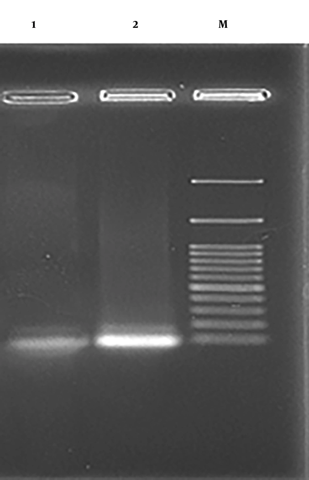
Figure 1.
PCR amplification of p28 gene. Lane M: size marker (100 - 3000 bp), lane 1: NTC, lane 2: p28 (84 bp).
4.2. Expression and Purification Analysis of the p28 Peptide
pET28a-p28 expression vector was constructed and expressed as a soluble and functional peptide in E. coli. Figure 2A demonstrates the 15% SDS-PAGE analysis of expressed p28 peptide at 3 kDa. Furthermore, SDS-PAGE and subsequent western blotting assays were used to confirm the purification of the p28 peptide (Figures 2B and 3).
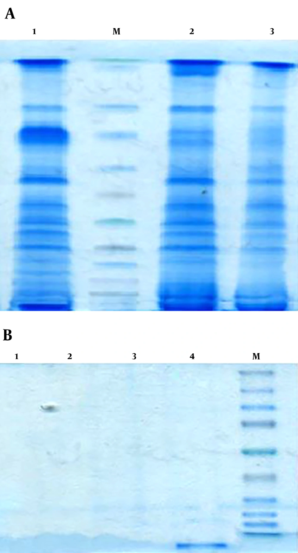
Figure 2.
A, SDS PAGE analysis of small scale for expression of the p28 peptide. Lanes 1: p28 peptide expression after the induction with 1 mM IPTG. The P28 peptide with 3 kDa molecular weight was overexpressed. Lane M: size marker (10 - 180 kDa). Lane 2, 3: pre-induced protein expression that IPTG was not added and then there was no overexpression and uninduced untransformed bacteria, respectively. B, purification of the p28 peptide using Ni-NTA purification system. Lane1: elution fraction 4, lane 2: elution fraction 3, lane 3: elution fraction 2, lane 4: elution fraction 1, 3 kDa peptide of p28 was purified, M: size marker (10 - 180 kDa).
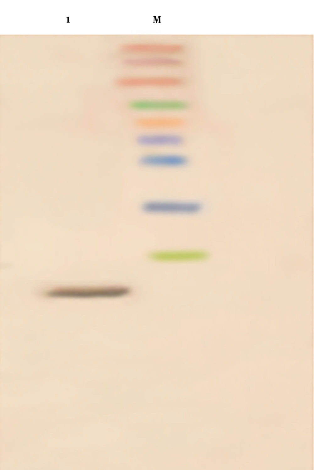
Figure 3.
Verification of the production of the p28 peptide by western blotting. Lane 1: the p28 peptide with 3kDa molecular weight, lane M: size marker.
Quantifications by Bradford assay showed a concentration of 0.05 mg/mL of purified p28 and a concentration of 0.03 mg/mL renatured p28 after dialysis.
4.3. MTT Assay
MTT assay was used for the determination of p28 cytotoxic effects in vitro. Target cells were treated with p28 peptide in a series of concentrations (0.5, 1, and 1.5 µM) for 24 hours. The MTT results are shown in Figure 4. The results suggest that, compared to the untreated groups, the treatment with p28 peptide had significant anti-proliferative effects on Raji cells in a dose-dependent manner (P < 0.0007), while its cytotoxicity on HEK-293 cells was insignificant (P > 0.9999).
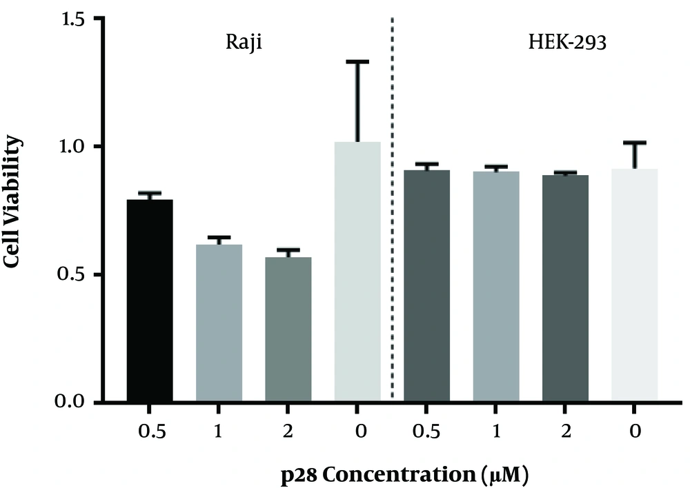
Figure 4.
The effect of p28 on the viability of Raji and HEK-293 cell lines. The cells were treated with p28 at concentrations of 0.5, 1, and 2 µM, and their viability was evaluated by MTT assay after 24 hours. The results show a statistically significant reduction in viability of Raji cells treated with 1 and 2 µM of p28 (P < 0.0007), but no significant reduction in viability of treated HEK-293 cells (P > 0.9999). (One-way ANOVA followed by Sidak post-hoc test was used for statistical analysis).
4.4. Apoptosis Assay
The pro-apoptotic effects of the p28 peptide on cancer cells investigated based on PE-annexin V apoptosis detection method. Raji and HEK-293 cells were treated with the peptide for a 24-hour period and then evaluated by a flow cytometry instrument (BD FACSCalibur, USA).
Apoptosis assay results are shown in Figure 5. The results suggest that treating the cells with p28 increases the apoptosis rate up to 25 ± 3% in Raji cells with a significant difference compared to untreated control cells (P < 0.0001). No significant effects on the apoptosis rate of HEK-293 normal cells treated with p28 peptide were observed in comparison to untreated control cells (P > 0.6).
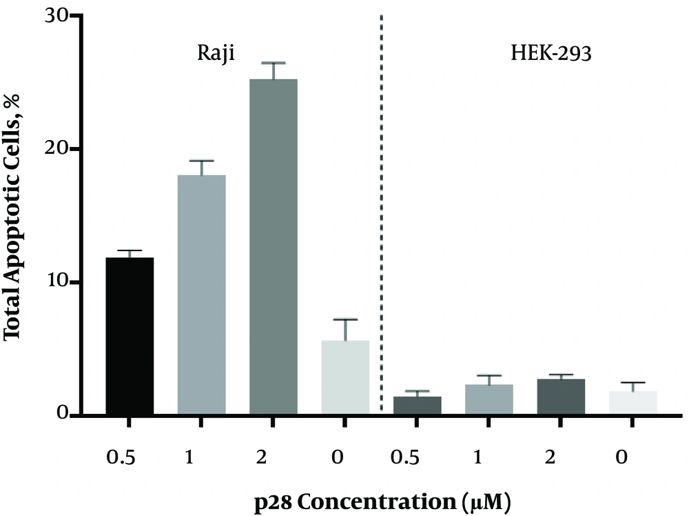
Figure 5.
Apoptosis was induced by p28 on Raji and HEK-293 cell. The cells were treated with p28 at concentrations of 0.5, 1, and 2 µM, and their apoptosis level was evaluated by PE-annexin V flow cytometry assay after 24 hours. The results show a statistically significant increase in the number of apoptotic Raji cells treated with 0.5, 1, and 2 µM of p28 (P < 0.0001). No significant changes were observed in apoptosis in treated HEK-293 cells (P > 0.6). (One-way ANOVA followed by Sidak post-hoc test was used for statistical analysis).
5. Discussion
One of the most important missions in the field of biomedical sciences is to search, find, and develop effective strategies for the treatment of cancer. To date, chemotherapeutic agents have been among the most common approaches for the treatment of various types of cancer. However, their lack of specificity in targeting cancer cells has brought numberless difficulties to the patients who are being treated with them (27). As an ongoing process, several scientific groups worldwide are carrying out large- and small-scale researches to discover and develop more efficient therapeutic agents. Consequently, a number of candidate drugs have been introduced (28). Targeted cancer therapy is described as the utilization of the agents that are involved in certain cellular signaling pathways or bound to specific molecular targets and inhibit the proliferation of cancer cells (29). Recently, peptide-based therapeutics have been considered promising candidates for cancer targeted therapy (15). These peptides are receiving attentions mainly due to their small molecular size, efficient tissue penetration, low toxicity, and non-immunogenic nature (30, 31).
Azurin, a bacterial protein found in P. aeruginosa, (2) can preferentially penetrate malignant cells, stabilize the tumor-suppressor p53 protein, and induce apoptosis in the target cells (5, 6). Nevertheless, azurin is potentially an immunogenic molecule and its employment as a therapeutic agent might not be reasonable (8). The p28, a peptide fragment of azurin, has shown specific cell penetration and anti-proliferative effects in cancer cells, similar to the whole protein; however, it does not provoke immunogenic responses (9, 17, 21). Several studies have reported that p28 forms a complex with either the DBD or NH2-terminal domain of p53 and elevates its intracellular concentration (18, 23). These studies emphasize that the p28-mediated elevation of p53 cellular levels occurs rather post-translationally and mainly to a reduction in the activity of proteasome (4, 6, 7, 18).
The p28 has shown to be one of the most promising anti-cancer peptides in preclinical studies (17). Thus it was the subject of clinical trials for the treatment of solid tumors. Results from phase I clinical trials revealed that p28 was highly efficient as a therapeutic agent in the treatment of p53+ metastatic solid tumors, indicating its promising potential of being used as an anti-cancer drug (26). In this study, we isolated p28 gene from its original source, cloned it in an expression system, and expressed its recombinant peptide in E. coli Bl21. We obtained pure functional p28 peptide by utilizing Ni-NTA chromatography column and dialysis membrane. By optimizing the expression conditions, we achieved the highest yields of p28 production. These processes were confirmed by SDS-PAGE, western blotting, and Bradford assays. Finally, to evaluate the apoptotic and cytotoxic effects of p28, we performed in vitro experiments using Raji and HEK-293 cell lines.
So far, the anticancer effects of p28 has been mainly studied on p53 wild-type cell lines such as MCF-7 with promising results (4, 10). Such cell lines are expected to undergo anti-proliferative effects of p28 readily. In the present study, we selected Raji as our cancerous cell line because of its mutant p53 status. The p53 protein in Raji cell line, has a mutation in its residue 213, while this mutation has no interference with p28 - p53 complex formation (23, 32). The cytotoxicity evaluation by MTT assay revealed that p28 peptide strongly inhibited the proliferation of Raji cells, while it had minimal effects on the non-cancerous HEK-293 cells. Apoptosis investigations were consistent with MTT assay results. The p28 induced apoptosis in the cancerous cells but not in the normal control cells.
5.1. Conclusions
In summary, despite the mutation in p53 protein in Raji cell line, it seems that there is no obstacle for the formation of p28 - p53 complex in these cells. Thus p28 can induce apoptosis in Raji cell line, suggesting its potential proficiency as an anticancer agent for the treatment of Burkitt’s lymphoma. However, in vivo anticancer effects of the p28 peptide on Burkitt’s lymphoma cells remains to be further investigated.