In the present study, the prevalence of atypical endometriosis was about 9% (25 out of 260), which is consistent with the results of a meta-analysis that reported a prevalence of 8% (
19). In this meta-analysis, five studies that in total, comprised 1109 patients with endometriotic lesions are reviewed, and a total of 89 cases of severe atypical endometriosis are identified (
19). In the present study, the mean age of typical and atypical endometriosis cases was about 30 years, in which patients in the EAOC group were significantly different concerning the variable of age (the mean age was 45 years). Another study conducted on a sample of patients with endometriosis in Iran with a mean age of 27.9 ± 6.1 years has reported similar results (
20). The higher mean age of patients in the EAOC group, compared to those in the endometriosis and atypical endometriosis groups, can be attributed to the long-lasting process of progression from endometriosis to atypical endometriosis and to carcinoma. Studies conducted by Mangili et al. (
21) and Akbarzadeh et al. (
18) reported a mean age of 55 ± 10 and 49.93 ± 9.36 years for patients with endometrioid ovarian cancer-associated endometriosis, respectively, which was lower than the mean age of patients with only endometrioid ovarian cancer (i.e., 62 ± 12 years) (
21). These findings are also consistent with those of the present study. Patient age, when diagnosing ovarian endometriosis, postmenopausal status, is a risk factor for the development of EAOC (
22).
Several studies have suggested that atypical endometriosis is a transition between benign endometriosis and EAOC (
2,
4,
7,
8). In 1925, Sampson was the first to describe the malignant transformation of endometriosis to ovarian carcinoma (
23). Moll et al. (
24) reported a chronological association between atypical endometriosis and ovarian carcinoma in a woman with clear cell carcinoma three years after ovarian cystectomy of an endometrioma with severe atypical changes.
Okamura and Katabuchi (
25,
26) presented evidence of a direct transition from the endometriotic gland to atypia to carcinoma in endometrioid carcinoma arising from an ovarian endometriotic cyst. In a study by Akbarzadeh et al. (
18), 25.4% (28 out of 110) of ovarian cancer cases had endometriosis. Twenty-three cases had typical endometriosis, 14 cases atypical endometriosis, and 19 cases, both typical and atypical endometriosis. In 11 cases, a transition from atypical endometriosis to carcinoma was observed (
18).
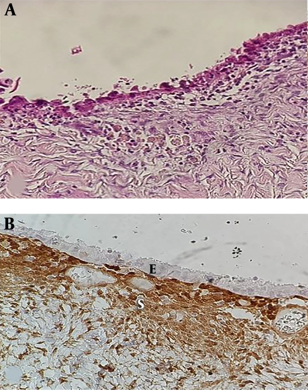
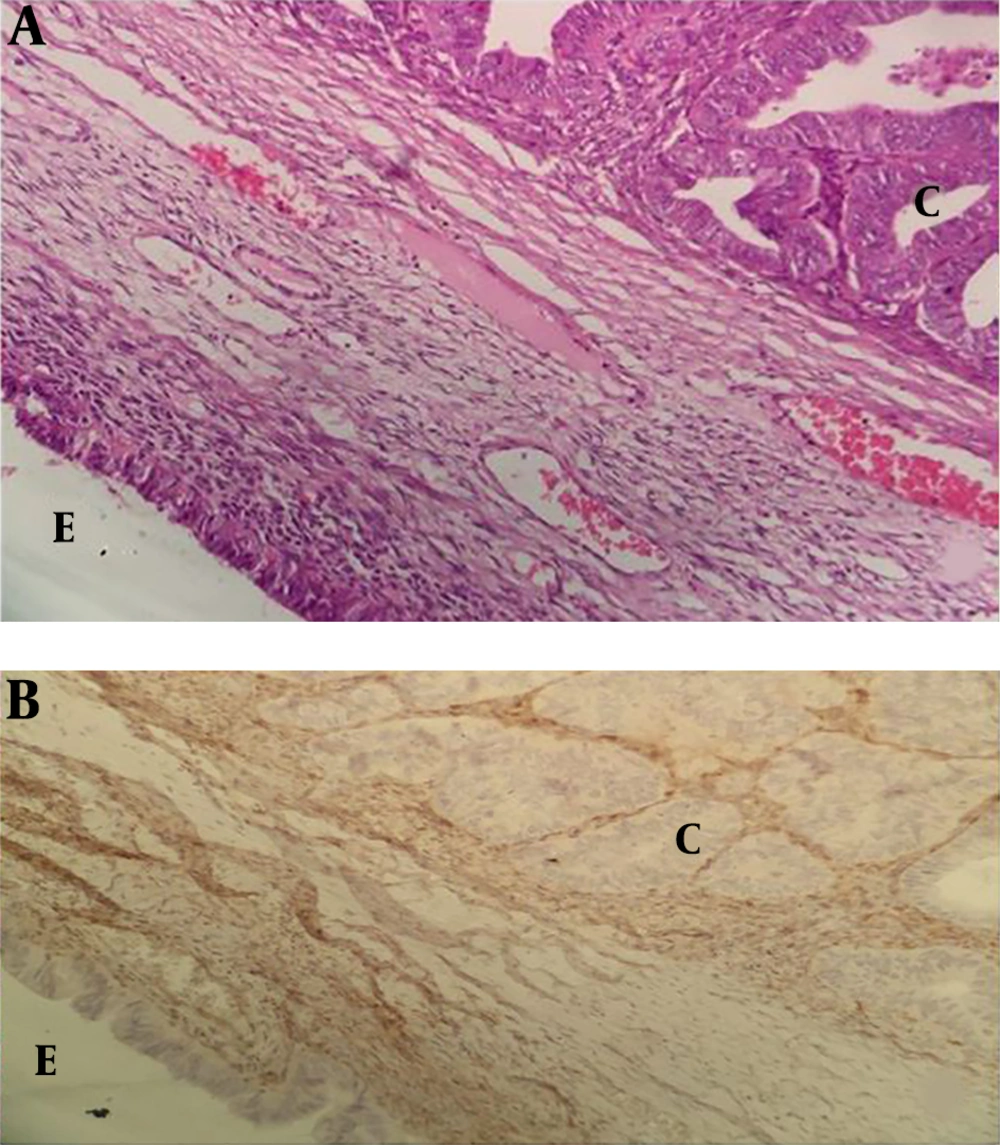
In the present study, 50% of the EAOC cases showed PTEN loss in epithelial cells of endometriosis. A significant difference was found concerning the PTEN loss between EAOC cases compared to both typical and atypical endometriosis. Seventy-five percent (6 of 8) of endometrioid carcinoma, 40% (4 of 10) of serous carcinoma, and 33.3% (2 of 6) of clear cell carcinoma had PTEN loss in both epithelial cells of carcinoma and adjacent endometriosis. Moreover, endometrioid carcinoma was found as the most common carcinoma, a result found in a study by Djordjevic et al. (
27) as well.
In all 12 PTEN loss cases, the pattern of PTEN loss in endometriosis adjacent to ovarian cancer was similar to ovarian cancer, which is in line with the study by Sato et al. (
16). They identified synchronous endometriosis in five cases of endometrioid and seven cases of clear cell carcinoma. Three out of five cases of endometriosis carcinoma with endometriosis had LOH of PTEN common to both the carcinoma and the endometriosis. Three out of seven cases of clear cell carcinoma with endometriosis had LOH of PTEN common to both the carcinoma and the endometriosis (
16). These findings revealed that the PTEN loss pattern is similar in ovarian cancer and atypical endometriosis adjacent to it, indicating that PTEN loss could be an early event in the tumor development pathway from endometriosis to ovarian cancer. Obata et al. (
15) detected LoH at 10q23.3 in 43% of the endometrioid carcinomas, and 28% of the serous carcinomas; however, they reported that LoH seemed to be infrequent in other histological subtypes of epithelial ovarian cancers. Djordjevic et al. (
27) evaluated PTEN loss in ovarian cancer and found PTEN loss in 64% (98 of 154) of all ovarian cancer. Out of 98 cases with PTEN loss, 75 (out of 100; 75%) were endometrioid carcinoma and 2 (out of 4; 50%) were clear cell carcinoma. In total, 40% (21 of 52) of other types of ovarian carcinoma with no PTEN loss were in serous carcinoma. With gene sequencing, PTEN sequencing abnormality was detected in 43% (66 out of 154) of all ovarian cancer and 51% (51 out of 100) of endometrioid carcinoma (
27).
Ballouk et al. (
28) reported that half of the atypical endometriotic cysts showed DNA aneuploidy, suggesting that these atypical endometriotic cysts have the potential to change to invasive epithelial malignancies. Martini et al. detected PTEN loss protein expression in 15% of endometriosis cases (
14). This finding suggests that inactivation of the PTEN is an early event in the malignant transformation of endometriosis and atypical endometriosis could be a precursor of ovarian carcinoma.
Ballouk et al. (
28) and Martini et al. (
14) also mentioned PTEN loss in atypical endometriotic cysts (
13). In the present study, 8% (2 of 25) of atypical endometriosis showed PTEN protein loss. However, there was no significant difference between typical endometriosis and atypical endometriosis. The following reasons can be considered as the possible explanations for this event: (A) transformation of any precursor cancerous lesion to carcinoma in the body organs is a long-standing process. As mentioned earlier, the mean age of patients with EAOC was significantly higher than patients with atypical and typical endometriosis, which suggests this point; (B) In this study all atypical changes in endomeriotic cysts of ovary were focal. Extensive sampling of endometriotic cysts is necessary to find more foci of atypical change; (C) The sample size of the present study was limited. Hence, studies with larger sample sizes are recommended to reveal the role of PTEN loss in the pathogenesis of atypical endometriosis. High-intensity expression of PTEN was only detected in typical endometriosis. None of the EAOC cases showed PTEN staining with 3+ intensity, and 83.3% (10 out of 12) of the cases with PTEN showed 1+ intensity. The results of a similar study by Sarmadi et al. (
13) revealed that the intensity of PTEN reaction was significantly higher in the group with proliferative endometrium than patients with hyperplastic endometrium and endometrioid endometrial carcinoma.
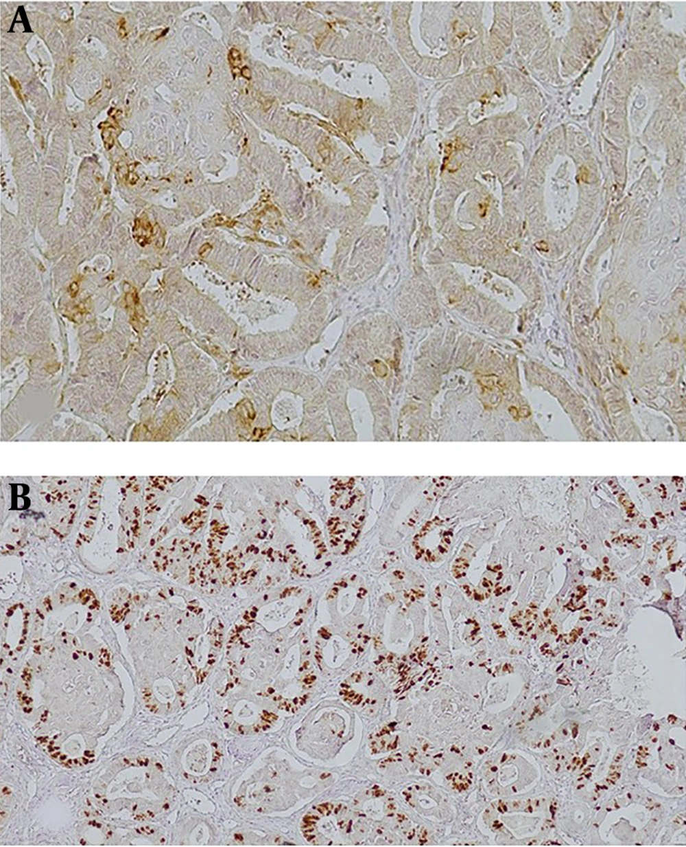
Several IHC studies reported an association between the ki67 index and biological behavior. Hence, it seems that the ki67 index can provide valuable prognostic information in some cancers (
7). Ogawa et al. (
7) detected a significant difference between the Ki-67 indices of typical endometriosis, atypical endometriosis, and ovarian cancer (2.7 ± 0.90, 9.9 ± 1.73, and 23.1 ± 3.29, respectively). Atypical endometriosis revealed a proliferative activity intermediate to those of typical endometriosis and EACO, which suggests its role as a precancerous lesion (
7). In the present study, 7.3% of typical endometriosis, 8% of atypical endometriosis, and 33.3% of EAOC had Ki67 staining in more than 50% of the epithelial cells.
Typical and atypical endometriosis had similarities in Ki 67 staining (P = 0.7), and both of them were different from EAOC. Several studies showed that ki67 indices in premalignant lesions and carcinoma in situ of the breast (
29) and biliary tract (
30) are lower than those associated with invasive carcinoma. Therefore, based on the results, premalignant lesions had a lower ki67 index than carcinoma.
In summary, the pattern of PTEN loss in endometriosis adjacent to ovarian cancer was similar to ovarian cancer. This result indicates PTEN loss could be an early event in the tumor development pathway from endometriosis to ovarian cancer, and atypical endometriosis can be considered as a precursor of ovarian carcinoma. Overall, these findings suggest that ovarian endometriosis is not as harmless as it seems because of its association with atypical endometriosis adjacent to ovarian cancer. Therefore, the follow-up of patients with atypical endometriosis is necessary.