Trends Adv Tech Med

Image Credit:Trends Adv Tech Med

A New Approach to Detect Proximal Caries Using Electrical Dental Parameters

Author(s):
Mohammad Amin Younessi HeraviMohammad Amin Younessi HeraviMohammad Amin Younessi Heravi ORCID1, Milad ShakeriMilad Shakeri2, Roya AmiriRoya Amiri3, Vahideh MotamedosanayeVahideh MotamedosanayeVahideh Motamedosanaye ORCID3,*
1Department of Medical Physics and Radiology, School of Medicine, North Khorasan University of Medical Sciences, Bojnurd, Iran
2Student Research Committee, Dental School, North Khorasan University of Medical Sciences, Bojnurd, Iran
3Department of Restorative Dentistry, Dental School, North Khorasan University of Medical Sciences, Bojnurd, Iran

Trends in Advanced Technologies in Medicine:Vol. 1, issue 1; e170005
Published online:May 04, 2026
Article type:Research Article
Received:Feb 03, 2026
Accepted:Feb 23, 2026
How to Cite:Younessi Heravi MA, Shakeri M, Amiri R, Motamedosanaye V. A New Approach to Detect Proximal Caries Using Electrical Dental Parameters. Trends Adv Tech Med. 2026;1(1):e170005. doi: https://doi.org/10.69107/tatm-170005

Abstract

Background:

Proximal caries remains difficult to detect during routine clinical examinations because of their anatomical location beneath contact areas.

Objectives:

This in vivo study aimed to evaluate electrical dental parameters, specifically electrical resistance and capacitance, at predefined anatomical locations and to assess their diagnostic performance for proximal caries detection.

Methods:

In this diagnostic observational study, 43 patients presenting with proximal caries in first molars were enrolled. Electrical resistance and capacitance were measured using a benchtop digital multimeter and a digital LC meter at four predefined tooth surfaces. Clinical and radiographic examinations served as reference standards for caries diagnosis. Diagnostic accuracy, sensitivity, and specificity of the electrical measurements were calculated and analyzed. Data were analyzed using the independent samples t-test in SPSS software, version 20.0.

Results:

Electrical resistance showed a significant reduction in teeth affected by proximal caries compared with intact teeth, particularly at the bucco-lingual surface and mesial-distal occlusal regions (P < 0.05). The highest diagnostic performance was achieved at the bucco-lingual surface, with an accuracy of 89.5%, sensitivity of 86.0%, and specificity of 90.7%. Electrical capacitance did not demonstrate a significant association with proximal caries at any measurement site.

Conclusions:

Electrical resistance measurements obtained under in vivo conditions can reliably differentiate proximal caries from intact teeth. Measurement at the bucco-lingual surface provides the highest diagnostic accuracy. Electrical resistance may serve as a non-invasive, radiation-free adjunctive diagnostic tool for proximal caries detection in clinical practice.

Clinical Relevance:

Electrical resistance assessment offers a practical and safe adjunct to conventional diagnostic methods for proximal caries, potentially improving early detection while reducing reliance on radiographic imaging.

1. Introduction

Proximal caries constitute one of the most diagnostically challenging forms of dental decay in routine clinical practice. Due to their location beneath interproximal contact areas, early proximal lesions often remain undetectable by visual and tactile examination and may progress significantly before clinical signs become apparent. Radiographic examination, particularly bitewing and periapical imaging, remains the conventional reference standard for proximal caries detection; however, its sensitivity for early enamel and shallow dentinal lesions is limited, and repeated exposure to ionizing radiation represents an inherent disadvantage (1, 2, 3).
To overcome these limitations, several adjunctive diagnostic modalities have been introduced, including laser fluorescence, near-infrared imaging, and ultrasound-based techniques. Although these methods have demonstrated acceptable diagnostic performance for occlusal and smooth-surface caries, their effectiveness in proximal regions remains inconsistent and highly dependent on anatomical constraints, lesion depth, and operator-related factors (4 - 11). Furthermore, the cost, technical complexity, and limited accessibility of some advanced imaging systems restrict their widespread clinical application.
Electrical-based diagnostic approaches offer a biologically plausible alternative for caries detection. Demineralization of enamel and dentin increases porosity and fluid content within the tooth structure, resulting in measurable alterations in electrical properties such as resistance, conductance, capacitance, and impedance (12, 13). Previous studies, primarily conducted under in vitro or ex vivo conditions, have consistently demonstrated a reduction in electrical resistance and impedance with increasing caries depth. Electrical Conductance Measurement (ECM) and Electrochemical Impedance Spectroscopy (EIS) have shown acceptable diagnostic accuracy; however, most investigations have focused on occlusal or smooth-surface lesions and were performed under controlled laboratory conditions (12, 13, 18).
Despite these promising findings, a critical gap remains in the literature regarding the in vivo clinical evaluation of electrical dental parameters specifically for proximal caries detection. The oral environment presents complex variables, including saliva, temperature, moisture, and anatomical limitations, that may significantly influence electrical measurements. Consequently, results derived from laboratory studies cannot be directly extrapolated to clinical settings. Moreover, the optimal anatomical locations for electrical measurements that provide the highest diagnostic accuracy for proximal lesions have not been systematically evaluated in clinical studies.
Therefore, the aim of the present in vivo diagnostic observational study was to evaluate electrical resistance and capacitance measurements at predefined anatomical locations on first molars and to assess their diagnostic performance for detecting proximal caries, using combined clinical and radiographic examinations as reference standards. By identifying the most reliable electrical parameters and measurement sites, this study seeks to clarify the clinical applicability of electrical diagnostics and address an unmet need in proximal caries detection.

2. Methods

Prior to electrical measurements, all selected teeth underwent scaling and prophylaxis to remove plaque and surface debris. Teeth were isolated using cotton rolls and saliva ejectors to minimize moisture contamination. The tooth surfaces were air-dried for 30 seconds using oil-free compressed air. All measurements were performed under standardized clinical conditions at room temperature, and excessive salivary contamination was avoided throughout the procedure.

2.1. Electrical Devices and Calibration

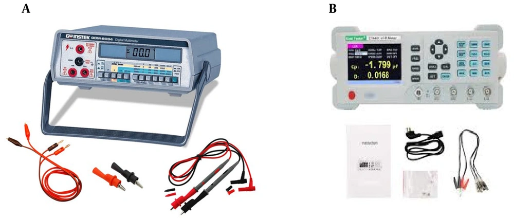
Electrical resistance was measured using a GDM-8034 benchtop digital multimeter, and electrical capacitance was assessed using a JLC-10 digital LC meter. Before each measurement session, both devices were calibrated according to the manufacturers’ instructions using standard reference components to ensure measurement accuracy and consistency. Measurements were repeated five times at each site, and the mean value was used for statistical analysis.

2.2. Measurement Locations and Anatomical Landmarks

Electrical measurements were obtained at four predefined anatomical locations:
from the occlusal center toward the buccal surface;
from the occlusal center toward the lingual surface;
at the midpoint of the bucco-lingual surface corresponding to the proximal contact area; and
at the mesial and distal aspects of the occlusal surface adjacent to proximal contacts.
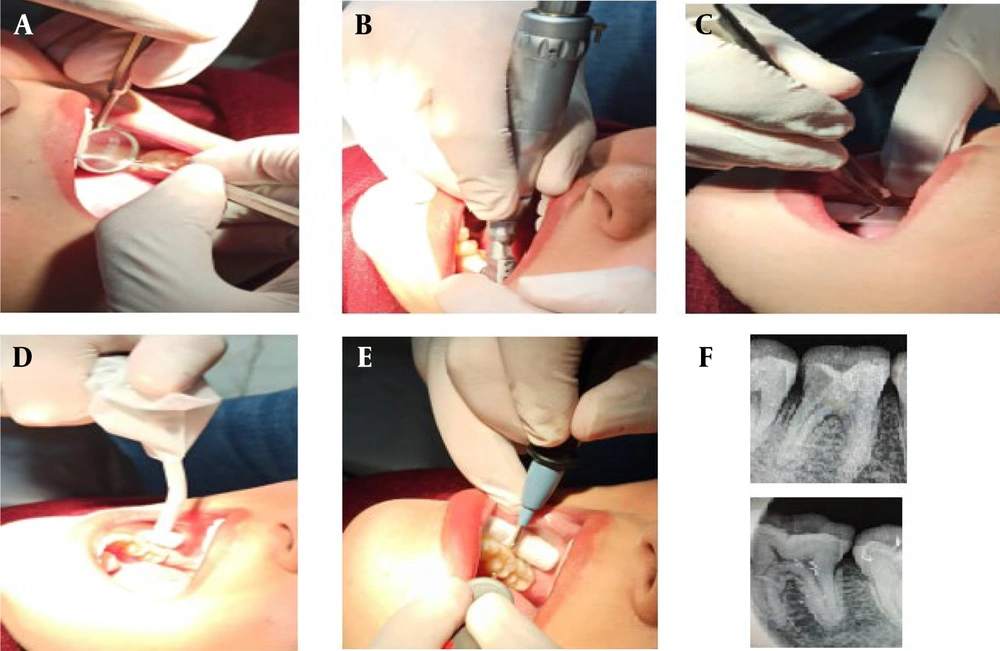
These locations were selected to represent clinically relevant regions associated with proximal caries development and to ensure reproducibility. The equipment setup, measurement setup, and probe positioning are shown in Figure 1 and Figure 2, respectively.
A, The GDM-8034 benchtop digital multimeter for measuring electrical resistance; B, the JLC-10 digital LC meter for measuring electrical capacitance.
Figure 1.

A, The GDM-8034 benchtop digital multimeter for measuring electrical resistance; B, the JLC-10 digital LC meter for measuring electrical capacitance.

A, Clinical examination and initial caries assessment; B, scaling of the target tooth; C, isolation of the tooth at the measurement site; D, air-drying of the tooth surface prior to measurement; E, measurement of dental electrical resistance and capacitance at different tooth surfaces using the designated device; F, representative periapical radiographs of intact and caries-affected teeth.
Figure 2.

A, Clinical examination and initial caries assessment; B, scaling of the target tooth; C, isolation of the tooth at the measurement site; D, air-drying of the tooth surface prior to measurement; E, measurement of dental electrical resistance and capacitance at different tooth surfaces using the designated device; F, representative periapical radiographs of intact and caries-affected teeth.

2.3. Radiographic Examination

Periapical radiographs were obtained using a standardized positioning technique and evaluated by an experienced restorative dentistry specialist. Radiographic assessment, in conjunction with clinical examination, served as the reference standard for caries diagnosis.

2.4. Data Analysis

Data analysis was conducted using IBM SPSS Statistics version 20.0 (IBM Corp., Armonk, NY, USA). Quantitative variables were expressed as mean ± standard deviation. Differences in electrical resistance and capacitance between intact and caries-affected teeth at each measurement location were analyzed using the independent samples t-test. The diagnostic performance of electrical resistance measurements was assessed by calculating sensitivity, specificity, and overall accuracy. Statistical significance was set at P < 0.05.

3. Results

A total of 43 patients were included in this study, comprising 18 males (41.9%) and 25 females (58.1%). The mean age, weight, and height of the participants were 36.11 ± 6.67 years, 71.18 ± 8.26 kg, and 168.33 ± 9.26 cm, respectively.
The agreement between clinical and radiographic diagnoses of proximal caries was evaluated. Out of 86 assessed teeth (43 intact and 43 caries-affected), a kappa coefficient of 0.812 was obtained (P < 0.001), indicating a high level of consistency between the two diagnostic methods. In intact teeth, agreement between clinical and radiographic findings was observed in 41 cases (95.3%), whereas agreement was observed in 38 caries-affected teeth (88.4%).

3.1. Electrical Parameter Measurements at Different Tooth Surfaces

The comparison of electrical resistance and capacitance values between intact and caries-affected teeth at the four predefined measurement locations is summarized in Table 1. Measurements obtained from the occlusal center toward the buccal and lingual surfaces did not reveal statistically significant differences in either electrical resistance or capacitance between intact and carious teeth (P > 0.05).
In contrast, a significant reduction in electrical resistance was observed at the center of the bucco-lingual surface in caries-affected teeth compared with intact teeth (P < 0.001). Similarly, electrical resistance measured at the mesial and distal aspects of the occlusal surface demonstrated a significant difference between intact and caries-affected teeth (P < 0.05). No statistically significant association was found between electrical capacitance and caries status at any of the evaluated measurement sites.
Table 1.Electrical Resistance and Capacitance Values at Different Tooth Surfaces a
VariablesIntactCaries-AffectedP-Value
Occlusal → Buccal
Resistance (MΩ)6.77 ± 1.455.42 ± 3.260.078
Capacitance (pF)1.44 ± 0.551.34 ± 0.380.118
Occlusal → Lingual
Resistance (MΩ)5.22 ± 1.084.55 ± 2.930.091
Capacitance (pF)1.55 ± 0.241.42 ± 0.610.221
Bucco-lingual surface (center)
Resistance (MΩ)10.96 ± 2.783.55 ± 1.16< 0.001
Capacitance (pF)2.23 ± 0.581.81 ± 0.740.089
Mesial and distal of occlusal
Resistance (MΩ)9.21 ± 2.184.93 ± 2.370.006
Capacitance (pF)1.06 ± 0.391.22 ± 0.460.105

a Values are presented as mean ± SD.

3.2. Diagnostic Performance of Electrical Resistance Measurements

The diagnostic accuracy, sensitivity, and specificity of electrical resistance measurements at the four evaluated locations are presented in Table 2. In all measurement locations, electrical resistance demonstrated a higher correct classification rate for caries-affected teeth compared with intact teeth.
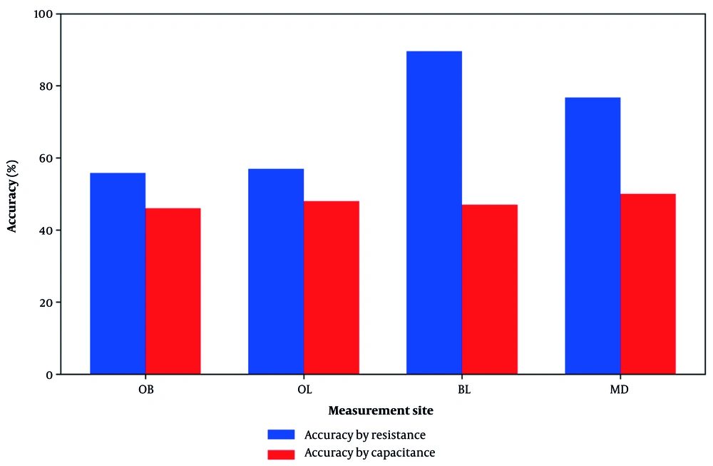
The highest diagnostic performance was achieved when measurements were obtained from the bucco-lingual surface, yielding an accuracy of 89.53%, sensitivity of 86.04%, and specificity of 90.70%. Measurements taken from the mesial and distal aspects of the occlusal surface showed moderate diagnostic accuracy, whereas measurements obtained from the occlusal center toward the buccal or lingual surfaces demonstrated lower discriminatory ability.
Table 2.Diagnostic Performance of Electrical Resistance Measurements at Different Tooth Surfaces
Measurement SiteAccuracy (%)Sensitivity (%)Specificity (%)Final Discrimination Rate (%)
Occlusal → Buccal55.8153.4858.1355.81
Occlusal → Lingual56.9751.1662.8056.97
Bucco-lingual surface89.5386.0490.7089.53
Mesial and distal of occlusal76.7474.4279.0776.74
A graphical comparison of diagnostic accuracy across the four measurement locations is illustrated in Figure 3. Electrical resistance measurements obtained from the bucco-lingual surface demonstrated the highest accuracy among all evaluated sites.
Comparison of diagnostic accuracy of electrical resistance and capacitance measurements across four measurement sites. OB, occlusal center to buccal end; OL, occlusal center to lingual end; BL, bucco-lingual surface (center); MD, mesial and distal sides of the occlusal surface.
Figure 3.

Comparison of diagnostic accuracy of electrical resistance and capacitance measurements across four measurement sites. OB, occlusal center to buccal end; OL, occlusal center to lingual end; BL, bucco-lingual surface (center); MD, mesial and distal sides of the occlusal surface.

4. Discussion

The present findings provide clinically relevant evidence supporting the diagnostic value of electrical resistance measurements for proximal caries detection under in vivo conditions. Among the evaluated measurement locations, the bucco-lingual surface demonstrated the highest diagnostic accuracy, sensitivity, and specificity. This observation is clinically significant, as this region anatomically corresponds to proximal contact areas where caries commonly initiate and progress. The results suggest that electrical resistance measurements obtained from this site are more sensitive to structural changes associated with proximal demineralization than measurements taken from the occlusal center toward buccal or lingual surfaces, where no statistically significant differences were observed.
In contrast, electrical capacitance did not show a significant association with proximal caries at any measurement site. This finding indicates that, under clinical conditions, resistance may represent a more robust and clinically relevant electrical parameter than capacitance for caries detection. This outcome aligns with prior laboratory-based studies that reported stronger correlations between caries depth and electrical resistance or impedance compared to capacitance alone (12 - 15).
When compared with other caries diagnostic modalities, the diagnostic accuracy achieved in this study is comparable to that reported for established techniques. Visual-tactile examination has demonstrated variable accuracy, largely dependent on clinician experience (1, 11). Radiographic methods typically achieve accuracies of approximately 85% - 88% for proximal caries detection but remain limited in identifying early enamel lesions and involve exposure to ionizing radiation (3, 15). Advanced optical techniques such as laser fluorescence and near-infrared imaging have shown high sensitivity, yet their performance in proximal regions may be affected by tooth morphology and surface conditions (7 - 10, 17). Ultrasound-based methods have also demonstrated promising accuracy; however, their availability and clinical practicality remain limited (4 - 6, 16).
The diagnostic accuracy of electrical resistance measurement observed in the present study, particularly at the bucco-lingual surface, falls within the range reported for these established techniques, while offering potential advantages such as low cost, absence of radiation exposure, and ease of clinical application. These characteristics suggest that electrical resistance measurement could serve as a valuable adjunctive diagnostic tool for proximal caries detection rather than a replacement for conventional methods.
Several limitations of the present study should be acknowledged. First, this investigation focused exclusively on first molar teeth, and the generalizability of the findings to other tooth types warrants further investigation. Moreover, although proximal caries were classified dichotomously as present or absent, the potential of electrical parameters to discriminate among different stages of caries progression was not evaluated. Lesion staging refers to the classification of carious lesions according to their depth and extent of tissue involvement, from early enamel demineralization to advanced dentinal lesions. Given that electrical resistance is influenced by the degree of mineral loss and microstructural alterations of dental tissues, its diagnostic performance may vary across different lesion stages. Consequently, future studies incorporating larger sample sizes and lesion staging are recommended to better define diagnostic thresholds and to further elucidate the clinical applicability of electrical resistance measurements.

4.1. Conclusion

The present in vivo study demonstrated that electrical dental parameters, particularly electrical resistance, can be effectively used for the detection of proximal caries under clinical conditions. A significant reduction in electrical resistance was observed in teeth affected by proximal caries compared with intact teeth, with the highest diagnostic accuracy achieved when measurements were obtained at the bucco-lingual surface and mesial-distal occlusal regions.
Among the evaluated electrical parameters, resistance showed superior diagnostic performance compared to capacitance, indicating that it represents a more clinically relevant indicator of proximal caries-related structural changes. The findings confirm that electrical resistance measurements retain diagnostic validity within the complex oral environment and are capable of distinguishing proximal caries with a level of accuracy comparable to conventional diagnostic modalities.
Given its non-invasive nature, absence of ionizing radiation, and relatively low cost, electrical resistance measurement may serve as a valuable adjunctive diagnostic tool for proximal caries detection in clinical practice. Further studies are recommended to evaluate its applicability across different tooth types and stages of caries progression, as well as to establish standardized diagnostic thresholds for routine clinical use.

Footnotes

References

  • 1.
    Twetman, Axelsson, Dahlén, Espelid, Mejàre, Norlund, et al. Adjunct methods for caries detection: A systematic review of literature. Acta Odontologica Scandinavica. 2013;71(3-4):388-97. [PubMed ID: 22630355]. https://doi.org/10.3109/00016357.2012.690448.
  • 2.
    Pretty. Caries detection and diagnosis. Comprehensive Preventive Dentistry. 2012:25-42. https://doi.org/10.1002/9781118703762.ch2.
  • 3.
    Ritter, Ramos, Astorga, Shugars, Bader. Visual-tactile versus radiographic caries detection agreement in caries-active adults. Journal of Public Health Dentistry. 2013. [PubMed ID: 23772747]. https://doi.org/10.1111/jphd.12024.
  • 4.
    Marotti, Heger, Tinschert, Tortamano, Chuembou, Radermacher, et al. Recent advances of ultrasound imaging in dentistry-a review of the literature. Oral Surgery, Oral Medicine, Oral Pathology and Oral Radiology. 2013;115(6):819-32. [PubMed ID: 23706922]. https://doi.org/10.1016/j.oooo.2013.03.012.
  • 5.
    Bozkurt, Tağtekin, Yanikoglu, Fontana, Gonzalez-Cabezas, Stookey, et al. Capability of an Ultrasonic System to Detect Very Early Caries Lesions on Human Enamel. Marmara Dental Journal. 2013;1(1):16-9. https://doi.org/10.12990/MDJ2013121.
  • 6.
    Matalon, Feuerstein, Calderon, Mittleman, Kaffe. Detection of cavitated carious lesions in approximal tooth surfaces by ultrasonic caries detector. Oral Surgery, Oral Medicine, Oral Pathology, Oral Radiology, and Endodontology. 2007;103(1):109-13. [PubMed ID: 17178503]. https://doi.org/10.1016/j.tripleo.2006.07.023.
  • 7.
    Teo, Ashley, Louca. An in vivo and in vitro investigation of the use of ICDAS, DIAGNOdent pen and CarieScan PRO for the detection and assessment of occlusal caries in primary molar teeth. Clinical Oral Investigations. 2013:1-8. [PubMed ID: 23793456]. https://doi.org/10.1007/s00784-013-1021-4.
  • 8.
    Çınar, Atabek, Odabaş, Ölmez. Comparison of laser fluorescence devices for detection of caries in primary teeth. International Dental Journal. 2013. [PubMed ID: 23550523]. [PubMed Central ID: PMC9374934]. https://doi.org/10.1111/idj.12024.
  • 9.
    Schwass, Leichter, Purton, Swain. Evaluating the efficiency of caries removal using an Er:YAG laser driven by fluorescence feedback control. Archives of Oral Biology. 2012. [PubMed ID: 23123070]. https://doi.org/10.1016/j.archoralbio.2012.09.017.
  • 10.
    DeBenedetto, Morais, Novaes, de Almeida Rodrigues, Braga, Mendes, et al. Comparing the reliability of a new fluorescence camera with conventional laser fluorescence devices in detecting caries lesions in occlusal and smooth surfaces of primary teeth. Lasers in Medical Science. 2011;26(2):157-62. [PubMed ID: 20157753]. https://doi.org/10.1007/s10103-010-0757-1.
  • 11.
    Bader, Shugars, Bonito. Systematic reviews of selected dental caries diagnostic and management methods. Journal of Dental Education. 2001;65(10):960-8. https://doi.org/10.1002/j.0022-0337.2001.65.10.tb03470.x.
  • 12.
    Verdonschot, Rondel, MCDNJM. Validity of electrical conductance measurements in evaluating the marginal integrity of sealant restorations. Caries Research. 1995;29(2):100-6. [PubMed ID: 7728822]. https://doi.org/10.1159/000262049.
  • 13.
    Huysmans, Longbottom, Pitts, Los, Bruce. Impedance Spectroscopy of Teeth with and without Approximal Caries Lesions-an in vitro Study. Journal of Dental Research. 1996;75(11):1871-8. [PubMed ID: 9003234]. https://doi.org/10.1177/00220345960750110901.
  • 14.
    Hope, Griffiths, Prior. Finding an Alternative to Formalin for Sterilization of Extracted Teeth for Teaching Purposes. Journal of Dental Education. 2013;77(1):68-71. https://doi.org/10.1002/j.0022-0337.2013.77.1.tb05445.x.
  • 15.
    Pretty. Caries detection and diagnosis: novel technologies. Journal of Dentistry. 2006;34(10):727-39. [PubMed ID: 16901606]. https://doi.org/10.1016/j.jdent.2006.06.001.
  • 16.
    Ng, Ferguson, Payne, Slater. Ultrasonic studies of unblemished and artificially demineralized enamel in extracted human teeth: a new method for detecting early caries. Journal of Dentistry. 1988;16(5):201-9. [PubMed ID: 3063732]. https://doi.org/10.1016/0300-5712(88)90070-X.
  • 17.
    Lussi, Hack, Hug, Heckenberger, Megert, Stich, et al. Detection of approximal caries with a new laser fluorescence device. Caries Research. 2006;40(2):97-103. [PubMed ID: 16508265]. https://doi.org/10.1159/000091054.
  • 18.
    Penta, Pirvu, Demetrescu. Electrochemical Impedance Spectroscopy (EIS) Investigation on Dental Hard Tissue Whitening Process Using Fluoride and Non-fluoride Carbamide Peroxide Gels. APCBEE Procedia. 2013;7:67-72. https://doi.org/10.1016/j.apcbee.2013.08.014.

Crossmark
Crossmark
Checking
Share on
Cited by
Metrics

Purchasing Reprints

  • Copyright Clearance Center (CCC) handles bulk orders for article reprints for Brieflands. To place an order for reprints, please click here (   https://www.copyright.com/landing/reprintsinquiryform/ ). Clicking this link will bring you to a CCC request form where you can provide the details of your order. Once complete, please click the ‘Submit Request’ button and CCC’s Reprints Services team will generate a quote for your review.
Search Relations

Author(s):

Related Articles