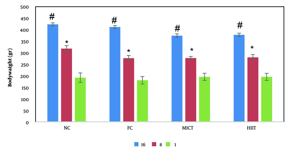
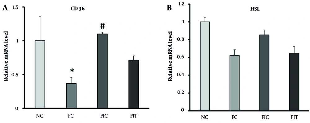
The results of the study showed that the consumption of a 20% fructose solution for 16 weeks did not lead to significant changes in body weight. However, fructose consumption significantly reduced the expression of CD36 and HSL genes in liver tissue. Previous studies have indicated that fructose consumption at different concentrations and durations can lead to an increase in body weight (
21). It is evident that high fructose intake can lead to metabolic disorders, and a long-term fructose-rich diet can induce MetS, which is usually associated with elevated glucose levels and IR (
23).
The increase in liver index in the fructose-receiving groups, although not significant compared to the control group, could be due to TG accumulation in this tissue. Additionally, the weight difference between the NC and FC groups could be explained by mechanisms of appetite suppression and energy homeostasis. It was observed that food intake was significantly reduced during the study period in the FC group, indicating that appetite control mechanisms were activated, balancing body weight. Consistent with this study, fructose-rich diets combined with inactivity did not lead to hepatic steatosis (
25), and body weight was controlled by energy balance in most studies using 124 to 201 g of fructose concurrently with a normal diet (
20).
Although HSL is a critical enzyme for lipolysis in target tissues of insulin and hormones involved in metabolism (
26), HSL gene expression in the FC group was decreased compared to the NC group, but not significantly. This could be justified by the fact that when blood sugar increases, there is no need to increase HSL gene expression to compensate and supply cellular energy. In this respect, interval and continuous training could not cause a significant increase in the expression of this gene compared with the FC group due to the increased cellular requirement for energy. Furthermore, this decrease could be attributed to the high levels of insulin in this group. Long-term fructose consumption was the leading cause of IR in the target tissues of this hormone, and insulin could thus prevent lipolysis in liver tissue by reducing the expression of its target genes, including this enzyme.
Accordingly, results showed that interval and continuous training in the groups receiving fructose could not bring significant changes to the hepatic expression of HSL compared with the FC group. Long-term fructose consumption along with interval and continuous training could moderate the serum levels of liver enzymes, glucose, and TGs, thereby preventing the development of metabolic disorders in these nutrients (
25,
27-
29). These results could be attributed to the improvement in the metabolic pathways of each nutrient (
5).
Fructose-rich diets accompanied by inactivity can stimulate hepatic lipogenesis, subsequently increasing intrahepatic triglyceride (IHTG) concentration and glucose levels (
6,
30). The molecular results in this study further established that, unlike HSL, CD36 gene expression significantly decreased in the liver tissue of rats with fructose consumption for 16 weeks. Receiving different concentrations of fructose over various periods leads to fat accumulation in the liver, manifesting as hepatic steatosis (
31), a condition that can progress to steatohepatitis following prolonged fructose intake. Despite this, some studies have found no significant changes in the expression of this gene after 60% fructose consumption for 28 days (
32).
Considering the transport of long-chain free fatty acids into liver cells and their subsequent storage or conversion to acetyl-CoA as one of the main functions of CD36 in the liver (
33,
34), the expression of this gene was reduced in the FC group compared to the NC group in this study. Indeed, this reduction could be validated as a protective response in liver cells with intrinsic mechanisms due to environmental factors during long-term fructose consumption (
35,
36). Additionally, CD36 gene expression was elevated in the exercise groups receiving the fructose solution compared with the FC and NC groups. Nevertheless, this rising trend was not significant in the FIT group. The discrepancy in results among the exercise groups in the present study suggests that metabolic pathways might be involved in the training protocols (
10,
37). Continuous training was effective in modulating CD36 gene expression in the liver (
38,
39).
The results showed that CD36 gene expression was incoherent with fasting blood glucose levels in the FC group, confirming the regulatory role of the liver in storing and utilizing different forms of lipids, compared with muscle tissue. The decrease in the expression of this gene in the present study could further provide a defense mechanism against the increasing free fatty acid load, due to the combined function of synthesis and reduction in fatty acid. However, the build-up of TGs in the liver is the initial stage of damage caused by hepatic steatosis, followed by inflammation, which could intensify with continued exposure. This could also be consistent with the development of steatohepatitis after high fructose consumption in this model (regardless of its dose), aligning with previous results (
25,
40).
Although continuous training significantly modulated the effect of long-term fructose intake, it could not bring the level of HSL gene expression closer to its expression level in the NC group. This observation also emphasizes the regulatory role of the liver, which does not rapidly alter its factors in a short period of exposure to damage, nor does it recover immediately after returning to normal conditions.
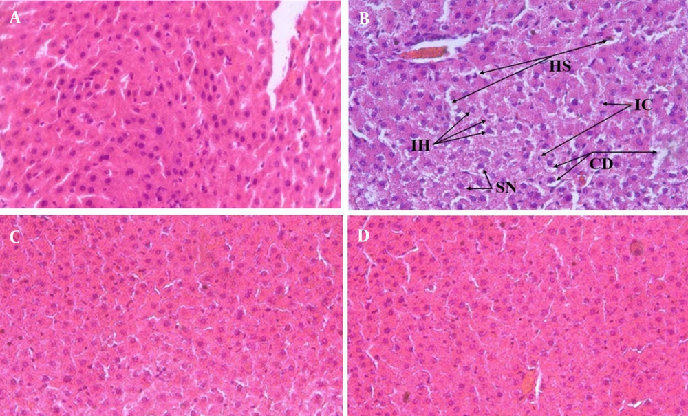
Interval and continuous training could moderate HSL gene expression, bringing it closer to normal levels. Additionally, exercise could reduce the size of fat droplets and normalize protein markers involved in lipogenesis and lipolysis (
41-
43), as observed in the FCT and FIT groups in terms of hepatic TG levels. In accordance with biochemical and molecular results, it is well established in this study and previous studies that fructose consumption does not cause obvious morphological changes in liver cells and fibrosis. Therefore, the presence of free fatty acids in the short term induces autophagic responses and impairs programmed cell death. The histological results of this study, similar to some previous findings, showed abnormal nuclear shapes and cellular disorganization in liver tissue in the FC group. However, in previous studies, no fructose-receiving animal model reports all the liver histological features of MetS in one study. These disorganizations approach the level of the NC group when combined with periodic and continuous exercise. Accordingly, it seems that the greater utilization of cellular reserves during exercise improves physiological function and liver tissue, preventing the progression of further metabolic disorders (
44-
49).
5.1. Conclusions
This study demonstrates that although prolonged high fructose intake can disrupt lipid metabolism, leading to elevated fasting blood glucose and IR, it may not significantly impact body weight or liver index. Moreover, both interval and continuous aerobic exercises were effective in mitigating the negative effects of fructose on metabolic parameters, improving insulin sensitivity and glucose regulation. Aerobic exercise also showed potential hepatoprotective effects by reducing inflammatory cells and enhancing hepatocyte structure. The findings suggest that aerobic exercise can serve as a viable non-pharmacological strategy to prevent and ameliorate early symptoms of metabolic disorders induced by fructose consumption. Further research is needed to uncover the precise molecular mechanisms involved and the impact of exercise on liver metabolism.