


1. Background
Cancer is one of the most deadly causes of death in humans. Unfortunately, despite human progress in various fields, the growth and impact of this disease are increasing day by day, a major part of which is due to changes in human lifestyle (1). The main problems with the treatment methods used for this disease, such as chemotherapy, are the high side effects and ineffectiveness of these treatments. In recent decades, mankind has tried to treat many diseases, including cancer, with fewer side effects by re-approaching nature and using herbal medicines (2). Temple figs are perennial and evergreen, belonging to the Ficus genus and Moraceae family (3). Among the therapeutic properties of temple figs, which have attracted the attention of researchers today, we can mention their antibacterial, antiviral, antifungal activities, and the inhibition of acetylcholinesterase. Also, the extracted compounds of this plant can be used to prevent and treat various diseases (4, 5).
Nanotechnology makes it possible to manufacture and design materials that have completely new properties and characteristics, with sizes ranging from 1 to 100 nanometers. The production of nanoparticles using green methods has gained a special place in research. Nanoparticles affect bacteria by damaging their proteins and destroying their cell walls.
2. Objectives
The aim was to study the synthesis of zinc nanoparticles in Ficus religiosa L leaf extract and investigate their anticancer properties on uterine cancer cells (Hela) and antibacterial activity.
3. Methods
The advantages of using plant materials for the biosynthesis of nanoparticles include the mechanism of metal ion uptake by plants and understanding the possible mechanism of metal nanoparticle formation in plants. To synthesize zinc oxide nanoparticles by a green method, firstly, the aqueous extract of the plant is prepared, and zinc acetate is used as the source. FTIR and SEM tests are performed for confirmation. The antibacterial effects of zinc oxide nanoparticles synthesized by the green method from the extract were studied on bacteria such as S. pyogenes, Staphylococcus aureus, E. faecalis, H. alvei, Proteus mirabilis, Streptococcus mutans, Streptococcus pneumoniae, and Bacillus cereus using the microdilution method.
3.1. Cancer Cell Cultivation
This study utilized health cancer cells (C115) and healthy hack (C10139) from the Tehran Pasteur Institute. The above cells were cultured using DMEM as the medium. To prepare the culture medium, deionized distilled water was first sterilized and cooled, with three-quarters of the final volume needed poured into a bottle. Full dissolution was awaited. NaHCO3 was then added according to the manufacturer's instructions. Since a pH of 7 is ideal for cell medium, but because FBS with acidic properties would be added to the environment, the pH of the environment was adjusted to be slightly alkaline, between 7.2 and 7.4. A pH meter and a pre-sterilized molar NaOH and HCl solution were used for this adjustment. In the next phase, the antibiotic solution was added, and its volume was brought up to 90% of the intended final volume. Since substances such as serum would eventually be added to the solution, the volume at this stage was considered less than the final amount. Sterile fetal bovine serum was added to the culture medium after filtration. The solution was then filtered using a syringe and a 0.22 μm syringe filter. This operation was performed under a laminar hood. Finally, the equivalent volume of the remaining fetal bovine serum was added to achieve the desired final volume. To store the culture medium, it was placed in glass containers with screw caps that had been previously sterilized and autoclaved, and stored in a refrigerator at 4°C (6, 7). To obtain the best results, sampling was performed in the logarithmic phase of cell culture. At the end of this test, light absorption at a wavelength of 400 - 630 nm was read with an ELISA reader, and the amount of light absorption was measured quantitatively. For tests of factors effective in cancer treatment, the positive or negative effects of the mentioned factors were examined by treating cancer cells with drugs or substances that cause intensification or reduction of their growth, and measuring their death or survival, growth, and proliferation using the MTT method. In this experiment, after culturing Hela cells, treatment and exposure to synthetic nanoparticles at doses of 0.1, 1, 10, 50, 100, 150, 200, 1000 µg/ml were performed, and MTT measurements were taken at 24, 48, and 72 hours (8). The results of this study were based on the average of three replicate experiments. The t-test and SPSS 21 software were used to analyze the data, and Excel was used to draw graphs. The significance level of the results was determined as P < 0.05.
4. Results
4.1. Antibacterial Activity
The results of this study showed that the highest diameter of the inhibition zone was at a concentration of 1024 micrograms per mL of zinc nanoparticles on S. pneumoniae (6 mm), while the lowest diameter of the inhibition zone was observed on P. mirabilis (1 mm) (Table 1). The FTIR results and the color of the nanoparticles, as shown in Images 1 and 2, confirmed the formation of zinc nanoparticles (Figures 1 and 2).
Table 1.Zone Diameter in Different Concentrations of Zinc Nanoparticles in, Ficus religiosa L Plants on Different Bacteria (mm)
| Strain Bacteria | 1024 Micrograms /mL | 512 Micrograms/mL |
|---|---|---|
| S. pyogenes | 3 | 1 |
| Staphylococcus aureus | 5 | 4 |
| E. faecalis | 3 | 1 |
| H. alvei | 2 | 2 |
| Proteus mirabilis | 1 | 0 |
| Streptococcus mutans | 2 | 1 |
| Streptococcus pneumoniae | 6 | 4 |
| Pseudomonas aeruginosa | 2 | 1 |
| Bacillus cereus | 2 | 1 |
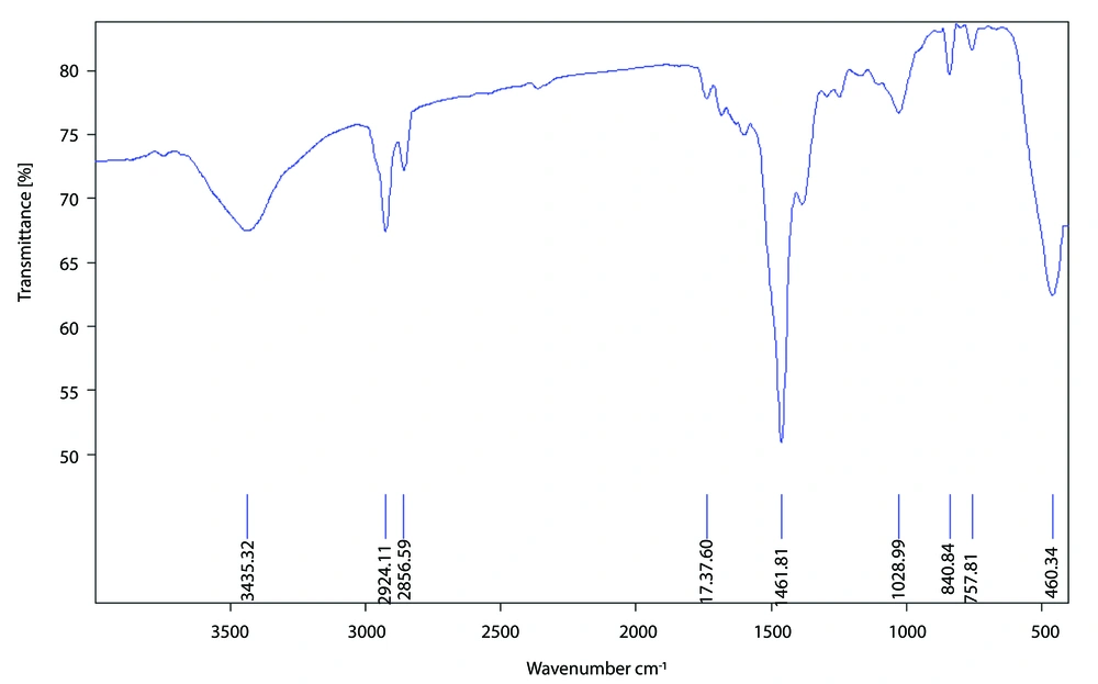
Figure 1.
FTIR spectrum of synthesized ZnO
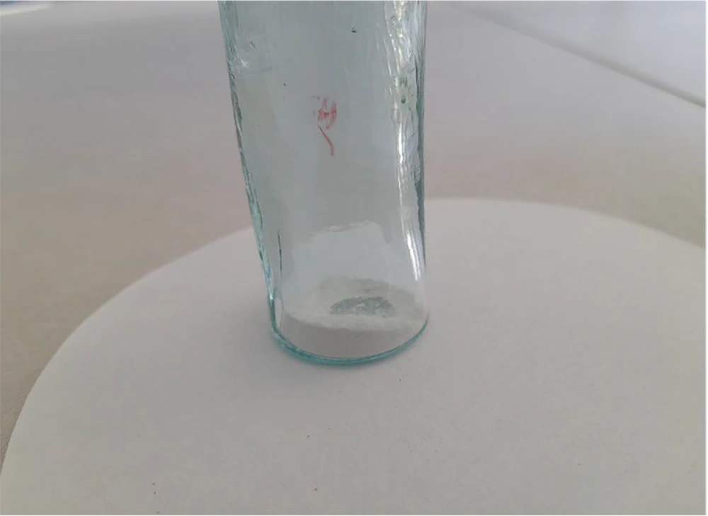
Figure 2.
Zinc nanoparticles produced
4.2. Anti-cancer Effects
To investigate the effect of synthetic zinc nanoparticles on the viability of Hela and healthy Hela cancer cells, the MTT assay was used. The effect of different doses of biological zinc nanoparticles was measured on cancer and normal cells at three time points: 24, 48, and 72 hours. The optical absorption results read by an ELISA reader from the MTT test were collected, then the survival rate of the cells was calculated, and their percentage of viability was measured (9). By observing the results in all three graphs, it was noted that only at 48 hours and at a dose of 1024 μg/mL did we witness the highest rate of cancer cell mortality, to the extent that this dose at this time point had a significant difference compared to the control group. While the survival rate of normal cells at this dose and time did not show a significant difference from the control group, numerically, at all three time points, it had the highest percentage of survival in the Heck cell lineage. Considering the highest survival rate of this dose in the Heck cell lineage and the lowest survival rate of cancer cells at this dose and time, the dose of 1024 μg/mL at 48 hours was identified as the most effective dose of these nanoparticles at a significant level of P < 0.001.
5. Discussion
In this study, the therapeutic effects of synthetic nanoparticles were investigated. The results indicated that zinc nanoparticles had a positive effect on the mortality of Hela cancer cells and pathogenic bacteria. So far, various anticancer effects of plants have been reported and proven. The study by Rahimi Kalateh Shah Mohammad on the antibacterial activity of zinc oxide nanoparticles synthesized by the green method from Hyssopus officinalis extract showed that zinc oxide nanoparticles at concentrations of 2000 and 1000 μg/mL had appropriate antibacterial activity, with the growth inhibition zone at a concentration of 2000 μg/mL in Escherichiacoli and Staphylococcus aureus being approximately equal to the control sample (10). In a study of the effect of ethanolic extract of Thymus vulgaris on prostate cancer in rats, its anticancer effects were particularly evident on the proliferation of the aforementioned cell masses, and these effects were also related to the dose used. In another study, its negative and inhibitory effect on epithelial carcinoma of the head was even observed in vivo (11). In a study by Saxena et al. in 2012, they were able to produce silver nanoparticles using fig extract (Ficus benghalensis). These nanoparticles showed good resistance against Escherichia bacteria (12). In the study by Mittal et al., they were able to synthesize silver nanoparticles, 25 - 40 nm in size, using Rhododendron dauricum flower extract (13).
5.1. Conclusions
According to our study, the tested synthetic nanoparticles show greater activity on the Hela cell line, indicating that this plant can be evaluated for potential promising anticancer activity.
