
1. Background
Trigger finger, a common condition, arises from a mismatch between the flexor tendon and its sheath, leading to pain, swelling, and irritation of the affected digit during finger movement (1). This condition occurs when the tendon's sliding movement is hindered by a channel in the first annular (A1) pulley, preventing the tendon from expanding naturally and returning to its original position (2). Trigger finger is most common in adults, with a 2 - 3% risk in the general population and up to 10% in patients with diabetes, making it one of the most common causes of hand pain (3). Trigger finger is often found in the dominant hand, with the middle and ring fingers being the most commonly affected. Women, especially middle-aged women, are affected six times more frequently than men (3, 4).
Ultrasound is playing an increasingly important role in the diagnosis of trigger finger. While clinical diagnosis is traditionally based on symptoms and physical examination, ultrasound is increasingly used to assist in diagnosis. Research shows that ultrasound can accurately measure the thickening of the affected sheath compared to the unaffected sheaths on the same hand. This offers a reliable alternative to directly measuring during surgery (5).
Symptoms of this disease range from a noticeable bump on the surface of the joint without movement restrictions in the early stages to complete immobility in the advanced stages. Nevertheless, there is no consensus in the literature regarding the definitive cause of the disease, and its etiology remains unidentified (2). While the precise cause of the disease is uncertain, factors like repetitive finger movements, injuries, underlying health conditions (like diabetes and rheumatoid arthritis), stress, and degenerative factors are linked to its development (3).
Treatment options for trigger finger include conservative and surgical approaches. Conservative treatment for mild to moderate trigger finger includes ice packs, splint extensions, nonsteroidal anti-inflammatory drugs (NSAIDs), physical therapy, joint immobilization, shock wave therapy, and corticosteroid injections (6-8). However, surgery is the preferred treatment for advanced trigger fingers and may involve open surgical release or percutaneous release of the A1 sheath (9).
Corticosteroid injections have been an effective treatment since their introduction in 1953 (10). However, there are complications associated with the common method of injecting the drug into the tendon sheath. Finding the correct anatomical position can be challenging; according to a study by Taras et al., 17% of injections are misplaced (11).
Imaging during injections offers several advantages, including increased accuracy, reduced discomfort, improved quality, longer duration of blocks, decreased medication usage, and fewer side effects. Ultrasound, fluoroscopy, and CT scans are commonly used for evaluating and administering injections in muscles, tendons, joints, and ligaments. Ultrasound assists in identifying the structure responsible for pain and enables specialists to visualize the needle in real-time, ensuring precise injections. Ultrasound is easily accessible, utilizes non-ionizing radiation, and poses lower risks than other imaging methods. Additionally, ultrasound displays the vessels at the injection site, which helps prevent intravascular injections and reduces injection discomfort. Ultrasound outperforms basic imaging in identifying arthritis, tissues, tendons, ligaments, muscles, and nerves (12-14).
According to Bodor et al., there was a high success rate one year after ultrasound-guided (USG) triamcinolone injection (15). In contrast, Cecen et al. found no significant difference in outcomes between USG Methylprednisolone acetate injection and blind guided (BG) Methylprednisolone acetate injection (16). The study by Mardani Kivi et al. (17) found that injecting 1 mL of 40 mg/mL methylprednisolone acetate under ultrasound guidance using two methods (intra-sheath and extra-sheath of the finger flexor tendon) for patients with trigger finger showed similar effectiveness. Injecting outside the sheath at the A1 pulley is as effective as injecting inside the sheath (17).
Therefore, this prospective study was conducted to further evaluate the difference between trigger finger treatment with corticosteroid injection by using ultrasound guidance versus without using ultrasound guidance to further evaluate and compare the clinical outcomes of corticosteroid injections administered with and without ultrasound guidance.
2. Methods
his prospective controlled study was conducted following approval from the local ethics committee, and informed consent was obtained from all participants before inclusion.
Patients with trigger finger at any Quinnell stage, except stage IV, were eligible to participate. Patients were classified according to Quinnell staging (I - III) (18). Stage IV patients were excluded due to the presence of fixed deformities requiring surgical intervention.
Only patients who had failed conservative management (splinting, NSAIDs, and exercise) were recruited. The sample size of 90 patients was determined based on feasibility and the number of eligible patients available during the study period.
2.1. Inclusion and Exclusion Criteria
Inclusion criteria consisted of a complete medical record and informed consent for participation. Exclusion criteria were as follows: Prior hand surgery, cervical radiculopathy with radiation of pain to the upper limbs, history of wrist or finger joint infections, congenital hand abnormalities, neurovascular disorders, use of immunosuppressive drugs or systemic corticosteroids, and previous fractures of the affected upper limb.
2.2. Study Groups and Intervention
Patients were randomized into two groups: The blind injection group (BGI) and the ultrasound-guided injection group (USGI). Both groups received corticosteroid injections consisting of 1 mL Depo-Medrol (40 mg/mL, methylprednisolone acetate) administered extra-sheath beneath the A1 pulley.
In the BGI group, patients were seated with the elbow flexed at 90°, the forearm supinated, and the application site disinfected twice. Using the palpation technique, the injection was administered at the distal palmar crease and the volar aspect of the metacarpophalangeal joint at the level of the A1 pulley.
In the USGI group, patients were placed in the same position. Injections were performed using an ALPINION ultrasound device equipped with a superficial linear probe (10 - 12 MHz). A distal-to-proximal approach was adopted, and the probe was aligned parallel to the flexor tendon. The corticosteroid was injected extra-sheath under the A1 pulley using a 2 mL syringe with a 23-gauge needle.
Following injection, patients were encouraged to perform active finger movements. They were informed that transient worsening of symptoms might occur within a few days after injection.
2.3. Outcome Assessment Clinical
Outcomes were assessed using the Visual Analogue Scale (VAS) and the Quick Disabilities of the Arm, Shoulder and Hand (QDASH) Questionnaire. Scores were recorded at baseline, and subsequently at 6 and 12 weeks post-injection. At the first-week follow-up, patients without clinical improvement received a repeat injection.
2.4. Statistical Analysis Descriptive
Statistics, including absolute and relative frequencies, percentages, means, and standard deviations, were used to summarize the data. Differences between groups were analyzed using the independent t-test for normally distributed continuous variables and the Mann-Whitney U test for non-normally distributed data. The chi-square test was employed for categorical variables. Statistical analysis was performed using SPSS software version 17.0 (SPSS Inc., Chicago, IL, USA), with significance set at P < 0.05.
3. Results
Results In this study, the number of 90 patients with trigger finger who referred to Baghiyyatollah al-Azam Hospital in Tehran were divided into two groups of Methylprednisolone acetate injection with ultrasound guidance (45 patients) and blind guidance (45 patients). Most of the patients in the study were female (71%). In most patients, the right hand was involved (68.7%). The demographic information of the patients is reported in Table 1, separately for the two groups.
In the comparison of Methylprednisolone acetate injection with ultrasound guidance (52.2 ± 12.01) and blind guidance (51.7 ± 13.9) in terms of age, the results of the t-test of two independent samples were not significant and the average age in the two groups was almost similar (P = 0.865).
Table 1.
Prevalence of Demographic Variables by Methylprednisolone Acetate Injection with and Without Ultrasound Guidance
| Variable | Total (n = 90) | USGI (n = 45) | BGI (n = 45) | P-Values |
|---|---|---|---|---|
| Gender | 0.64 | |||
| Male | 26 (28.9%) | 14 (31.1%) | 12 (26.7%) | |
| Female | 64 (71.1%) | 31 (68.9%) | 33 (73.3%) | |
| Involved hand | 0.23 | |||
| Right | 57 (68.7%) | 30 (75.0%) | 27 (62.8%) | |
| Left | 26 (31.3%) | 10 (25.0%) | 16 (37.2%) |
Abbreviation: USGI, ultrasound-guided injection; BGI, blind injection group.
In the comparison of gender distribution between the USGI and BGI groups, the chi-square test showed no significant difference (P = 0.64). The distribution of affected hands is shown as the number of patients by involved hand. Most patients had the right hand affected, with no significant difference between the groups (P = 0.23). (Table 1).
In the comparison of Methylprednisolone acetate Injection with and without ultrasound guidance in terms of disease severity and pain, independent t-test and Mann-Whitney test results were not significant (P < 0.05). In USGI, the average severity of the disease was higher in grade 3, but the average pain was lower in grade 3.
In the comparison of Methylprednisolone acetate Injection with and without ultrasound guidance according to the mean level of QDASH, the independent t-test results were not significant (P < 0.05). However, in USGI, the average QDASH was higher (Table 2).
Table 2.
Average Severity of Disease and Pain and QDASH by Methylprednisolone Acetate Injection with and Without Ultrasound Guidance
| Variable | USGI a | BGI a | P-Values |
| Severity of disease | |||
| Time 0 (Baseline) | 3.04 ± 0.64 | 2.91 ± 0.56 | 0.29 |
| Time 6 weeks | 1.84 ± 1.11 | 1.80 ± 0.97 | 0.85 |
| Time 12 weeks | 0.80 ± 0.89 | 0.51 ± 0.66 | 0.11 |
| Pain | |||
| Time 0 (Baseline) | 2.13 ± 1.65 | 1.71 ± 1.72 | 0.29 |
| Time 6 weeks | 0.80 ± 1.19 | 0.53 ± 0.92 | 0.35 |
| Time 12 weeks | 0.09 ± 0.35 | 0.044 ± 0.21 | 0.63 |
| QDASH | |||
| Time 0 (Baseline) | 30.8 ± 13.3 | 28.8 ± 14.7 | 0.50 |
| Time 6 weeks | 9.44 ± 9.9 | 7.57 ± 8.7 | 0.35 |
| Time 12 weeks | 1.77 ± 4.2 | 0.86 ± 2.5 | 0.21 |
Abbreviation: USGI, ultrasound-guided injection; BGI, blind injection group; QDASH, Quick Disabilities of the Arm, Shoulder, and Hand.
aValues are expressed as mean ± SD.
As shown in Figure 1, the average severity of disease was comparable between the USGI and BGI groups. Figure 2 illustrates the mean pain levels in both groups, which were not significantly different. Additionally, Figure 3 demonstrates the QDASH scores, showing similar functional outcomes between groups.
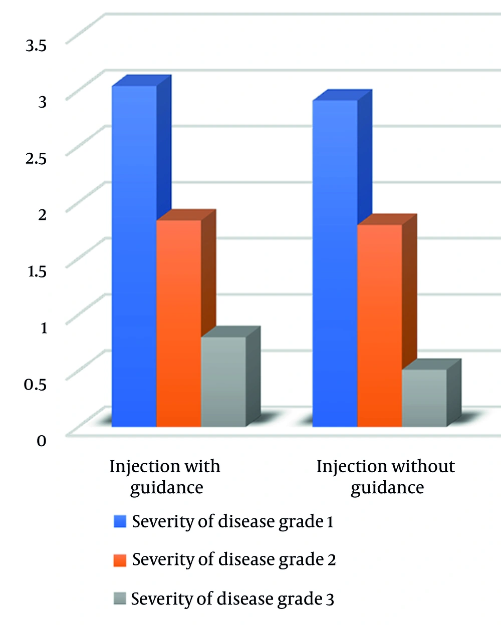
Figure 1.
Average severity of disease at Baseline, 6 weeks, and 12 weeks in USGI and blind injection groups (Abbreviation: USGI, ultrasound-guided injection).
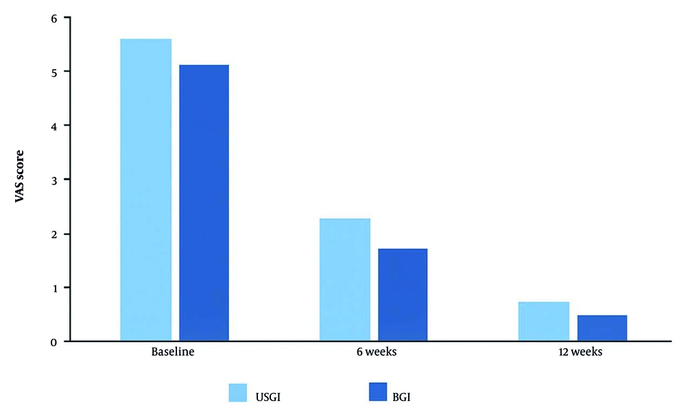
Figure 2.
Average pain VAS at Baseline, 6 weeks, and 12 weeks in USGI and blind injection groups. (Abbreviation: VAS, Visual Analogue Scale; USGI, ultrasound-guided injection).
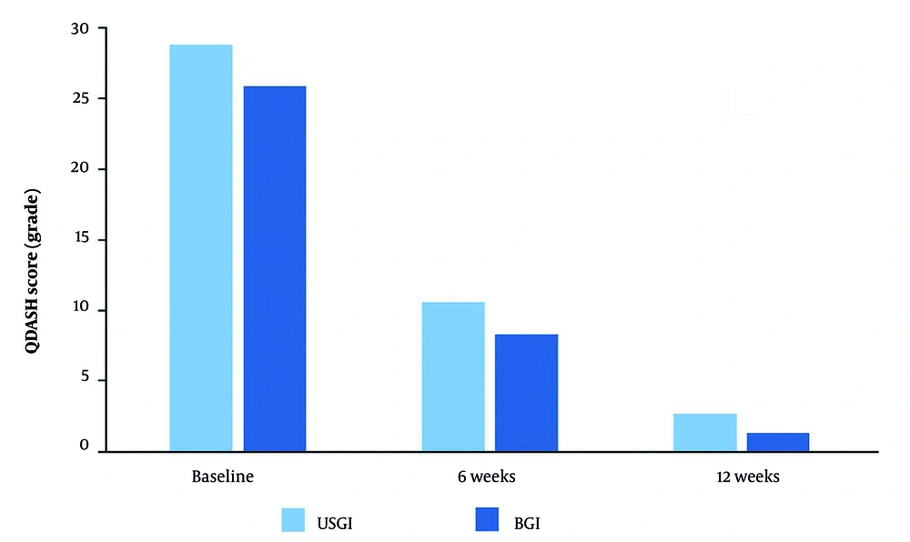
Figure 3.
Average QDASH scores at Baseline, 6 weeks, and 12 weeks in USGI and blind injection groups (Abbreviation: QDASH, Quick Disabilities of the Arm, Shoulder, and Hand; USGI, ultrasound-guided injection).
4. Discussion
Trigger finger is a common condition resulting from a mismatch between the flexor tendon and its sheath, leading to pain, swelling, and functional limitations. Corticosteroid injections are widely used to reduce pain and improve hand function in patients with trigger finger as well as in other musculoskeletal disorders (19, 20). Image-guided injections, such as USG injections, have been reported to provide increased accuracy and reduced discomfort compared to blind injections (21).
In the present study, we compared the effectiveness of corticosteroid injections administered with and without ultrasound guidance. Our results showed no significant differences between the USG and BGI groups in terms of disease severity, pain, or QDASH scores at follow-up. While the average severity of the disease in grade 3 was slightly higher in the USG group, the reported pain in this grade was lower. Similarly, QDASH scores were slightly higher in the USG group, but these differences were not statistically significant. These findings indicate that, in our patient population, both injection methods are similarly effective for treating trigger finger.
These results are consistent with previous studies. Cecen et al. reported no significant difference between USG and blind corticosteroid injections, with only a small proportion of patients requiring a second injection, and no local complications were observed (16). Likewise, Shinomiya et al. found that true intra-sheath injections did not show any clear advantage over extra-sheath injections, and the injection site had no significant effect on treatment outcomes (22).
Some studies have suggested potential benefits of USG injections in specific contexts. For instance, Polat et al. reported that USG-guided corticosteroid injections combined with orthosis use improved early symptom relief and functional recovery compared to orthosis alone (23). Similarly, Tunçez et al. found that USG-guided injections could shorten recovery time and facilitate an earlier return to work, especially in the first few weeks post injection (24). However, these findings relate to specific combined interventions or early recovery periods and do not contradict our observation that, in general, ultrasound guidance did not provide a significant advantage over blind injections in our study population.
Overall, our findings suggest that while ultrasound guidance can be a useful technical tool for improving injection accuracy and identifying coexisting pathologies (e.g., tendon sheath effusions, ganglion cysts), its routine use may not necessarily lead to superior clinical outcomes compared to blind injections in patients with trigger finger. Both methods remain effective, and selection may depend on available resources, practitioner experience, and patient preference.
Our study has several limitations. First, the sample size was relatively small, which may limit the generalizability of the findings. Second, the study was not fully blinded, as it was not conducted in a triple-blinded manner. Third, the follow-up period was relatively short (12 weeks), and therefore the long-term efficacy of the interventions could not be assessed. Future studies with larger sample sizes, rigorous blinding, and longer follow-up are needed to confirm these findings and provide more comprehensive insights into the effectiveness of USG versus blind corticosteroid injections for trigger finger.
4.1. Conclusion
The results of this study showed that the efficiency of injection was evaluated with and without ultrasound guidance, which indicated that both methods are efficient. Therefore, people who are not able to inject with ultrasound guidance can also use blind guidance. To obtain more objective results, more objective indicators are needed in future experiments.