
1. Background
Globally, colorectal cancer (CRC) is among the most common causes of cancer-related mortality and is the fourth leading cause of death among women and the second among men (1). Treatment strategies for colorectal cancer vary according to tumor type and disease stage and may include chemotherapy, immunotherapy, radiation therapy, surgery, and targeted therapy (1).
Koome is a traditional dairy product found in rural areas around Naein, Isfahan Province, Iran. It is made from barley or wheat flour, fresh oregano, and sheep milk. According to traditional beliefs, it is used as a remedy for bone, leg, and back pain. The price of Koome is comparable to that of mutton at any given time. Among traditional dairy products, Koome is notable for its stability and prolonged storage period. Koome has strong potential for the isolation of probiotic strains, and dietary intake of Lactobacillus-based microbial supplements may confer beneficial health effects (2).
2. Objectives
Research over the last decade has shown that Lactobacillus strains derived from dairy products exhibit antitumor activity by inducing apoptosis (3, 4). Despite growing interest in the potential anticancer properties of Lactobacillus, the available evidence remains insufficient, and further studies are needed to clarify its therapeutic efficacy. Therefore, the present study evaluated the effects of supernatants isolated from Koome on the HCT116 colorectal carcinoma cell line. Cell viability was assessed using the MTT assay at various concentrations and time intervals. Apoptosis in these cells was subsequently evaluated using flow cytometry.
3. Methods
3.1. Sampling
A total of 25 traditional Koome cheese specimens were obtained by random sampling from different areas around Naein, Isfahan, Iran. Sampling was performed in sterile containers under strictly aseptic conditions. The samples were transported under cold conditions to the microbiology laboratory of Zabol University.
3.2. Sample Preparation and Microbial Culture
Ten grams of each Koome sample was suspended in 90 mL of bacteriological peptone diluent. After 60 minutes, 5 mL of the prepared suspension was mixed with 100 mL of MRS culture medium for the cultivation and isolation of Lactobacillus species. Bacterial cultures were incubated in MRS broth (Merck, Germany) under anaerobic conditions for 24 and 48 hours at 42 - 45°C. Bacterial growth was assessed by measuring optical density at 600 nm. Subsequently, to isolate individual colonies, samples were streaked onto MRS agar using a sterile inoculation needle and incubated at 37°C for 48 hours. Biochemical tests, including oxidase testing, Gram staining, catalase testing, and motility assessment, were then performed on the colonies. Each bacterial culture (10 mL) was centrifuged at 4000 × g for 5 minutes. After removal of the supernatant, the cell pellets were gently washed with phosphate-buffered saline (PBS). The growth rate of the isolates was then measured using a spectrophotometer (Eppendorf, Germany) at an optical density of 600 nm (5). The isolated probiotic Lactobacillus species were confirmed by PCR using species-specific primers, according to the method previously described by Kwon et al. (6).
The number of viable cells was calculated by spread plating serial dilutions of the culture onto MRS agar in triplicate. After incubation, bacterial counts were calculated using the following equation:
In this equation, N represents the concentration of viable cells per mL; ∑C represents the total number of colonies obtained from all plates; n1 represents the total number of plates retained from the first dilution; n2 represents the total number of plates retained from the second dilution; and d represents the dilution factor used for the first dilution.
The cell-free supernatant was obtained by centrifuging the incubated culture at 15 000 rpm for 15 minutes. Different concentrations of cell-free supernatant were prepared at 25, 50, 100, 150, 200, and 250 μg/mL. Dilutions were prepared using DMEM F-12 + Glutamax. The cell-free supernatant was filtered through a presterilized 0.22-μm nitrocellulose membrane. The supernatants were divided into small aliquots and stored at -20°C until use.
3.3. Cell Culture
The HCT116 colorectal carcinoma cell line was obtained from the Pasteur Institute Cell Bank. HCT116 cells were cultured in DMEM F-12 + Glutamax (BIO-IDEA, Iran; Cat No. BI 1027) supplemented with 10% fetal bovine serum (FBS) (Gibco; Cat No. 10270 - 106) and 1% Pen Strep 100× (penicillin-streptomycin; 10 000 units/mL penicillin and 10 000 μg/mL streptomycin) (BIO-IDEA, Iran; Cat No. BI 1036) in T25 flasks. The flasks were incubated at 37°C under 5% CO2 and 90% humidity.
3.4. Trypan Blue Exclusion Test
The proportion of viable cells was determined by staining cells with trypan blue. Viable cells were impermeable to the dye and exhibited clear cytoplasm, whereas dead cells absorbed the dye and exhibited blue cytoplasm. Viable (colorless) and nonviable (dead) cells were quantified using a Neubauer hemocytometer. Cell viability was calculated using the following formula:
Cell viability (%) = number of viable cells ÷ number of total cells × 100
Cell counts are presented as mean ± standard deviation (SD).
3.5. MTT Assay
The MTT (microculture tetrazolium test) assay was performed to assess the inhibitory potential of supernatants derived from Lactobacillus isolated from Koome on the growth of the HCT116 cancer cell line and a normal cell line. Cells were seeded in 96-well plates at a density of 10 000 cells per well, except for the 12th column, which served as the untreated control. Each well received 100 μL of DMEM F-12 + Glutamax, and the plates were transferred to the incubator. After 24 hours, the culture medium was removed, 100 μL of fresh culture medium was added to each well, and the cells were treated with 25 - 1000 μM of supernatants derived from Lactobacillus isolated from Koome, followed by incubation for 24, 48, and 72 hours. The medium was then discarded and replaced with fresh DMEM F-12 + Glutamax and 0.5 mg/mL MTT solution purchased from Bio-Idea Company (Cat No. BI1017). The plates were incubated at 37°C for 4 hours. After removal of the medium, 50 μL of dimethyl sulfoxide (DMSO) was added to solubilize the formazan crystals formed during the experiment. Optical density was measured using an ELISA reader (Organon-Teknika, Netherlands) at 540 nm. The percentage of viable cells was determined according to the following formula (7, 8):
3.6. Flow Cytometry Assay
To evaluate the ability of supernatants derived from Lactobacillus isolated from Koome to induce apoptosis in the HCT116 colorectal carcinoma cell line, compared with the cell population in the negative control, cells were stained with Annexin V-FITC and propidium iodide. The Annexin V-FLOUS staining kit (Roche; Cat No. 11858777001) was used. A volume containing 100 000 cells was calculated and added to each well of a 6-well plate, after which 2 mL of DMEM F-12 + Glutamax was added to each well before incubation. After 24 hours, in the first three wells (controls), the old culture medium was replaced with fresh medium in triplicate. In wells 4, 5, and 6, the old medium was removed and replaced with the IC50 concentration of the bacterium. After 48 hours, the plates were removed from the incubator, and the cells were passaged to obtain cell pellets. The cell pellet from each well was transferred to a sterile 15-mL Falcon tube. The tubes were centrifuged for approximately 5 minutes at 140 g, after which the cells were resuspended in PBS and centrifuged again. Then, 500 μL of 1× binding buffer was added to each cell pellet and mixed thoroughly. For each Falcon tube, a 4-mL flow cytometry tube was used. The first tube contained unstained cells. Tubes 2 and 4 received 5 μL of Annexin V-FITC, and 3 μL of propidium iodide was added to tubes 3 and 4. Samples were incubated for 15 minutes at 25°C in the dark and then analyzed using a BD FACSCalibur flow cytometer (9). Data were analyzed using FlowJo software.
3.7. Statistical Analysis
SPSS version 18 was used for statistical analyses. Values are reported as mean ± SD. Each experiment was performed three times (n = 3), and statistical significance was assessed using one-way ANOVA, with P < 0.05 considered statistically significant. Microsoft Excel 2013 was used to generate selected charts. For statistical analyses, depending on the normality of the variables, paired t tests were used for data with a completely normal distribution, and the nonparametric Wilcoxon test was used for data with an abnormal distribution.
4. Results
4.1. Isolation of Lactobacillus
Colonies that appeared on MRS medium and were related to Lactobacillus were round, convex, semitransparent, soft, and cream-colored (10).
4.2. MTT Analysis
Treatment of HCT116 cells with various concentrations (25, 50, 100, 150, 200, and 250 μg/mL), assessed using the MTT assay, yielded IC50 values of 165.29 at 24 hours, 150.907 at 48 hours, and 130.863 at 72 hours. The results also showed that 250 μg/mL of Lactobacillus supernatants isolated from Koome led to the greatest reduction in cell proliferation, which was statistically significant (P < 0.001).
The findings of this study show that Lactobacillus supernatants isolated from Koome exhibit cell-killing activity compared with that of the control group, which did not receive any treatment. Based on these results, cell-killing activity appears to be dose dependent; that is, the number of dead cancer cells increased with increasing concentrations of supernatants derived from Lactobacillus isolated from Koome (Figure 1).
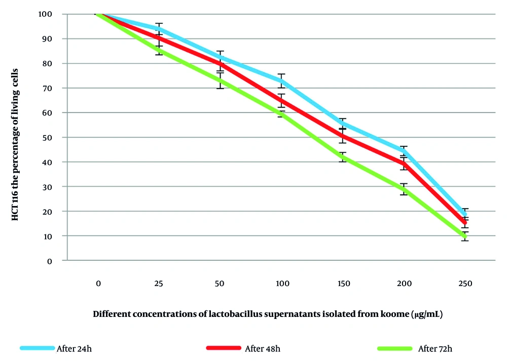
Figure 1.
The survival of HCT116 colorectal tumor cells after treatment with Lactobacillus supernatants isolated from Koome for 24, 48, and 72 hours.
4.3. Apoptosis Analysis
Apoptosis was evaluated by flow cytometry using Annexin V and propidium iodide staining. The percentage of cells in each quadrant is presented in the representative dot plots: the lower left quadrant (Q4) corresponds to viable cells (untreated cells), the lower right quadrant (Q3) represents apoptotic cells, the upper right quadrant (Q2) represents nonviable late apoptotic cells, and the upper left quadrant (Q1) represents necrotic cells (Figure 2).
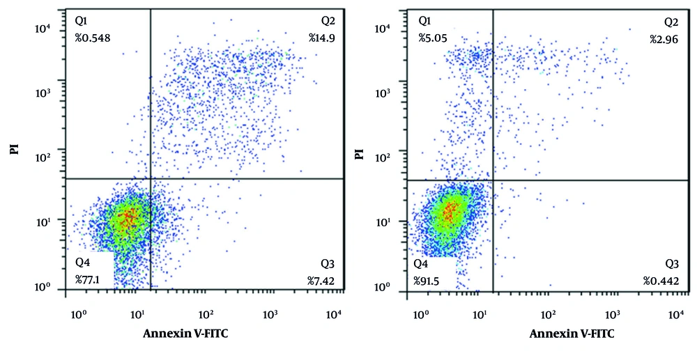
Figure 2.
Effect of Lactobacillus supernatants isolated from Koome on apoptosis in HCT116 colorectal cancer cells. Group 1: control group, untreated HCT116 cells. Group 2: HCT116 cells 48 hours after treatment with 150.907 μg/mL of Lactobacillus supernatants from Koome.
The flow cytometry analysis, as shown in Figure 2, reported apoptosis and necrosis percentages of 0.442% and 5.05%, respectively, in the control group. In the group treated with a concentration of 150/907, the apoptosis and necrosis rates were 7.42% and 0.548%, respectively. These results demonstrate that supernatants derived from Lactobacillus cultures can suppress the growth of HCT116 colorectal cancer cell lines. Our findings showed that the supernatant of lactobacilli derived from Koome significantly inhibited the growth of HCT116 colorectal cancer cells in a dose- and time-dependent manner while also triggering apoptosis (Figure 2) (P < 0.05).
5. Discussion
Recently, various studies have examined the cytotoxic effects of probiotics on different cancer cell lines. Probiotics comprise a diverse range of microorganisms, and most probiotic bacteria belong to the Lactobacillus genus. In addition, Lactobacillus probiotics are known to have beneficial roles in colorectal cancer treatment (11). Probiotics may also be considered anticancer agents without side effects (12). Accordingly, many studies have investigated the cytotoxic effects of probiotic bacteria. In one study evaluating the anticancer effects of several heat-killed Lactobacillus species and soluble bacterial components, such as polysaccharides, the results indicated that these probiotics induced apoptosis (3).
In our previous study, we reported that Lactobacillus supernatants from Sistani yellow kashk exerted cytotoxic effects on the U87MG glioblastoma brain tumor cell line by triggering apoptotic pathways (13).
Soltan Dallal et al. showed that Lactobacillus acidophilus and Lactobacillus casei effectively suppressed the malignant phenotypes of CaCo-2 colorectal cancer cells (14).
The another study indicated that Lactobacillus casei cell extract upregulated the BAX gene, downregulated the BCL2 gene, and triggered programmed cell death in the HT29 and HEC293 colon cancer cell lines.
Isazadeh et al. demonstrated that Lactobacillus acidophilus supernatant inhibited the growth of the Caco-2 colorectal cancer cell line through apoptosis induction and increased the survival rate of colon cancer patients (15).
Nami et al. reported that 21 Lactobacillus strains isolated from dairy products, such as milk, cheese, and yogurt, in Kermanshah, Iran, exhibited inhibitory effects on KB and OSCC oral cancer cells. They showed that Lactiplantibacillus plantarum had significant probiotic activity by reducing oral cancer cell proliferation, highlighting its potential to prevent tumor progression and support patient outcomes (16).
Our findings are consistent with those reported by Alaa Hadi-Al-Ward et al., who demonstrated that liposomal daunorubicin significantly reduced the viability of HCT116 colorectal cancer cells in a dose-dependent manner and exhibited stronger antiproliferative effects than the free drug. The authors further showed that treatment was associated with downregulation of PI3K gene expression and induction of cell cycle arrest, highlighting the importance of apoptosis-related pathways in colorectal cancer inhibition. These observations support the concept that biologically active agents can suppress HCT116 cell growth through modulation of key signaling pathways involved in cell survival and proliferation (17).
Consistent with our findings, Mora-Guzmán et al. demonstrated that Tournefortia mutabilis leaf extract exerted significant antiproliferative and proapoptotic effects on MCF-7 cancer cells. Their study showed that growth inhibition was associated with activation of caspases 3, 6, and 9, indicating involvement of the intrinsic apoptotic pathway. In agreement with these observations, the inhibitory effects of Lactobacillus-derived metabolites on HCT116 cells observed in the present study further support the potential of natural bioactive compounds as promising agents for colorectal cancer management (18).
The purpose of the present study was to evaluate the apoptotic effects of Lactobacillus supernatants isolated from Koome on the HCT116 cancer cell line. Overall, triggering programmed cell death is a common strategy in cancer therapy. The cell death pathway can include activation of proapoptotic events that begin with mitochondrial membrane permeabilization by Bax and Bak proteins, cytochrome c release, activation of caspase 9, and subsequent activation of caspase 3 (19). In addition, Bcl2 and Bclxl proteins, by localizing to the surface of the endoplasmic reticulum, mitochondria, and nucleus, prevent Bax and Bak protein assembly and exhibit antiapoptotic activity (20). In this study, flow cytometry was used to evaluate apoptosis and necrosis induced by the IC50 concentration of Lactobacillus supernatants. The results indicated that Lactobacillus supernatants isolated from Koome induced apoptosis in HCT116 colorectal cancer cells.
5.1. Conclusions
This study demonstrated that Lactobacillus supernatants isolated from Koome can effectively suppress cell growth and trigger apoptosis in cultured colon cancer cells, highlighting their potential as a therapeutic strategy.