
1. Context
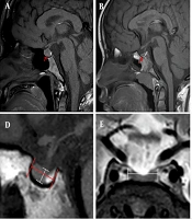
The pituitary gland serves a critical function. It is located in the sella turcica of the sphenoid bone and has 2 functionally active parts: the anterior and posterior lobes. Most pituitary hormones are secreted by the anterior lobe and are regulated by hormones released from the hypothalamus.
Pituitary gland size may vary widely. In some cases, only a thin layer of glandular tissue is present on the floor of the pituitary fossa. Normal pituitary growth and development depend on age-related changes in endocrine gland activity and the individual’s physiological status. Accordingly, pituitary height and volume vary by age and sex and reach their maximum size under conditions such as puberty and pregnancy (1, 2). Pituitary gland size and shape may also be associated with several conditions, including various pathologies, endocrine gland disorders, idiopathic intracranial hypertension, schizophrenia, and poor growth and development in children (3).
Magnetic resonance imaging is an accurate diagnostic modality for evaluating the pituitary gland (4, 5). High-resolution 3-dimensional MRI enables a more accurate volumetric assessment of pituitary gland shape and size (6). The anterior and posterior pituitary lobes can be identified on MRI images. The posterior lobe usually generates a high signal on T1-weighted images because of neurosecretory granules in the neurohypophyseal region.
Recent advances in MRI technology have greatly improved the diagnostic accuracy for detecting oncological, neural, and endocrine gland pathologies over the past 2 decades. Brain MRI of the hypothalamic-pituitary complex is an important tool for the comprehensive assessment of this region (7). Currently, pituitary MRI is commonly requested for children with growth retardation and endocrine gland disorders. Despite the frequent clinical use of MRI for this purpose, identifying pituitary gland abnormalities can be difficult. Radiology reports on pituitary gland size are mainly based on subjective judgment (8). Nonetheless, pituitary gland size can help differentiate normal from pathological cases.
Most available studies on pituitary gland size have been conducted in adults, and studies in children are limited (1, 8-12, 13). Moreover, existing studies have reported widely variable results. For instance, Berntsen et al. (1) reported that mean midsagittal and maximum sagittal pituitary height were significantly greater in females than in males. They found no significant differences in sagittal pituitary height or volume among different age groups in either males or females. Lamichhane et al. (12) demonstrated evidence of a growth spurt in adolescents aged 10 to 14 years, which was more prominent in females than in males. Their findings indicated more pronounced pituitary gland growth and development in adolescents, particularly females, and showed that pituitary dimensions did not significantly change after 20 years of age (12).
Given differences in pituitary gland size among males and females, age groups, and races, population-specific studies are required to determine normal pituitary gland size across age groups within each population. This information can be highly useful for detecting pathologies and differentiating them from normal variations. Because no published data were available on pituitary gland dimensions in children from Zahedan, this study aimed to assess pituitary gland dimensions in 2- to 18-year-old Iranians residing in Zahedan, Iran, using MRI and to provide descriptive data from this clinical sample.
2. Evidence Acquisition
2.1. Study Design and Setting
This cross-sectional study was conducted among Iranian children and adolescents aged 2 to 18 years who underwent brain MRI at a radiology center in Zahedan, Iran, in 2024. The study protocol was approved by the university ethics committee (IR.ZAUMS.REC.1403.131).
2.2. Eligibility Criteria
The inclusion criteria were brain MRI scans of patients aged 2 to 18 years with chief complaints of seizures, headache, or growth retardation; mild to moderate periventricular leukomalacia changes on MRI; and no significant change in pituitary gland volume compared with the normal population.
The inclusion criterion was no significant change in pituitary gland volume compared with the reported range for asymptomatic children.
The exclusion criteria were poor-quality images; endocrine abnormalities; a history of asphyxia, breech delivery, or preterm birth (< 35 weeks); long-term use of antiepileptic medications; other abnormalities in the pituitary gland or hypothalamic region (such as cysts, adenoma, invasive sellar tumors, and partially empty sella); previous or current pituitary gland disorders; previous or current hormonal therapy such as thyroxine, gonadal or adrenal steroids (except corticosteroid use within the 7 days before MRI); any psychiatric disorder or use of antipsychotic medications; intracranial surgery or radiation; and MRI findings suggestive of increased intracranial pressure or perinatal insult. Patients with MRI findings of increased intracranial pressure or perinatal injury were excluded from the study.
The screening process flowchart is shown in Figure 1.
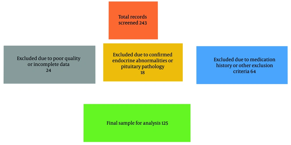
Figure 1.
Flowchart of the screening process
2.3. Sample Size
The sample size was calculated as 125 patients according to a previous study (8), with alpha = 0.05, beta = 0.2, and d = 0.07.
2.4. Data Collection
Written informed consent was obtained from the parents for the use of their children’s MRI images for research purposes. All MRI images were acquired using a 1.5-Tesla GE Healthcare MRI scanner (Signa HD). T1-weighted and T2-weighted midsagittal and coronal views were evaluated on an LCD monitor (IZO RadiForce MX215, Japan) with a resolution of 1024 × 1280 pixels using Metric V10.1 Marco PACS software.
Eligible MRI images were selected by convenience sampling, and pituitary dimensions were measured as follows (Figures 2-4):
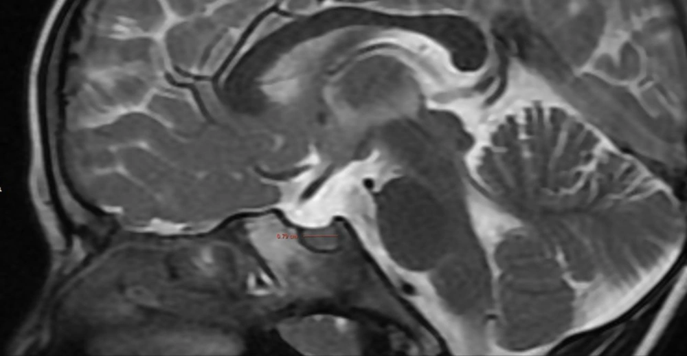
Figure 2.
Measurement of the anteroposterior diameter of the pituitary gland on the midsagittal T2-weighted image
Pituitary volume was calculated in cubic millimeters using the following formula:
Craniocaudal dimension × anteroposterior dimension × transverse dimension × 0.52
This factor was obtained from the equation used to calculate the volume of a sphere or a cube:
The anteroposterior diameter of the pituitary gland was defined as the distance, in millimeters, from the anterior border of the gland (closest to the tuberculum sellae) to the posterior border (near the dorsum sellae) on the midsagittal view in which the pituitary stalk was visible.
Pituitary height was defined as the maximum distance, in millimeters, between the most superior convexity of the gland (or its flat upper border) and the line of the sellar floor (sella turcica) on the midsagittal view in which the pituitary stalk was visible.
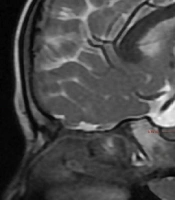
The transverse diameter was defined as the maximum distance, in millimeters, between the two lateral borders of the gland at its widest part on the coronal view between the two cavernous sinuses.
Patients’ age and sex were also recorded.
All measurements were performed independently by two trained radiologists with 5 and 8 years of experience in neuroradiology who were blinded to the patients’ age and sex.
To assess intraobserver reliability, 20 randomly selected images were remeasured by the first radiologist after a 4-week interval. For interobserver reliability, the measurements obtained by the two radiologists were compared. The intraclass correlation coefficient (ICC) was calculated for both assessments, showing excellent agreement (ICC > 0.92 for all parameters).
2.5. Statistical Analysis
Measures of central tendency and dispersion were reported for the measured variables. Data were analyzed using SPSS version 26 (SPSS Inc., IL, USA).
Datasets generated and analyzed during the current study are not publicly available because of patient confidentiality and ethical restrictions but are available from the corresponding author upon reasonable request.
3. Results
The brain MRI images of 137 patients aged 2 to 18 years were evaluated, including 79 males (57.7%) and 58 females (42.3%). The mean age was 7.78 ± 4.48 years in boys and 7.12 ± 4.06 years in girls. The overall mean age was 7.55 ± 4.31 years. Table 1 presents the sex distribution in the two age groups of 2- to 10-year-olds and 10- to 18-year-olds.
Table 1.
Sex Distribution in the 2 Age Groups of 2- to 10-Year-Olds and 10- to 18-Year-Olds
| Age Group (y) and Sex | Mean ± SD |
|---|---|
| 2 - 10 | |
| Male | 5.48 ± 2.69 |
| Female | 5.33 ± 2.37 |
| Total | 5.42 ± 2.54 |
| 10 - 18 | |
| Male | 13.65 ± 1.94 |
| Female | 13.31 ± 2.09 |
| Total | 13.53 ± 1.97 |
Table 2 presents the mean pituitary gland volume overall and by age group and sex. As shown, pituitary volume increased with age and was larger in females than in males.
Table 2.
Mean Pituitary Gland Volume Overall and by Age Group and Sex a
| Category | Pituitary Volume (mm3) |
|---|---|
| Overall | 201.5 ± 117.39 |
| 2 - 10-year-olds | 151.2 ± 62.19 |
| 10 - 18-year-olds | 342.93 ± 120.94 |
| Males | 195.4 ± 98.1 |
| Females | 209.9 ± 139.9 |
a Values are expressed as mean ± SD.
The mean pituitary volume was significantly larger in the 10- to 18-year age group than in the 2- to 10-year age group (342.93 ± 120.94 vs. 151.2 ± 62.19 mm3; P < 0.001; 95% CI, 179.1 to 204.3). Furthermore, the mean pituitary volume was significantly greater in females than in males (209.9 ± 139.9 vs. 195.4 ± 98.1 mm3; P = 0.04; 95% CI, 1.2 to 28.8).
Table 3 shows the mean pituitary gland height overall and by age group and sex. As shown, pituitary height increased with age and was greater in females than in males.
Table 3.
Mean Pituitary Gland Height Overall and by Age Group and Sex a
| Category | Pituitary Height (mm) |
|---|---|
| Overall | 4.35 ± 1.32 |
| 2 - 10-year-olds | 3.78 ± 0.801 |
| 10 - 18-year-olds | 5.94 ± 1.19 |
| Males | 4.28 ± 1.25 |
| Females | 4.45 ± 1.4 |
a Values are expressed as mean ± SD.
Pituitary height was significantly greater in the older age group (5.94 ± 1.19 mm) than in the younger age group (3.78 ± 0.81 mm; P < 0.001). Although the mean height was greater in females (4.45 ± 1.4 mm) than in males (4.28 ± 1.25 mm), the difference was not statistically significant (P = 0.09).
Table 4 presents the mean anteroposterior diameter of the pituitary gland by age group and sex. As indicated, the mean anteroposterior diameter of the pituitary gland increased with age. This variable did not differ between males and females.
Table 4.
Mean Anteroposterior Diameter of the Pituitary Gland by Age Group and Sex
| Category | Mean ± SD | P Value (95% CI) |
|---|---|---|
| 2 - 10-year-olds | 6.66 ± 1.13 | Reference |
| 10 - 18-year-olds | 8.64 ± 1.53 | P < 0.001 (1.53 to 2.43) |
| Males | 7.17 ± 1.37 | Reference |
| Females | 7.19 ± 1.71 | P = 0.91 (-0.68 to 0.72) |
Independent-samples t-tests were used to compare means between two independent groups, such as boys versus girls and the 2- to 10-year-old versus 10- to 18-year-old age groups. A P value < 0.05 was considered statistically significant. All tests were 2-tailed.
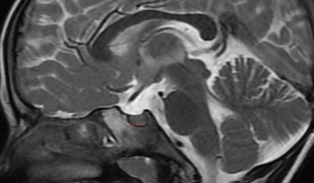
Figure 3.
Measurement of the height of the pituitary gland on the midsagittal T2-weighted image
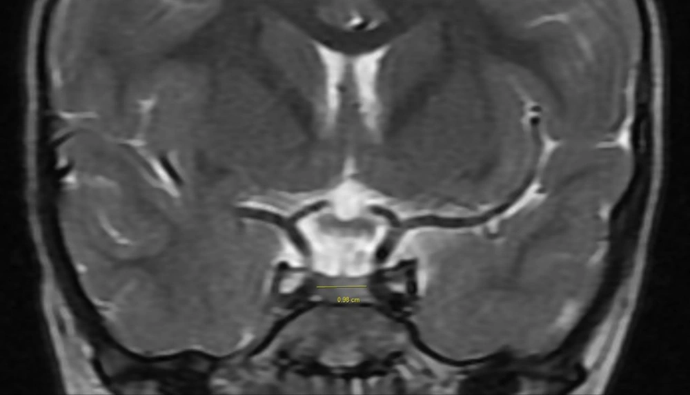
Figure 4.
Measurement of the transverse diameter of the pituitary gland on the coronal T2-weighted image
3.1. Discussion
The mean pituitary height (4.35 ± 1.32 mm) and volume (201.5 ± 117.39 mm3) in this study were lower than those reported in a Turkish population by Akşan and Sarı (8), which may be attributable to racial, nutritional, or methodological differences. In their study of a Turkish population, the mean pituitary volume was 436.7 ± 165.4 mm3 in females and 378.8 ± 65.5 mm3 in males. They also reported that pituitary height was 5.7 ± 1.17 mm in females and 5.4 ± 0.86 mm in males, with a maximum height of 8 mm in females and 7.5 mm in males. They added that although pituitary volume in boys was larger than in girls during infancy, this pattern was reversed after approximately 6 years of age. They suggested 3-dimensional MRI for direct measurement of pituitary volume in children before puberty. Differences between their results and the present findings may be due to racial or methodological differences.
In the present study, the mean pituitary volume was 151.2 ± 62.19 mm3 in 2- to 10-year-old children and 342.93 ± 120.94 mm3 in 10- to 18-year-olds, indicating an increase in pituitary volume with age. The same finding was reported by Saba et al. (14) in a study conducted in Kerman, Iran. Evidence shows that pituitary volume and size increase with age, owing to normal growth and development and increased secretion of pubertal hormones. Fink et al. (15), in a study conducted in Australia, showed a linear increase in pituitary volume in children with age and reported that this increase continued until the end of puberty. Their results were consistent with the current findings. Argyropoulou and Kiortsis (4), in a study conducted in Greece, reported a significant increase in pituitary volume in children during puberty as a result of sex hormone release and body growth and development.
The mean pituitary volume was 195.4 ± 98.1 mm3 in males and 209.9 ± 139.9 mm3 in females. The mean pituitary volume was larger in females than in males, which was consistent with the findings of Khadgawat et al. (9) in India and Lamichhane et al. (12) in Nepal. Nonetheless, sex-related differences in pituitary gland size and volume have been reported in the literature, which may be due to differences in the timing of puberty and the secretion of sex hormones in males and females. An older study also reported, on average, a larger pituitary volume in females than in males (16), which may be due to earlier onset of puberty in females and the release of sex hormones. Although some differences often exist in absolute values across studies because of racial, geographical, or imaging differences, the overall trend of pituitary gland growth and development appears to be almost the same across populations.
The mean pituitary height was 3.78 ± 0.81 mm in 2- to 10-year-olds and 5.94 ± 1.19 mm in 10- to 18-year-olds. Pituitary height increased with age. The mean pituitary height was 4.28 ± 1.25 mm in boys and 4.45 ± 1.4 mm in girls. Pituitary height was greater in girls than in boys. The same finding was reported by Takano et al. (17) in Japan, which may be due to physiological and hormonal differences between males and females.
The mean anteroposterior diameter of the pituitary gland was 6.66 ± 1.13 mm in 2- to 10-year-olds and 8.64 ± 1.53 mm in 10- to 18-year-olds. This variable also increased with age. The mean anteroposterior diameter of the pituitary gland was 7.17 ± 1.37 mm in boys and 7.19 ± 1.71 mm in girls; these values were almost the same. According to the results, the mean anteroposterior diameter of the pituitary gland is influenced by growth and development and increases with age. Elster et al. (18), in a study conducted in the United States, showed that this parameter significantly increased with age, which was in line with the present findings. Hanaoka et al. (19), in Japan, found no significant difference in the mean anteroposterior diameter of the pituitary gland between males and females, which was consistent with the present findings. Puyana et al. (20), in the United States, showed that variations in pituitary gland size and volume across populations can be due to differences in genetic and environmental factors. Therefore, direct comparison of study findings should be performed with caution. Bonczar et al. (21), in their systematic review and meta-analysis, showed that the mean pituitary height was 5.64 mm and the mean pituitary length was 9.98 mm. Asian females had the largest pituitary gland volume, and European males had the smallest. Pituitary gland dimensions increased with age. Measuring pituitary gland size and volume can help detect related disorders, such as hypopituitarism, pituitary hyperplasia, and pituitary tumors. It is also useful for assessing normal growth and development in children (22). In patients treated for pituitary disorders, measurement of pituitary volume and dimensions can help monitor treatment response. For instance, a reduction in pituitary volume in patients treated for pituitary tumors or an increase in volume in those receiving growth hormone therapy can indicate a positive response to treatment (23).
The present results can be used as basic data for future studies on differences in pituitary gland size and volume across populations and age groups (24).
This study had a cross-sectional design, which does not allow causal relationships to be determined. Future longitudinal studies are required to assess changes in pituitary size and volume over time. Determination of normal ranges for pituitary dimensions according to age, sex, ethnicity, and race is also recommended. The relationship between pituitary size and volume and serum hormone levels should also be investigated.
This study had a cross-sectional design, which does not allow causal relationships to be determined. Additionally, the use of a clinical sample of children with headache, seizure, or growth retardation limits the generalizability of the findings to the general pediatric population. Therefore, the results should be interpreted as descriptive data from this specific sample rather than definitive normative values. Future longitudinal studies are required to assess changes in pituitary size and volume over time.
4. Conclusions
The present results indicated that pituitary volume, height, and diameter increased with age in this clinical sample of Iranian children. Pituitary volume was significantly greater in girls than in boys, whereas anteroposterior diameter was similar between the sexes. These descriptive findings may serve as preliminary comparative data for radiologists and clinicians when evaluating pediatric brain MRI scans in similar clinical settings in southeastern Iran. Assessing pituitary volume, rather than height alone, may better reveal subtle sex- and age-related differences.