
1. Background
Brain tumors represent a major global health challenge and encompass a diverse array of neoplasms affecting the central nervous system (CNS). These tumors are classified according to histological origin, location, and molecular characteristics, with primary brain tumors arising from neural or glial cells and secondary tumors resulting from metastases (1). Recent global burden analyses indicate that the age-standardized incidence of primary malignant and nonmalignant CNS tumors is approximately 5 - 7 per 100,000 population, with gliomas accounting for approximately 30% - 40% of all cases and showing a slight upward trend in low- and middle-income countries (2, 3). Among these, posterior fossa lesions (PFLs), including tumors originating from the cerebellum, brainstem, and fourth ventricle, pose unique diagnostic and therapeutic challenges because of their proximity to critical structures, including the brainstem and CSF pathways (4). PFLs constitute approximately 50% - 70% of pediatric brain tumors but are less common in adults, in whom they represent 15% - 20% of intracranial neoplasms (5, 6).
The posterior fossa, also known as the infratentorial compartment, is a confined space bounded superiorly by the tentorium cerebelli and inferiorly by the foramen magnum; it contains the cerebellum, pons, and medulla oblongata (7). Lesions in this region, whether neoplastic (eg, medulloblastomas, astrocytomas, and ependymomas) or nonneoplastic (eg, hemorrhages and strokes), often disrupt normal CSF dynamics, leading to hydrocephalus, a pathological accumulation of CSF that enlarges the ventricular spaces and increases intracranial pressure (ICP) (8). Hydrocephalus in patients with PFLs primarily results from obstruction of CSF outflow at the level of the fourth ventricle or the cerebral aqueduct (aqueduct of Sylvius) or from impaired CSF absorption secondary to inflammation or hemorrhage (9). This complication is reported in 70% - 90% of patients with posterior fossa tumors (PFTs) at presentation and contributes substantially to morbidity and mortality if left unmanaged (9-11).
Clinically, hydrocephalus manifests with symptoms such as headache, nausea, vomiting, altered consciousness, and ataxia, often exacerbated by the mass effect of the lesion itself (5). In children, PFLs such as pilocytic astrocytomas and medulloblastomas predominate, with hydrocephalus occurring in up to 80% of cases preoperatively (12). In adults, common etiologies include metastatic tumors, hemangioblastomas, and vascular events such as cerebellar hemorrhages or infarcts, with reported hydrocephalus rates of 30% - 60% (10). Histologically, astrocytomas, including pilocytic astrocytomas, and medulloblastomas are among the most common posterior fossa lesions; epidemiological studies report that astrocytomas account for approximately 25% - 30% of adult PFLs (13). The 2021 World Health Organization classification emphasizes molecular markers, such as IDH mutations in gliomas and SHH/WNT pathway alterations in medulloblastomas, which influence prognosis and treatment (1).
Surgical resection remains the cornerstone of PFL management, with the goal of achieving gross total removal while preserving neurological function (5). However, postoperative hydrocephalus persists or develops in 10% - 40% of cases, necessitating interventions such as VP shunting or endoscopic third ventriculostomy (ETV) (9, 10). VP shunting, which diverts CSF from the ventricles to the peritoneal cavity, is effective but is associated with complications such as infection (5% - 15%), obstruction (20% - 30%), and overdrainage leading to subdural hematoma (14, 15). Predictive factors for shunt dependency include younger age (< 3 years), midline tumor location, incomplete resection, and the severity of preoperative hydrocephalus (8, 16).
Despite advances in neuroimaging, including magnetic resonance imaging (MRI) for tumor characterization and molecular profiling, gaps remain in understanding hydrocephalus incidence and shunt requirements across diverse populations, particularly in resource-limited settings (2, 9). Iranian studies report that brain tumors are the ninth and tenth most common cancers in men and women, respectively, with an increasing incidence from 4.5 to 6.2 per 100,000 population between 2015 and 2020; PFLs contribute substantially to neurological mortality (17, 18). Local data indicate that astrocytomas and medulloblastomas are prevalent, with hydrocephalus occurring in 40% - 50% of surgical cases (19). However, comprehensive analyses of postoperative outcomes in Iranian cohorts are scarce, underscoring the need for region-specific evidence to optimize management protocols.
Although several studies have investigated hydrocephalus associated with posterior fossa tumors, limited data are available regarding the overall incidence of hydrocephalus and permanent shunt dependency across both tumorous and nontumorous posterior fossa lesions. PFLs encompass a broad spectrum of neoplastic and nonneoplastic entities with distinct etiologies and natural histories, but they share anatomical confinement within the infratentorial compartment and a common potential to obstruct CSF pathways at the level of the fourth ventricle or aqueduct of Sylvius. This shared anatomical and hydrodynamic consequence supports their initial aggregation in a single cohort to describe overall hydrocephalus rates and CSF diversion requirements in routine clinical practice. Nevertheless, vascular lesions, benign tumors, and high-grade malignancies may differ substantially in their propensity to cause persistent CSF obstruction and in their response to surgical decompression. These differences were therefore considered in interpreting the findings and are addressed as a key study limitation.
2. Objectives
This study evaluated the incidence of hydrocephalus and the need for CSF diversion in 69 patients with PFLs who underwent surgery at Ayatollah Kashani Hospital, Shahrekord, Iran, between 2019 and 2024. By examining demographic, histopathological, and clinical characteristics, we also assessed factors associated with hydrocephalus and shunt dependency. These findings may improve the understanding of postoperative hydrocephalus and inform future studies aimed at optimizing its management in this high-risk population.
3. Methods
3.1. Study Design and Setting
This retrospective observational study used a census sampling approach. No formal sample size calculation was performed. All consecutive eligible patients with PFLs who underwent surgery at Ayatollah Kashani Hospital between March 2019 and March 2024 were included to maximize the available sample size and minimize selection bias.
3.2. Participants and Eligibility Criteria
All consecutive patients with a documented diagnosis of PFL who underwent surgical intervention at Ayatollah Kashani Hospital between March 2019 and March 2024 were eligible for inclusion. Patients were excluded if they had 1) incomplete medical records or insufficient follow-up data, defined as less than 6 months of postoperative follow-up; 2) a history of previous CSF diversion procedures or permanent VP shunt placement before admission; or 3) undergone repeat surgery for recurrent lesions during the study period.
After receiving written approval from the Research Deputy of Shahrekord University of Medical Sciences, the principal investigator attended the study site. An official introduction letter was submitted to the administration of Ayatollah Kashani Hospital, the study objectives were explained to the relevant authorities, and formal permission was obtained from hospital management. The approved permissions were subsequently presented to the heads of the respective departments. After completion of all required authorizations and coordination, eligible patients were identified according to the predefined inclusion criteria, and data were extracted from their medical records.
3.3. Data Collection
Baseline demographic characteristics, including age and sex; histopathological diagnosis; and lesion characteristics, including size, location, and type, were recorded. Patients were followed for 6 months postoperatively to assess the development of hydrocephalus and the need for CSF diversion. Additional information, including the degree of hydrocephalus and CSF status before surgery, was recorded in the questionnaires. Patient position during surgery, extent of resection (complete or subtotal [> 90% lesion removal]), biopsy results, postoperative hydrocephalus, time to VP shunt placement, and complications were extracted from the medical records and entered into checklists (Table 1).
Table 1.
Distribution of Demographic and Clinical Characteristics According to Hydrocephalus Status a
| Variables | With hydrocephalus | Without hydrocephalus | Total Number |
|---|---|---|---|
| Gender | |||
| Male | 19 (46.3) | 22 (53.7) | 41 |
| Female | 13 (46.4) | 15 (53.6) | 28 |
| Age (y) | |||
| < 16 | 3 (100.0) | 0 (0.0) | 3 |
| 16 - 59 | 17 (58.6) | 12 (41.4) | 29 |
| ≥ 60 | 12 (32.4) | 25 (67.6) | 37 |
| Type of lesion | |||
| Pilocytic astrocytoma | 3 (100.0) | 0 (0.0) | 3 |
| Diffuse astrocytoma | 2 (50.0) | 2 (50.0) | 4 |
| Ischemic stroke | 8 (34.8) | 15 (65.2) | 23 |
| Intracerebral hemorrhage | 10 (43.5) | 13 (56.5) | 23 |
| ICH + IVH | 0 (0.0) | 1 (100.0) | 1 |
| IVH + ICH | 1 (100.0) | 0 (0.0) | 1 |
| Breast cancer metastasis | 2 (100.0) | 0 (0.0) | 2 |
| Medulloblastoma | 3 (100.0) | 0 (0.0) | 3 |
| Colon cancer metastasis | 1 (100.0) | 0 (0.0) | 1 |
| Meningioma | 1 (33.3) | 2 (66.7) | 3 |
| Lung cancer metastasis | 0 (0.0) | 2 (100.0) | 2 |
| Testicular cancer metastasis | 0 (0.0) | 1 (100.0) | 1 |
| Schwannoma | 1 (50.0) | 1 (50.0) | 2 |
| Duration of hospitalization (d) | |||
| ≤ 13 | 16 (37.2) | 27 (62.8) | 43 |
| > 13 | 16 (61.5) | 10 (38.5) | 26 |
a Values are expressed as No. (%). ICH indicates intracerebral hemorrhage.
3.4. Study Outcomes
The primary outcomes were the incidence of hydrocephalus associated with PFLs and the requirement for permanent VP shunt placement during the 6-month follow-up period. Secondary outcomes included the timing of hydrocephalus (preoperative versus postoperative), the need for temporary CSF diversion (external ventricular drainage or ventriculostomy), time to permanent shunt placement, postoperative complications, and associations between demographic and lesion-related characteristics, including age, sex, lesion type, lesion location, and severity of hydrocephalus, and shunt dependency.
3.5. Outcome Definitions
Hydrocephalus was defined as ventricular enlargement on computed tomography (CT) or MRI accompanied by compatible clinical manifestations of increased intracranial pressure. Preoperative hydrocephalus was defined as hydrocephalus documented by clinical and radiological assessment before the index posterior fossa surgery. De novo postoperative hydrocephalus was defined as newly developed hydrocephalus after surgery in patients without evidence of hydrocephalus before the operation. Postoperative hydrocephalus was defined as new or worsening ventricular dilatation with compatible clinical symptoms after surgical treatment. Hydrocephalus diagnosed within 30 days after surgery was considered early postoperative hydrocephalus, whereas cases detected after 30 days were classified as delayed postoperative hydrocephalus.
Temporary CSF diversion was defined as external diversion of CSF using an external ventricular drain (EVD) or ventriculostomy for the acute management of hydrocephalus. Permanent CSF diversion was defined as VP shunt insertion for persistent hydrocephalus. Shunt dependency was defined as the requirement for permanent VP shunt placement because hydrocephalus persisted or progressed despite surgical treatment of the posterior fossa lesion and/or temporary CSF diversion during follow-up.
3.6. Diagnostic Criteria
Hydrocephalus was diagnosed based on clinical manifestations suggestive of increased intracranial pressure, such as headache, nausea, vomiting, decreased level of consciousness, or papilledema, in combination with radiological evidence of ventricular enlargement on CT or MRI, as documented in the medical records. Permanent shunt dependency was defined as the requirement for VP shunt placement because of persistent or progressive hydrocephalus that did not resolve after treatment of the posterior fossa lesion or after temporary CSF diversion, as determined by the treating neurosurgical team during follow-up.
3.7. Statistical Analysis
Data were analyzed using Stata software, version 17.0 (StataCorp, College Station, TX, USA). Descriptive statistics were used to summarize patient characteristics and outcome variables. Categorical variables were compared using the chi-square test or Fisher exact test, as appropriate. Continuous variables were compared using the independent t-test or Mann-Whitney U test, depending on the distribution. To quantify the strength of associations between potential predictors and permanent shunt dependency, unadjusted odds ratios (ORs) with 95% confidence intervals (CIs) were calculated using univariate logistic regression. Multivariable logistic regression was not performed because the small number of outcome events (n = 13) precluded reliable adjustment for multiple covariates; a rule of thumb requires at least 10 - 15 events per predictor variable. Therefore, the reported associations are unadjusted and should be considered exploratory and hypothesis-generating. A 2-sided P value < 0.05 was considered statistically significant.
3.8. Ethical Considerations
This study was conducted in accordance with the principles of the Declaration of Helsinki. Written informed consent was obtained from all patients or their legal guardians before inclusion. Patients were informed that they could withdraw from the study at any time without affecting their medical care. All information extracted from medical records was treated confidentially and used solely for research purposes.
4. Results
4.1. Demographic and Clinical Characteristics
In total, 69 patients with PFLs who were referred to Kashani Hospital in Shahrekord and underwent surgical intervention between 2019 and 2024 were enrolled. Of these, 41 (59.4%) were male and 28 (40.6%) were female. Overall, 32 patients (46.4%) developed hydrocephalus. The incidence of hydrocephalus was nearly identical by sex, occurring in 19 of 41 male patients (46.3%) and 13 of 28 female patients (46.4%).
Regarding age distribution, hydrocephalus occurred in all 3 patients (100%) younger than 16 years, 17 of 29 patients (58.6%) aged 16 - 59 years, and 12 of 37 patients (32.4%) aged 60 years or older. Analysis by lesion type showed that hydrocephalus developed in 32 patients. Among these cases, the most frequent etiology was intracerebral hemorrhage, observed in 10 patients (31.3% of hydrocephalus cases), followed by ischemic stroke in 8 patients (25.0%), pilocytic astrocytoma in 3 patients (9.4%), medulloblastoma in 3 patients (9.4%), diffuse astrocytoma in 2 patients (6.3%), breast cancer metastasis in 2 patients (6.3%), colon cancer metastasis in 1 patient (3.1%), schwannoma in 1 patient (3.1%), and meningioma in 1 patient (3.1%).
Among patients with hydrocephalus, headache (40.6%) and decreased level of consciousness (34.4%) were the most frequently reported symptoms. Nausea and vomiting were observed in 2 patients (6.3%), whereas imbalance with vomiting, headache with decreased level of consciousness, headache with vomiting, hearing loss with facial palsy, and nausea and vomiting with headache were each reported in 1 patient (3.1%).
Hospitalization duration was dichotomized at the mean value of 13 days into 2 categories: ≤ 13 days and > 13 days. Based on this classification, the incidence of hydrocephalus did not differ significantly between the 2 groups (P > 0.05).
4.2. Incidence of Hydrocephalus Before and After Surgical Intervention
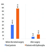
Among the 69 patients with PFLs, 32 (46.4%) developed hydrocephalus. Of these, 28 (40.6% of all patients and 87.5% of patients with hydrocephalus) had hydrocephalus before the initial surgery, whereas 4 (5.8% of all patients and 12.5% of patients with hydrocephalus) developed hydrocephalus after surgery (Figure 1).
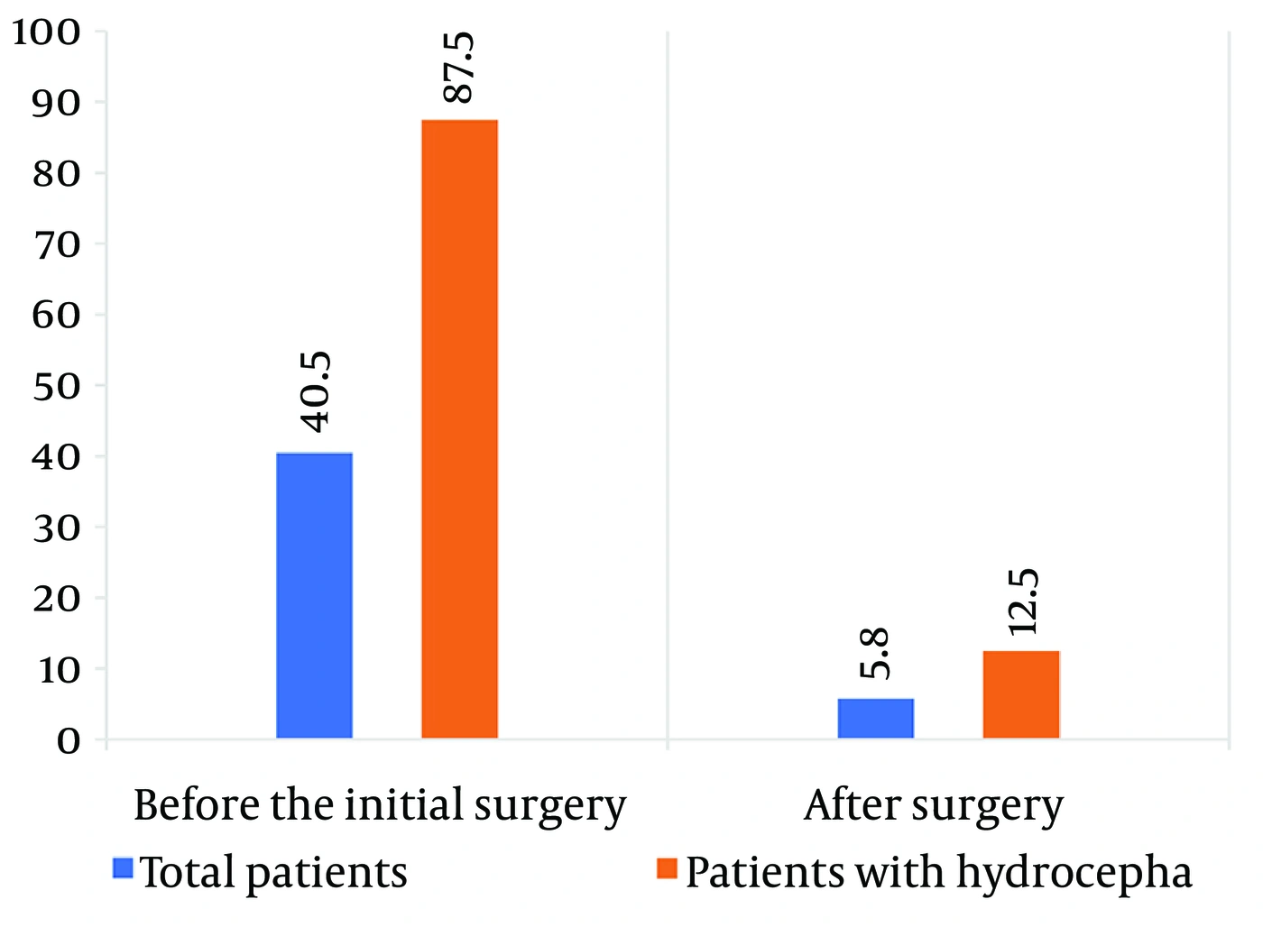
Figure 1.
Incidence of Hydrocephalus Before and After Surgical Intervention
4.3. Cerebrospinal Fluid Diversion and Permanent Shunt Placement
Overall, 26 patients (81.25% of patients who developed hydrocephalus) required either temporary or permanent CSF diversion. Of these 26 patients, 12 (46.1% of patients who underwent CSF diversion and 37.5% of patients with hydrocephalus) required both temporary and permanent shunting; 13 (50.0% of patients who underwent CSF diversion and 40.6% of patients with hydrocephalus) received temporary diversion only via an EVD or ventriculostomy; and 1 patient (3.8% of patients who underwent CSF diversion and 3.1% of patients with hydrocephalus) received a permanent shunt without prior temporary diversion. Thus, permanent VP shunting was required in 13 of 32 patients with hydrocephalus (40.6%) and 13 of 69 patients overall (18.8%).
4.4. Interventions in Patients With De Novo Postoperative Hydrocephalus
Among the 4 patients who developed de novo hydrocephalus after surgery, 1 patient (25%) required permanent VP shunt placement. This patient was a 10-year-old boy with a cerebellar pilocytic astrocytoma who presented with headache; the interval from symptom onset to definitive shunt insertion was 12 days. One patient (25%), a 41-year-old woman with a cerebellopontine angle (CPA) schwannoma who presented with decreased level of consciousness and facial palsy, did not require shunt placement.
The remaining 2 patients were managed with temporary CSF diversion only. One was a 68-year-old man with decreased level of consciousness due to a cerebellar cerebrovascular accident (CVA), with a treatment duration of 10 days. The other was a 66-year-old woman with decreased level of consciousness due to cerebellar intracerebral hemorrhage (ICH), also with a treatment duration of 10 days.
5. Discussion
In this retrospective cohort of 69 patients undergoing surgery for PFLs, the overall incidence of hydrocephalus was 46.4%, with 87.5% of hydrocephalus cases present preoperatively and 12.5% developing de novo postoperatively. Permanent VP shunting was ultimately required in 18.8% of the entire cohort and 40.6% of patients with hydrocephalus. These findings indicate that permanent shunting was not required in most patients with hydrocephalus; however, the results should be interpreted in the context of this small, heterogeneous cohort.
These overall figures represent an average across a heterogeneous mix of pathologies. The rate of permanent shunting was higher in patients with neoplastic lesions (32.0%) than in those with vascular lesions (11.4%), a difference that is clinically plausible given the more durable CSF obstruction often associated with progressive tumors. Consequently, the finding that permanent shunting was avoided in most patients should not be interpreted as equally applicable to every subgroup, particularly pediatric patients with malignant posterior fossa tumors, in whom the risk of persistent hydrocephalus may be higher.
The predominance of preoperative hydrocephalus (87.5%) aligns with previous reports in pediatric populations, in which rates of 70% - 90% have been described (9, 10). However, the low rate of de novo hydrocephalus (12.5%) in our series is lower than the 25% - 30% reported in pediatric-dominated cohorts (21, 23). This discrepancy may be related to the adult predominance of our cohort (mean age > 50 years), as adults are less prone to CSF absorption failure following tumor resection (20). The high proportion of nonneoplastic lesions, particularly intracerebral hemorrhage (31.3%) and ischemic stroke (25.0%), further reflects the acute obstructive pathophysiology observed in adult PFLs, which may resolve after surgical decompression without permanent CSF diversion (7).
In our cohort, hydrocephalus occurred in all 3 patients younger than 16 years; however, the very small pediatric subgroup precludes meaningful inference regarding pediatric shunt dependency. Previous systematic reviews have identified very young age, particularly age < 3 years, as a predictor of permanent shunting (10, 22). This association has been attributed to narrower subarachnoid spaces and immature CSF absorption capacity in young children, which may predispose them to persistent ventriculomegaly after lesion resection (8). Similarly, midline tumor location has been associated with shunt requirement in the literature (12, 16).
Our results reinforce the principle that preoperative hydrocephalus alone should not automatically mandate prophylactic permanent shunting. In this series, 81.25% of patients with hydrocephalus required some form of CSF diversion, but temporary EVD alone was sufficient in half of those who underwent diversion. This observation supports a stepwise management strategy involving initial EVD for acute CSF diversion, followed by conversion to permanent shunting only when weaning fails or symptoms persist. Such an approach may minimize unnecessary shunt placement and reduce the risk of shunt-related complications such as infection (5% - 15%) and mechanical failure (20% - 30%) (14, 24). Outcomes after shunt placement in our cohort were also consistent with evidence that VP shunting, when indicated, can provide durable CSF control (15, 25).
The added value of the present study lies in its evaluation of hydrocephalus across a heterogeneous cohort of PFLs and its practical implications for predicting postoperative CSF diversion requirements.
5.1. Study Limitations
This study has several limitations. First, its retrospective, single-center design may have introduced selection and information bias and may limit the generalizability of the findings. Second, the relatively small sample size, particularly within individual pathological subgroups, precluded multivariable logistic regression and robust subgroup analyses. Consequently, the reported associations should not be interpreted as independent predictors, but rather as exploratory findings. Third, the short postoperative follow-up period of 6 months may have underestimated late-onset hydrocephalus or delayed shunt dependency. In addition, ETV was not systematically evaluated as an alternative to shunting because it was not routinely performed at our institution during the study period. Finally, the heterogeneous study population included a broad spectrum of PFLs with distinct pathophysiological characteristics, which may have influenced the observed rates of hydrocephalus and CSF diversion.
Future prospective, multicenter studies with larger cohorts, stratified analyses, and extended follow-up are needed to validate these findings and to develop risk-stratification models that can identify patients most likely to benefit from different CSF diversion strategies. Comparative effectiveness research in adult populations is also warranted.
5.2. Conclusions
In this mixed cohort of 69 patients with PFLs, hydrocephalus was present in nearly half of the cases, with most hydrocephalus already evident preoperatively. De novo postoperative hydrocephalus developed in only a small fraction of patients. Temporary external ventricular drainage alone was sufficient in half of those who required CSF diversion, whereas permanent VP shunting was ultimately required in 18.8% of the entire cohort and 40.6% of patients who developed hydrocephalus. The presence of preoperative hydrocephalus should therefore not automatically be equated with permanent shunt dependency. However, the likelihood of shunt dependency may vary across lesion types and should be interpreted according to the specific pathology, patient age, and radiological severity of hydrocephalus. Given the single-center retrospective design and small sample size, these observations require confirmation in larger prospective multicenter studies.