
1. Background
Atherosclerosis is a progressive, chronic inflammatory disease characterized by lipid accumulation, endothelial dysfunction, immune cell infiltration, and vascular remodeling, ultimately leading to plaque formation in the arterial wall and driving the development of most cardiovascular diseases (CVDs) globally (1). The disease process arises from complex interactions among metabolic, inflammatory, and genetic factors, with both innate and adaptive immune responses contributing to lesion initiation and progression (1). Traditional risk factors, including hypertension, dyslipidemia, smoking, obesity, and a sedentary lifestyle, markedly increase susceptibility to atherosclerotic CVDs, which remain the leading cause of morbidity and mortality worldwide (1). Despite the widespread implementation of pharmacologic therapies, such as statins, antihypertensives, and antiplatelet agents, that reduce events, residual cardiovascular risk persists, highlighting the need for novel mechanistic insights and modifiable targets (2). In recent years, epigenetic regulation of cardiovascular pathways has emerged as a pivotal mechanism in both the development and modulation of atherosclerosis.
Among epigenetic regulators, microRNAs (miRNAs), a class of small non-coding RNAs (~ 18 - 22 nucleotides), are recognized as critical post-transcriptional modulators of gene expression that orchestrate key cellular processes, including inflammation, apoptosis, angiogenesis, and endothelial function (3). miRNAs bind to complementary sequences in target messenger RNAs (mRNAs), leading to translational repression or mRNA degradation and thereby shaping phenotypic outcomes in health and disease (3, 4). In the cardiovascular context, dysregulated expression of specific miRNAs has been associated with plaque instability, endothelial dysfunction, and progression of atherosclerotic lesions in both experimental and clinical settings (5).
Two miRNAs of particular relevance to vascular homeostasis and atherosclerotic pathology are miR-126 and miR-146a. miR-126 is predominantly expressed in endothelial cells and endothelial progenitor cells (EPCs), where it promotes angiogenesis, maintains vascular integrity, and facilitates endothelial repair by targeting negative regulators of the phosphoinositide 3-kinase (PI3K)/Akt/endothelial nitric oxide synthase (eNOS) pathway (5, 6). Specifically, miR-126 enhances vascular endothelial growth factor (VEGF)-mediated signal transduction and stabilizes interendothelial junctions, thereby preserving endothelial barrier function and attenuating plaque development (5, 7). In contrast, miR-146a serves as a master regulator of innate immune signaling by targeting components of the Toll-like receptor (TLR) pathway, including TLR4, TNF receptor-associated factor 6 (TRAF6), and interleukin-1 receptor-associated kinase 1 (IRAK1), thereby dampening nuclear factor-kappa B (NF-κB)-mediated proinflammatory responses (8, 6). In atherosclerosis models, elevated miR-146a has been associated with reduced inflammatory cytokine expression and decreased lesion burden, underscoring its anti-inflammatory potential (9, 10).
Although the biological importance of miR-126 and miR-146a in atherogenesis is established, the molecular mechanisms regulating their expression in response to lifestyle interventions, particularly exercise, remain incompletely understood (4, 7). This mechanistic gap is especially relevant because physical activity is a cornerstone nonpharmacological strategy for preventing and managing CVDs, including atherosclerosis. Regular aerobic exercise improves endothelial function, enhances nitric oxide bioavailability, reduces systemic inflammation, optimizes lipid metabolism, and modulates vascular shear stress, collectively conferring an atheroprotective phenotype (5, 11). Despite these recognized benefits, the precise molecular pathways by which exercise induces these systemic and vascular adaptations remain incompletely delineated.
Emerging evidence indicates that exercise influences the expression of specific miRNAs within cardiovascular tissues and in the circulation, suggesting that miRNAs may mediate exercise-induced cardiometabolic adaptation (12, 10). In the HERITAGE Family Study, 20 weeks of endurance training in previously sedentary adults significantly altered the circulating profile of multiple cardiovascular-related miRNAs, including increases in circulating miR-126 - 3p and miR-146a-5p, supporting an association between aerobic exercise and miRNA regulation (8, 7). These findings suggest that exercise may modulate vascular health not only through classical physiological adaptations but also via epigenetic regulation of gene networks underlying endothelial function and inflammation. Indeed, other investigations have shown that sustained moderate-intensity aerobic training induces durable changes in circulating miRNA profiles, including miRNAs associated with endothelial repair and immune modulation (13).
More specifically, miR-126 has been shown to increase in response to both acute and chronic aerobic exercise, with associated benefits for angiogenesis, endothelial repair, and vascular function (7). Mechanistically, exercise-stimulated miR-126 enhances VEGF signaling and mobilizes EPCs, contributing to improved neovascularization and plaque stability in disease models (7, 13). Similarly, miR-146a has been linked to exercise-induced anti-inflammatory effects, whereby its upregulation corresponds with reduced systemic inflammation and lower circulating levels of proinflammatory biomarkers such as IL-6 and TNF-α (14, 8). These miRNAs therefore reflect key biological processes modulated by physical activity, namely angiogenesis and immune regulation, which are central to both the pathogenesis and amelioration of atherosclerotic disease.
Despite this growing body of evidence, most existing research has important limitations. First, the current evidence base is largely derived from studies in healthy subjects or animal models, creating uncertainty as to whether exercise-induced miRNA responses observed in these settings accurately reflect those occurring in patients with established atherosclerosis.
Second, studies that include CVD populations often rely on mixed-gender cohorts or focus predominantly on males, thereby obscuring potential sex-specific molecular responses to exercise (15). This is a critical gap because women exhibit distinct cardiovascular risk profiles, disease progression patterns, and responses to both pharmacological and behavioral interventions compared with men (15). Understanding how exercise modulates cardioprotective miRNAs in women with atherosclerosis is therefore essential for developing tailored interventions that optimize cardiovascular health across populations.
A further gap in the existing evidence concerns the influence of different aerobic exercise protocols on miRNA regulation. In particular, the relative effects of continuous and interval-based aerobic training on the expression of miR-126 and miR-146a have not been sufficiently investigated in patients with atherosclerosis. HIIT and MICT represent two widely studied aerobic paradigms with distinct physiological demands and potentially differential effects on molecular adaptations (16). Some evidence suggests that interval training may elicit more robust increases in endothelial-related signals, possibly due to greater fluctuations in shear stress and metabolic strain, whereas continuous training promotes sustained improvements in baseline cardiovascular function (16). Despite growing interest in exercise-mediated miRNA regulation, comparative data on the effects of different aerobic training approaches on circulating and tissue-specific miRNAs related to vascular recovery and inflammatory control remain scarce, particularly in women with established atherosclerosis.
Another limitation of the current literature is that studies have generally been confined to specific tissues or selected cellular compartments when assessing exercise-induced changes in miRNA expression. There remains a need to assess system-wide miRNA regulation in response to exercise, including circulating (serum and plasma) levels that may reflect both cardiovascular and peripheral tissue responses. Circulating miRNAs serve not only as biomarkers of disease state and therapy response but also as functional mediators of intercellular communication, encapsulated in exosomes or protein complexes that facilitate cross-talk among tissues (4). This capacity for systemic signaling positions miRNAs as attractive targets for modulating disease progression and for monitoring the efficacy of therapeutic strategies such as exercise.
Addressing these gaps is particularly relevant in regions such as Iran, where cardiovascular diseases, particularly atherosclerosis, pose substantial public health challenges, yet molecular studies in clinical populations remain sparse. To date, no Iranian study has provided a direct comparison of continuous and interval aerobic training with respect to their influence on circulating miR-126 and miR-146a levels in women affected by atherosclerosis. Such data are crucial for both local and global efforts to refine exercise prescriptions that optimize cardiovascular outcomes in women, a historically underrepresented group in cardiovascular research.
2. Objectives
The present study aimed to improve understanding of the epigenetic adaptations induced by different aerobic exercise modalities and to clarify the roles of miR-126 and miR-146a as mediators of exercise-induced vascular improvements in women with atherosclerosis. We hypothesized that both modalities would favorably modulate circulating levels of these miRNAs, with interval exercise potentially producing greater upregulation of miR-126 and miR-146a because of its distinct physiological demands. Elucidating these effects will provide mechanistic insight into nonpharmacologic strategies for mitigating atherosclerotic progression and may help tailor exercise recommendations to maximize cardiovascular benefits in women.
3. Methods
3.1. Study Design and Participants
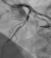
This study was a prospective randomized controlled clinical trial with a pre- and post-test design that aimed to investigate the effects of MICT and HIIT on inflammatory markers and circulating microRNAs in women with atherosclerosis. The target population comprised women aged 30 years or older with clinically confirmed atherosclerosis residing in Shadegan, Khuzestan Province, Iran. Atherosclerosis was diagnosed by a cardiologist based on medical history, clinical evaluation, vascular imaging findings, and/or documented evidence of atherosclerotic cardiovascular disease (Figures 1-4).
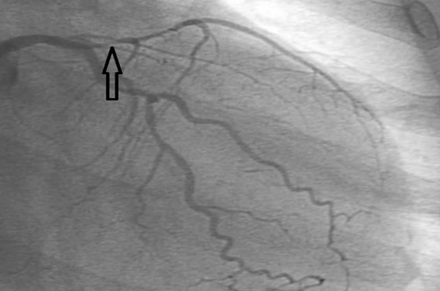
Figure 1.
Coronary angiography showing significant stenosis of the proximal left anterior descending
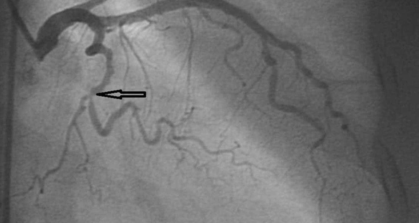
Figure 2.
Significant stenosis of the proximal LCX (left circumflex coronary artery).
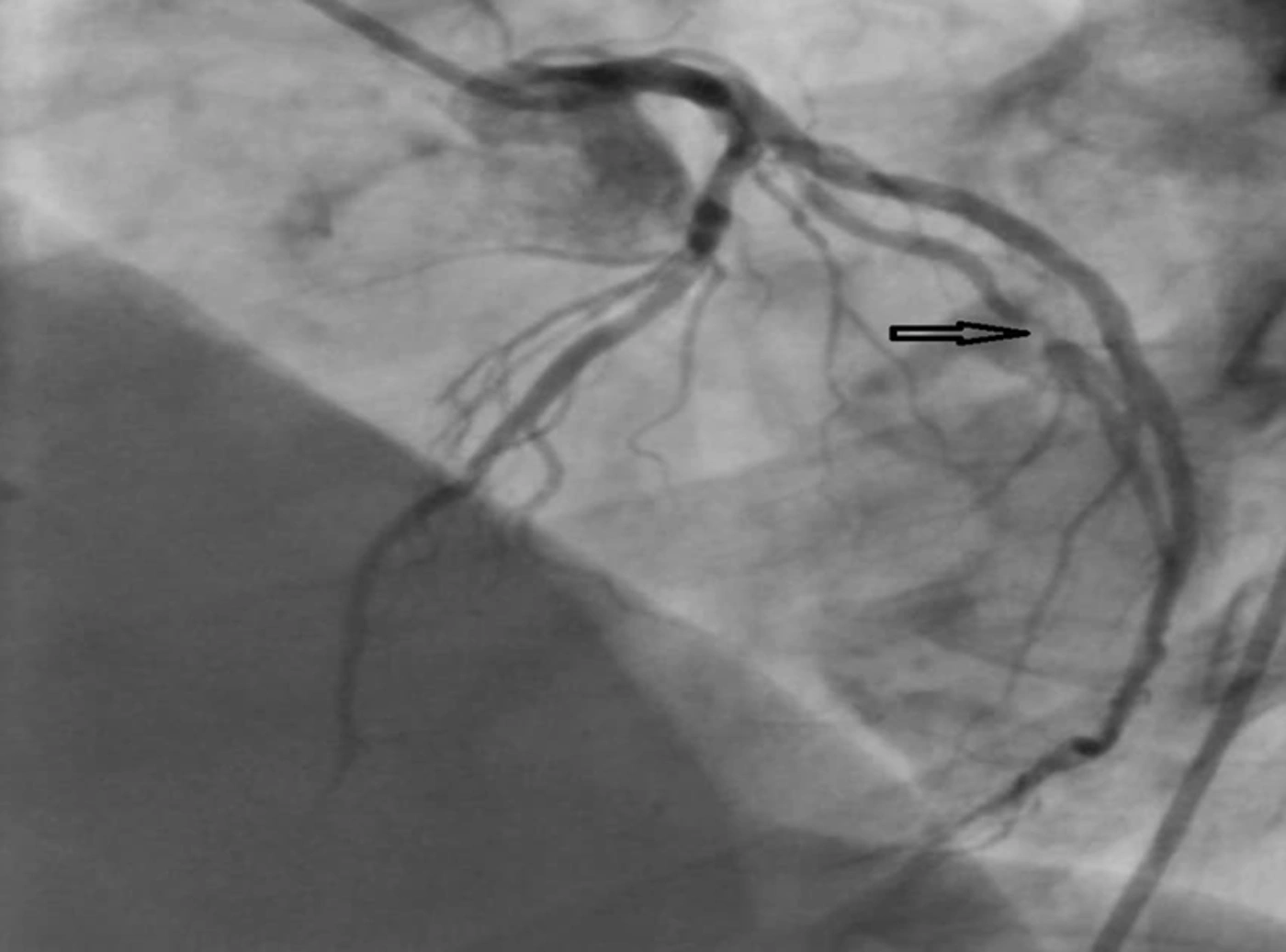
Figure 3.
Significant stenosis of the mid-diagonal artery.
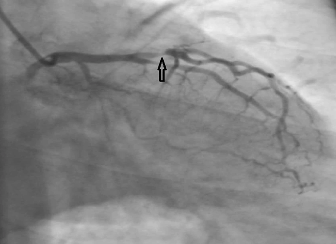
Figure 4.
The proximal LAD stenosis demonstrated in a complementary angiographic projection (arrow)
A total of 58 women were assessed for eligibility. Thirteen participants were excluded because they did not meet the inclusion criteria or declined to participate. Ultimately, 45 eligible participants were recruited through purposive sampling. The sample size was estimated using G*Power software based on an expected moderate effect size (f = 0.25), a statistical power of 80%, and a significance level of α = 0.05. To account for potential attrition, an additional 10% was included, resulting in a final target sample size of 45 participants. Participants were then randomly allocated in equal numbers to the control, MICT, and HIIT groups using a 1:1:1 allocation ratio (n = 15 per group) (Figure 5). The randomization sequence was generated by an independent researcher who was not involved in participant recruitment, outcome assessment, intervention delivery, or data analysis. Randomization was performed using a computer-generated block randomization sequence with equal allocation to the three groups.
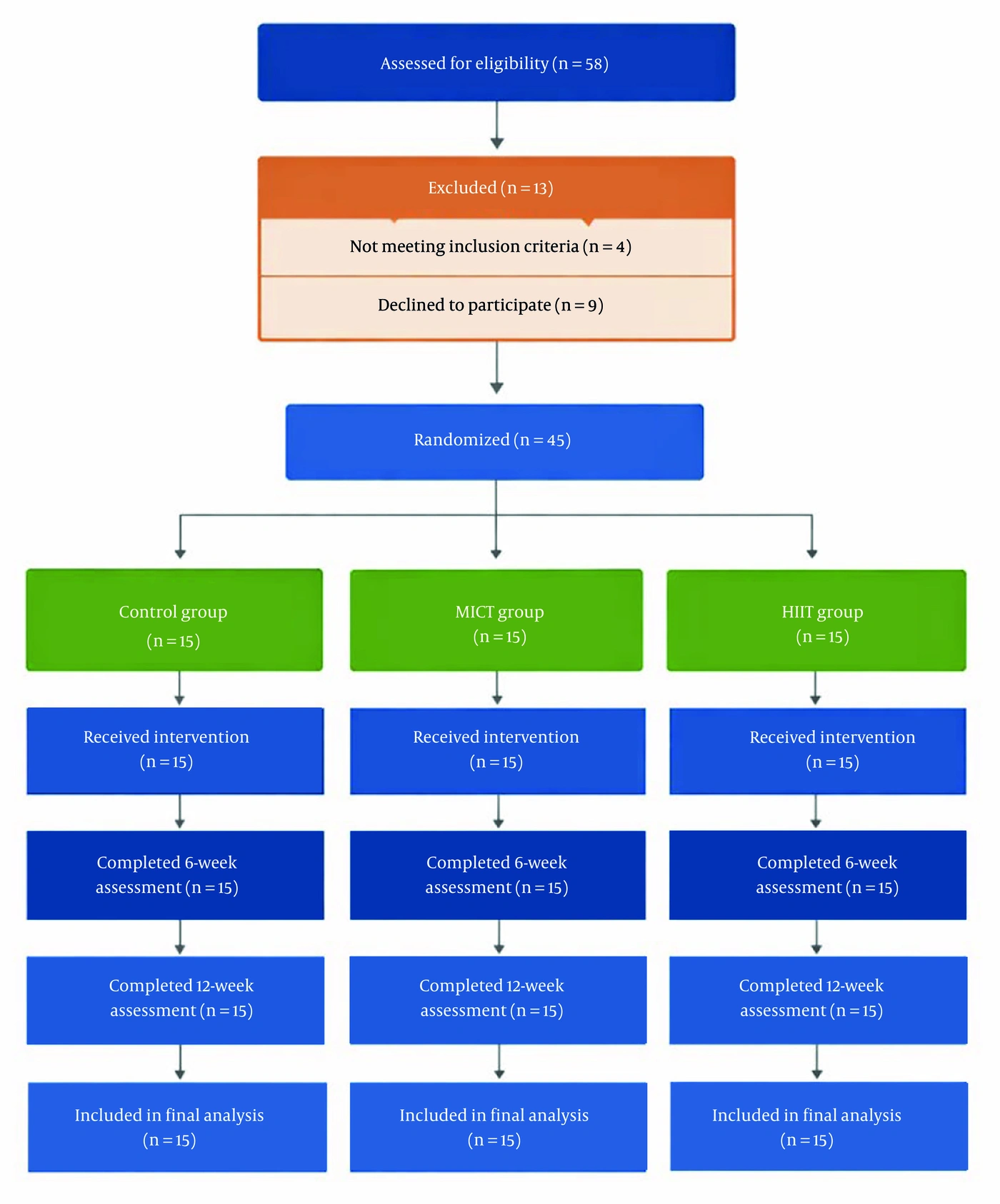
Figure 5.
CONSORT flow diagram showing participant screening, eligibility assessment, exclusions, randomization, follow-up assessments (6 and 12 weeks), and inclusion in the final analysis for the control, MICT, and HIIT groups.
Allocation concealment was ensured using sealed, opaque envelopes prepared by an independent researcher who was not involved in participant recruitment or outcome assessment. Participant enrollment was performed by the principal investigator, whereas group assignment was conducted by an independent research assistant after completion of baseline assessments. All participants completed baseline assessments before randomization and then entered the intervention period in their assigned groups. Outcome assessments were conducted at 6 and 12 weeks after baseline. No loss to follow-up occurred during the intervention period, and all randomized participants were included in the final analysis. All participants had confirmed atherosclerotic cardiovascular disease; the requirement of at least one risk factor was applied for baseline characterization and did not serve as a diagnostic criterion.
Baseline cardiovascular assessments included electrocardiography and echocardiography. Blood sample analyses and laboratory assessments were performed by laboratory personnel who were blinded to group allocation. Statistical analyses were also conducted using coded group assignments to minimize potential bias. The study protocol was approved by the Research Council of Islamic Azad University, Shoushtar Branch (Reference No: IR.SSRC.REC.1403.026). The study was also registered in the Iranian Registry of Clinical Trials (IRCT) under the registration code IRCT20240719062472N1 and received approval on April 19, 2025. Written informed consent was obtained from all participants before enrollment. All procedures were conducted in accordance with the ethical standards of the Helsinki Declaration and relevant institutional regulations.
3.2. Inclusion and Exclusion Criteria
Eligible participants were women aged 30 years or older with a body mass index (BMI) greater than 25 kg/m2 who had not engaged in regular physical activity during the preceding six months. Participants had clinically confirmed atherosclerosis diagnosed by a cardiologist based on medical history, vascular imaging findings, and/or documented evidence of atherosclerotic cardiovascular disease (17). Diagnostic confirmation included at least one of the following: carotid artery plaque detected by Doppler ultrasonography, coronary artery disease documented by angiography or physician diagnosis, peripheral arterial disease, or previous clinical evidence of atherosclerotic vascular involvement (1). In addition, all participants were required to present at least one atherosclerotic risk factor according to the American Heart Association/American College of Cardiology guidelines (17). Risk factor assessment included paraclinical evaluations for diabetes mellitus, dyslipidemia, and renal insufficiency, as well as laboratory measurements of blood urea nitrogen (BUN), creatinine (Cr), and monitoring of glomerular filtration rate (GFR). Baseline cardiovascular assessments included electrocardiography and echocardiography, and additional cardiovascular evaluations were performed when clinically indicated (18). Medication status, including antihypertensive, lipid-lowering, and antiplatelet therapies, was documented at baseline, and all prescribed medications remained stable throughout the intervention period under cardiologist supervision. Exclusion criteria included active smoking, unstable angina, decompensated heart failure, myocardial infarction within the previous four weeks, complex ventricular arrhythmias, and orthopedic or neurological limitations that could interfere with participation in exercise training.
3.3. Exercise Testing and Training Protocol
Before the exercise intervention, all participants underwent a maximal oxygen uptake (VO2peak) assessment using a Metamax II gas analyzer (Germany) and a customized treadmill ramp protocol. During the test, treadmill speed remained constant while the incline increased in 2% increments until volitional exhaustion or achievement of physiological termination criteria. VO2peak was defined by the presence of at least one of the following criteria: a plateau in oxygen consumption despite increased workload, a respiratory exchange ratio greater than 1.05, attainment of age-predicted maximal heart rate calculated using the Karvonen formula, or voluntary exhaustion (19). Heart rate was continuously monitored throughout testing and training sessions using a chest-strap heart rate monitoring system.
Exercise sessions were conducted at the Cardiac Rehabilitation Center of Shadegan under the supervision of an exercise physiologist, a trained nurse for emergency support, and a cardiologist. Interventions were performed three times per week for 12 consecutive weeks. Participants in the control group were instructed to maintain their usual lifestyle and refrain from participating in structured exercise programs during the intervention period.
Participants in all study groups were instructed to maintain their habitual dietary patterns and lifestyle behaviors throughout the study period and to refrain from engaging in additional structured exercise outside the prescribed intervention.
In addition, participants were not informed about the specific molecular outcomes (miR-126 and miR-146a) to minimize expectation-driven behavioral changes.
Medication regimens remained unchanged throughout the intervention period under cardiologist supervision.
Moderate-intensity continuous training (MICT) sessions consisted of 41 minutes of continuous treadmill exercise at 50 - 70% of HRpeak (approximately 50 - 60% VO2peak), including a 5-minute warm-up and a 3-minute cool-down (19). HIIT sessions consisted of 33 minutes of exercise beginning with a 5-minute warm-up at 50 - 70% HRpeak, followed by four 4-minute high-intensity intervals performed at 85 - 95% HRpeak (approximately 80 - 90% VO2peak), interspersed with three 3-minute active recovery periods at 50 - 70% HRpeak, and ending with a 4-minute cool-down at 50 - 70% HRpeak. Exercise intensity was progressively adjusted weekly to maintain the prescribed training stimulus (19).
Attendance and adherence to exercise sessions were recorded throughout the intervention period. Participants who attended at least 90% of scheduled sessions were considered adherent to the protocol. Heart rate responses were continuously monitored during exercise sessions to ensure that participants achieved the prescribed intensity ranges. Occasional missed sessions due to personal or medical reasons were rescheduled within the same week whenever possible. No major protocol deviations occurred during the intervention period.
All exercise sessions were supervised, and participants were monitored for cardiovascular symptoms, exercise intolerance, abnormal blood pressure responses, arrhythmias, musculoskeletal complaints, and other adverse events. No serious cardiovascular complications, exercise-related injuries, or adverse events requiring discontinuation of participation were observed during the study.
3.4. Clinical Assessments and Blood Sampling
Baseline cardiovascular assessments were conducted 48 hours before the intervention and included transthoracic echocardiography (GE Vivid S5, General Electric, USA), standard 12-lead electrocardiography (Schiller Cardiovit AT-101, Switzerland), resting blood pressure measurement, and VO2peak evaluation. Circulating miR-146a and miR-126 were defined as the primary outcomes of this study, whereas cardiovascular and physiological parameters were considered secondary outcomes. These secondary variables were primarily used for participant characterization, cardiovascular safety assessment, and exercise prescription.
Venous blood samples were collected from the antecubital vein at three predefined time points: baseline, 48 hours after completion of the first 6 weeks of intervention, and 48 hours after the final exercise session of the 12-week intervention period. Blood samples were collected into EDTA-containing tubes under fasting conditions between 8:00 and 10:00 AM to minimize circadian variation. Samples were centrifuged at 3000 rpm for 10 minutes at 4°C, and plasma was immediately aliquoted and stored at −80°C until molecular analysis. All circulating miRNA analyses were performed using plasma samples.
The predefined primary outcomes of the study were circulating expression levels of miR-146a and miR-126. Total RNA, including small RNA fractions, was extracted from plasma samples using the miRNA Extraction Kit (Gene Tech Pooyan Co., Iran) according to the manufacturer’s instructions. The extraction protocol was based on selective binding of RNA molecules to silica-based spin columns under optimized chaotropic salt and ethanol conditions, ensuring efficient recovery of small non-coding RNAs.
RNA quantity and purity were assessed using NanoDrop spectrophotometry, and samples with A260/A280 ratios between 1.8 and 2.1 were included for downstream analysis. Reverse transcription was performed using a miRNA-specific cDNA synthesis kit supplied by Gene Tech Pooyan Co.
Quantitative real-time PCR (qRT-PCR) was performed using SYBR Green chemistry on a MIC Real-Time PCR System (Switzerland) with reagents provided by Gene Tech Pooyan Co. U6 small nuclear RNA (U6 snRNA) was used as the endogenous reference gene for normalization of circulating miRNA expression levels. No exogenous spike-in RNA control was included. All samples were processed under identical pre-analytical conditions to minimize technical variability. Relative miRNA expression levels were calculated using the comparative 2−ΔΔCt method. All reactions were performed in duplicate, and mean Ct values were used for statistical analysis. Samples with Ct variation greater than 0.5 between replicates were reanalyzed to ensure data reliability (Table 1).
Table 1.
Primer Sequences Used for qRT-PCR Analysis
| Oligo name | Primer Type | Oligo Sequence (5′→3′) |
|---|---|---|
| miR-146a-5p | Forward | TGAGAACTGAATTCCATGGGTT |
| miR-126 | Forward | TCGTACCGTGAGTAATAATGCG |
| Universal Primer | Reverse | GTGCAGGGTCCGAGGT |
| U6 snRNA | Forward | CTCGCTTCGGCAGCACA |
| U6 snRNA | Reverse | AACGCTTCACGAATTTGCGT |
Primer sequences for miR-146a, miR-126, and U6 are presented in Table 1. Amplification specificity was confirmed by melting curve analysis, and no-template controls were included in each run to exclude contamination or nonspecific amplification. PCR efficiency for all targets was within the acceptable range of 90 - 110%.
3.5. Statistical Analyses
In the present study, a linear mixed model (LMM) was used to compare changes from baseline to 6 and 12 weeks of intervention among the different study groups. For this purpose, the factors time (baseline, 6 weeks, and 12 weeks) and group (control, MICT, and HIIT), as well as the time × group interaction, were defined in the model. When a significant interaction effect was observed for time × group, the Kruskal–Wallis test followed by the DSCF post hoc test was applied to determine the source of differences in intervention-induced changes across the different time points. Within-group changes, in the case of a significant main effect of time, were analyzed using the Bonferroni post hoc test. All statistical analyses were performed using Jamovi, and the level of statistical significance was set at P < 0.05.
4. Results
All participants completed the 12-week intervention and follow-up assessments, and no withdrawals occurred during the study. Attendance rates were 95.6% and 96.8% in the MICT and HIIT groups, respectively. Continuous heart-rate monitoring confirmed that participants consistently achieved the prescribed training intensity ranges throughout the intervention period. Occasional missed sessions due to personal or medical reasons were rescheduled whenever possible, and no major protocol deviations were recorded. No serious adverse events, cardiovascular complications, exercise-related injuries, or cases of exercise intolerance requiring discontinuation were observed in any group.
Cardiovascular and physiological variables, including echocardiographic findings, electrocardiographic assessments, resting blood pressure, and VO2peak, were collected for participant characterization, cardiovascular safety monitoring, and exercise prescription purposes and were not predefined efficacy endpoints; therefore, detailed analyses of these variables are not presented in the current report.
The increase in miR-146a-5p occurred earlier with HIIT than with MICT. Linear mixed-model analysis demonstrated a significant interaction effect between time and type of intervention (F = 35.5, P = 0.0001, η = 0.53), indicating that changes in mean miR-146a-5p values across study time points varied by intervention type (Figure 6). At each time interval, both interventions resulted in a significant increase in miR-146a-5p compared with the control group (P < 0.001). The greatest significant increase was observed after 12 weeks of HIIT and MICT; however, no significant difference was found between the two exercise intensities.
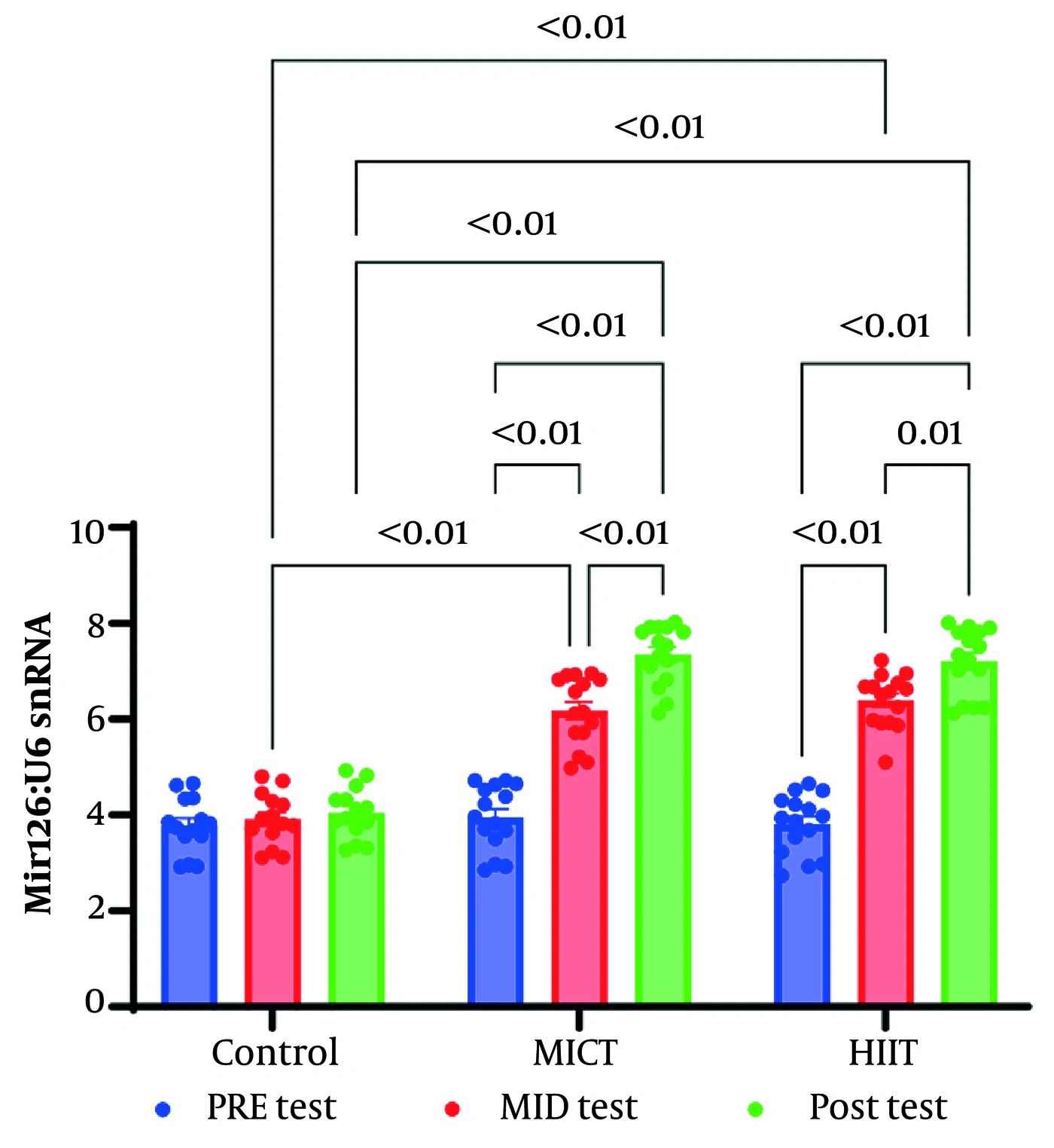
Figure 6.
Mean miR-146a-5p levels in the control group and the different exercise-intensity groups at baseline, 6 weeks, and 12 weeks.
Changes during the first and second 6-week periods of both HIIT and MICT similarly resulted in significant increases in miR-146a-5p (P < 0.001). Despite the absence of a significant difference between exercise intensities, the increase in miR-146a-5p during the first 6 weeks of HIIT showed the highest level of significance, suggesting earlier adaptation. During the second 6 weeks, the slope of increase was smaller, although the upward trend remained stable. In contrast, the increase observed with MICT during the first 6 weeks was slower, whereas during the second 6 weeks the upward slope became steeper, suggesting that greater adaptation in this training model occurred during the latter phase of the intervention (Figure 7). Furthermore, changes from baseline to 6 weeks and subsequently to 12 weeks across the different interventions are presented in Table 2.
Table 2.
Results of the Bonferroni Post Hoc Test for Within-Group Comparisons of miR-146a-5p Changes a
| Groups | Time (wk) | Group | Difference | SE | t | df | Pbonferroni |
|---|---|---|---|---|---|---|---|
| Control | |||||||
| Pre test | 6 | Control | -0.0213 | 0.193 | -0.110 | 126 | 1.000 |
| Pre test | 12 | Control | -0.0538 | 0.193 | -0.279 | 126 | 1.000 |
| 6 (wk) | 12 | Control | -0.0325 | 0.193 | -0.169 | 126 | 1.000 |
| MICT | |||||||
| Pre test | 6 | MICT | -1.1960 | 0.199 | -13.807 | 126 | < 0.001 |
| Pre test | 12 | MICT | -2.7500 | 0.203 | -7.011 | 126 | < 0.001 |
| 6 (wk) | 12 | MICT | -1.5540 | 0.199 | -7.802 | 126 | < 0.001 |
| HIIT | |||||||
| Pre test | 6 | HIIT | -1.8821 | 0.206 | -9.129 | 126 | < 0.001 |
| Pre test | 12 | HIIT | -2.9536 | 0.203 | -12.296 | 126 | < 0.001 |
| 6 (wk) | 12 | HIIT | -1.0714 | 0.206 | -5.197 | 126 | < 0.001 |
a Difference values were calculated as baseline minus follow-up measurements. Negative values therefore indicate increased biomarker expression during the intervention, whereas positive values indicate decreased expression.
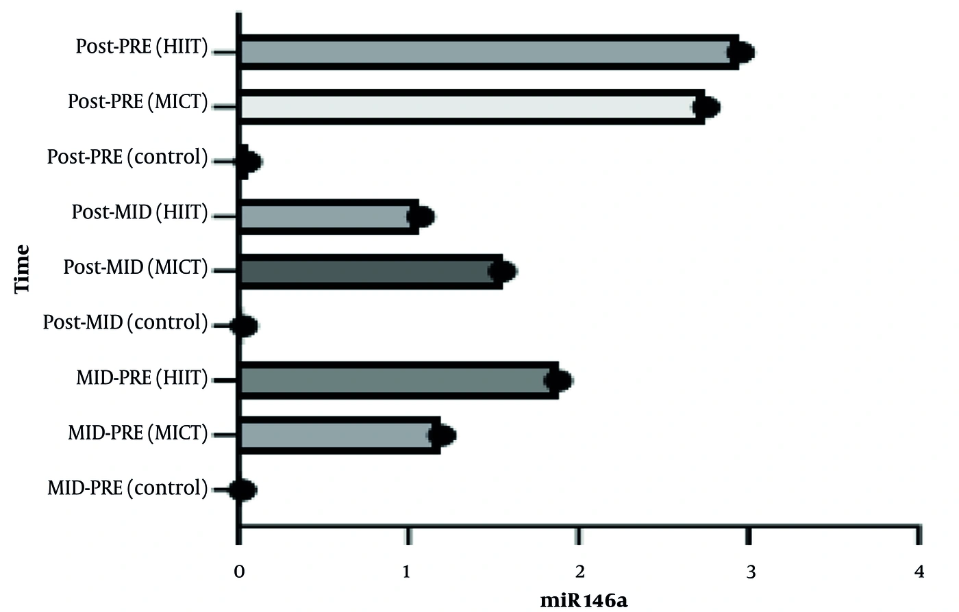
Figure 7.
Between-group and within-group changes in circulating miR-146a levels across baseline, 6 weeks, and 12 weeks following MICT and HIIT interventions.
HIIT increased miR-126 earlier than MICT. Linear mixed-model analysis showed a significant interaction effect between time and type of intervention (F = 33.1, P = 0.0001, η = 0.52), indicating that mean changes in miR-126 across study time points varied according to the type of intervention (Figure 8). Post hoc analyses showed that both HIIT and MICT significantly increased miR-126 at each time interval compared with the control group (P < 0.001). Twelve weeks of HIIT and MICT produced similar increases in miR-126 expression. Likewise, the first and second 6-week periods of HIIT and MICT similarly increased miR-126 expression (P < 0.001). However, a greater increase in miR-126 was observed during the first 6 weeks with HIIT, whereas a larger increase was observed during the second 6 weeks with MICT (Figure 9). The trend of mean changes in miR-126 across the different interventions is presented in Table 3.
Table 3.
Results of the Bonferroni Post Hoc Test for Within-Group Comparisons of miR-126 Changes a
| Groups | Time (wk) | Difference | SE | t | df | pbonferroni |
|---|---|---|---|---|---|---|
| Control | ||||||
| Pre test | 6 | -0.1329 | 0.233 | -0.569 | 123 | 1.000 |
| Pre test | 12 | -0.2629 | 0.233 | -1.126 | 123 | 0.969 |
| 6 (wk) | 12 | -0.1300 | 0.233 | -0.557 | 123 | 1.000 |
| MICT | ||||||
| Pre test | 6 | -2.2253 | 0.225 | -9.870 | 123 | < 0.001 |
| Pre test | 12 | -3.3887 | 0.225 | -14.435 | 123 | < 0.001 |
| 6 (wk) | 12 | -1.1633 | 0.225 | -5.160 | 123 | < 0.001 |
| HIIT | ||||||
| Pre test | 6 | -2.5827 | 0.225 | -11.455 | 123 | < 0.001 |
| Pre test | 12 | -3.3927 | 0.225 | -15.048 | 123 | < 0.001 |
| 6 (wk) | 12 | -0.8100 | 0.225 | -3.593 | 123 | 0.017 |
a Difference values were calculated as baseline minus follow-up measurements. Negative values therefore indicate increased biomarker expression during the intervention, whereas positive values indicate decreased expression.
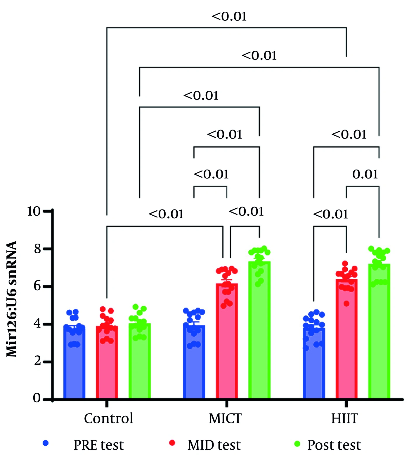
Figure 8.
Mean miR-126 levels in the control group and the different exercise-intensity groups at baseline, 6 weeks, and 12 weeks.
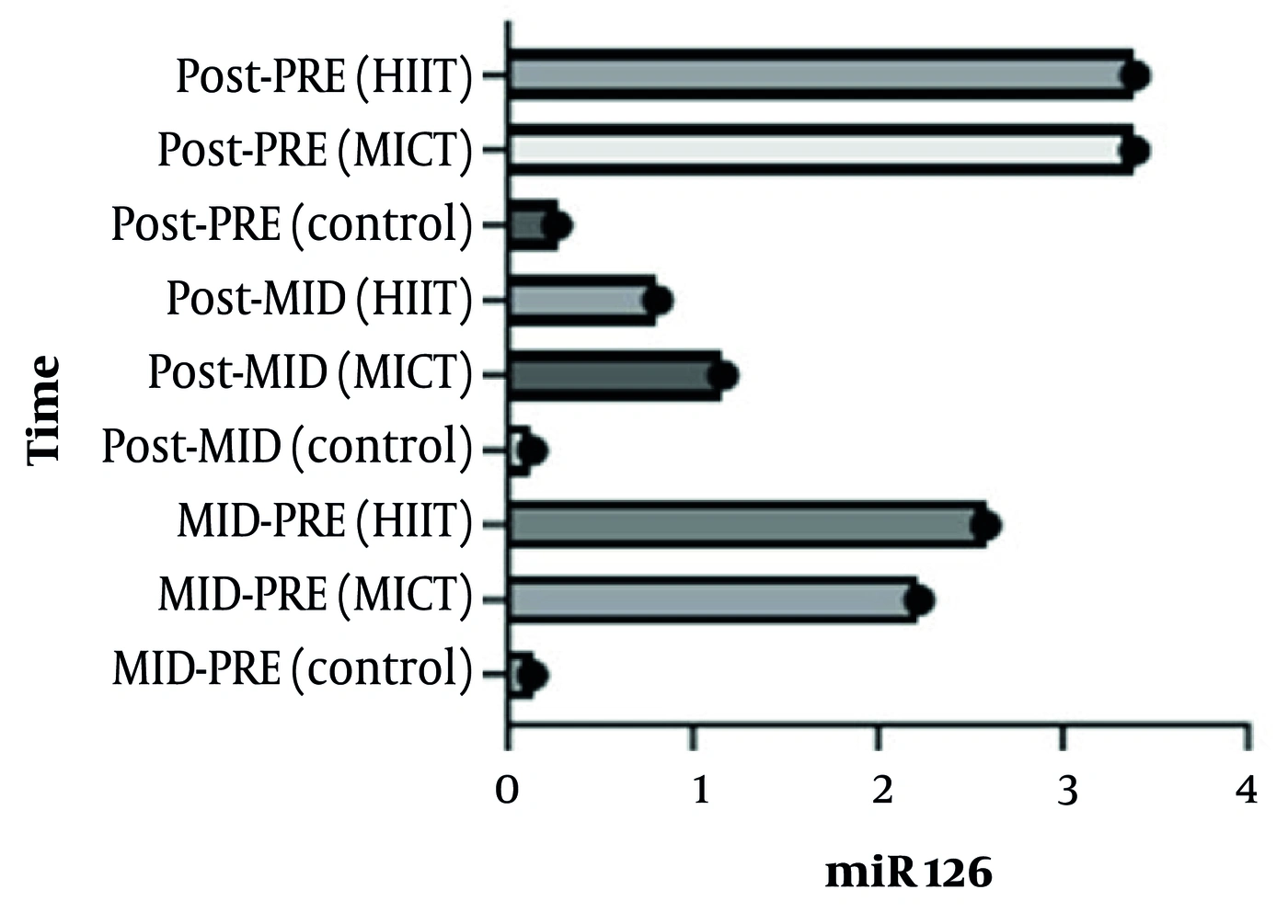
Figure 9.
Between-group and within-group changes in circulating miR-126 levels across baseline, 6 weeks, and 12 weeks following MICT and HIIT interventions.
The clinical findings of the present study indicate that both intensities of aerobic exercise were implemented without adverse events and may be considered effective strategies for patients with atherosclerosis. Furthermore, when rapid improvement within a shorter period is desired, HIIT may represent the superior option. Although the selection of exercise modality may vary according to patients’ individual conditions, previous investigations in cardiac patients have shown that high-intensity interval training is not only more efficient in several physiological aspects, such as producing greater improvements in cardiorespiratory endurance and body composition, but is also considered a more appealing training modality than moderate-intensity continuous training.
5. Discussion
Regardless of exercise modality, participation in a 12-week aerobic training intervention was associated with marked increases in circulating miR-146a and miR-126 levels in women with atherosclerosis. These findings support the concept that aerobic exercise can beneficially influence epigenetic regulators implicated in vascular homeostasis and atherosclerotic disease progression, further emphasizing its therapeutic value as a nonpharmacological intervention.
The magnitude of change observed in both miRNAs suggests that aerobic exercise may exert beneficial effects by modulating epigenetic networks implicated in atherosclerotic pathophysiology.
Notably, the temporal pattern differed between modalities: HIIT elicited faster initial upregulation, particularly for miR-126, whereas MICT showed a steeper increase in the latter half, especially for miR-146a. This discussion contextualizes these miRNA alterations within the broader framework of exercise-induced vascular adaptations, explores their mechanistic links to endothelial function and vascular repair, and addresses implications for sex-specific CVD management, limitations, and future directions.
Exercise-induced miRNA modulation represents a pivotal mechanism for attenuating atherosclerotic progression, as miRNAs orchestrate post-transcriptional regulation of genes involved in endothelial function, angiogenesis, and immune responses. miR-126, predominantly expressed in endothelial cells, promotes vascular integrity by targeting negative regulators of the PI3K/Akt/eNOS pathway, enhancing VEGF-mediated signaling and EPC mobilization (7). In the present study, the 89% upregulation observed with HIIT is consistent with findings from Schmitz et al. (12), who reported dose-dependent increases in miR-126 following progressive HIIT in healthy adults and attributed this effect to shear stress fluctuations that stimulate endothelial repair (12). Similarly, Schmitz et al. (20) demonstrated that HIIT elevated miR-126 levels in association with improvements in the microvascular glycocalyx, thereby reducing endothelial permeability and atherogenic risk (20). The accelerated initial response to HIIT observed here may stem from its high metabolic demand, which amplifies shear stress and hypoxic signaling, as evidenced by Yuan et al. (21), who noted miR-126 upregulation in response to moderate-to-high-intensity aerobic exercise and correlated this response with improved vascular homeostasis (21).
Conversely, the delayed but equivalent maximal effect of MICT on miR-126 suggests a cumulative adaptation, potentially mediated by sustained nitric oxide bioavailability and lipid optimization, as described by Ke et al. (22) in their review of exercise-modulated miRNAs in atherosclerosis (22). This phase-related difference aligns with Van Guilder (23), who highlighted miR-126’s role in exercise-induced vascular remodeling, whereby interval protocols such as HIIT trigger rapid EPC recruitment, while continuous training may promote longer-term plaque stability (23). In women specifically, Kashef (24) found that concurrent training elevates miR-126, improving cardiovascular function in postmenopausal cohorts, mirroring our demographic and emphasizing sex-specific responsiveness that may be influenced by estrogen decline (24). The 75% increase with MICT observed here exceeds reports from Improta Caria et al. (26), who observed modest increases in miR-126 in hypertensive patients, but aligns with Feng et al. (25), in which HIIT and MICT acutely improved arterial stiffness in hypertensive patients, with miR-126 implicated in endothelial recovery (25, 26).
In parallel with miR-126, miR-146a upregulation (58% in HIIT and 67% in MICT) positions this miRNA as a key regulator of innate immune signaling, targeting TLR pathway components such as TLR4, TRAF6, and IRAK1, thereby dampening NF-κB-mediated responses (27). Afzal et al. (28) corroborated this in their systematic review, showing that aerobic exercise upregulates miR-146a in patients with heart failure with preserved ejection fraction (HFpEF) and coronary artery disease (CAD) and linking this response to enhanced vascular protection (28). The HIIT-driven early surge observed here is consistent with Balchin et al. (29), who reported increases in miR-146a after resistance exercise in rheumatoid arthritis, attenuating pathological signaling (29). However, Mayr et al. (30) reported decreases in miR-146a after combined endurance-strength training in heart failure, suggesting context-dependent responses; our focus on aerobic training may explain this divergence, as Yuan et al. (21) emphasized miR-146a sensitivity to exercise intensity in CVD models (30, 21).
The interplay between these miRNAs further illustrates the atheroprotective effects of exercise. For miR-126, effects mediated through SIRT1 and superoxide dismutase-2 modulation reduce reactive oxygen species (ROS) (7), consistent with the temporal patterns observed in this study. Soplinska et al. (31) reviewed endurance exercise-induced miRNA alterations and linked miR-146a and miR-126 to cardiac protection (31). In atherosclerosis, Letonja et al. (32) identified miR-146a and miR-126 as biomarkers that correlate with vascular health, reinforcing our findings (32). Yang et al. (33) further connected exercise-induced miRNAs to fibrosis in CVD, with miR-146a mitigating plaque instability (33).
These results are particularly relevant for women with atherosclerosis, a population underrepresented in CVD research. Sex-specific differences in miRNA responses, influenced by hormonal factors, may amplify exercise benefits; for example, Eyileten et al. (34) noted miR-126 fluctuations during ultra-endurance exercise, with potential sex-related differences (34). Sanchis-Gomar et al. (35) highlighted miR-146a and miR-126 in exercise-induced cardiac remodeling, suggesting their utility as biomarkers for residual risk reduction beyond statins (35). Equivalent endpoints between MICT and HIIT imply flexibility in exercise prescription, with HIIT potentially better suited to time-constrained individuals seeking more rapid adaptations, as Dlouha et al. (36) observed miR-146a reductions after myocardial infarction with physical activity (36). However, Barber et al. (37) cautioned that exercise volume modulates miRNA profiles, aligning with our dose-response observations (37).
5.1. Study Limitations
Despite these insights, several limitations warrant consideration. The sample size (n = 45) limits generalizability, although effect sizes were large.
The exclusive recruitment of women represents an important limitation, as sex-based biological differences—including hormonal milieu, endothelial function, and epigenetic regulation—may modulate circulating miRNA responses to exercise interventions. Consequently, the present findings may not be directly generalizable to male patients with atherosclerosis.
Reliance on circulating miRNAs may not fully capture tissue-specific changes, as Wang et al. (38) noted discrepancies between plasma and cardiac miR-146a (38). Another methodological limitation of the present study is the absence of an exogenous spike-in control (e.g., cel-miR-39) for normalization of circulating miRNA extraction efficiency. This was related to the use of a standardized commercial SYBR Green-based assay workflow in which spike-in controls are not routinely incorporated. Although U6 snRNA was used as an endogenous reference gene and all pre-analytical procedures were strictly standardized, the lack of an exogenous normalization control may have introduced technical variability into the qRT-PCR measurements.
Another limitation of this study is the lack of systematic monitoring of several lifestyle-related confounding factors that may influence circulating microRNA expression. Dietary intake, particularly consumption of anti-inflammatory or proinflammatory foods, was not quantitatively assessed during the intervention period. Similarly, sleep quality and duration, psychological stress levels, and unstructured physical activity outside the prescribed exercise program were not objectively measured. Although participants were instructed to maintain their habitual diet and lifestyle and to avoid additional structured exercise, the absence of formal monitoring or validated questionnaires for these variables may have introduced residual confounding.
The 12-week duration precluded assessment of long-term outcomes, echoing calls from Ardinal et al. (39) for extended trials on miR-146a in atherosclerosis (39). Future research should integrate mechanistic studies, such as miRNA knockdown in animal models (40), and explore lncRNA interactions (21). Incorporating diverse populations, including men for sex comparisons, and assessing plaque burden via imaging could enhance translational value.
5.2. Conclusions
In conclusion, this study supports the role of exercise as an epigenetic modulator, upregulating miR-146a and miR-126 to promote vascular resilience in women with atherosclerosis. By bridging rapid HIIT adaptations with sustained MICT gains, these findings support personalized protocols in CVD rehabilitation, potentially reducing morbidity through miRNA-mediated endothelial enhancement. These findings also pave the way for miRNA-targeted therapies, further strengthening the role of exercise in addressing residual risk.