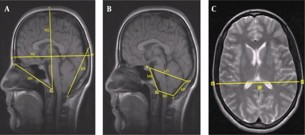
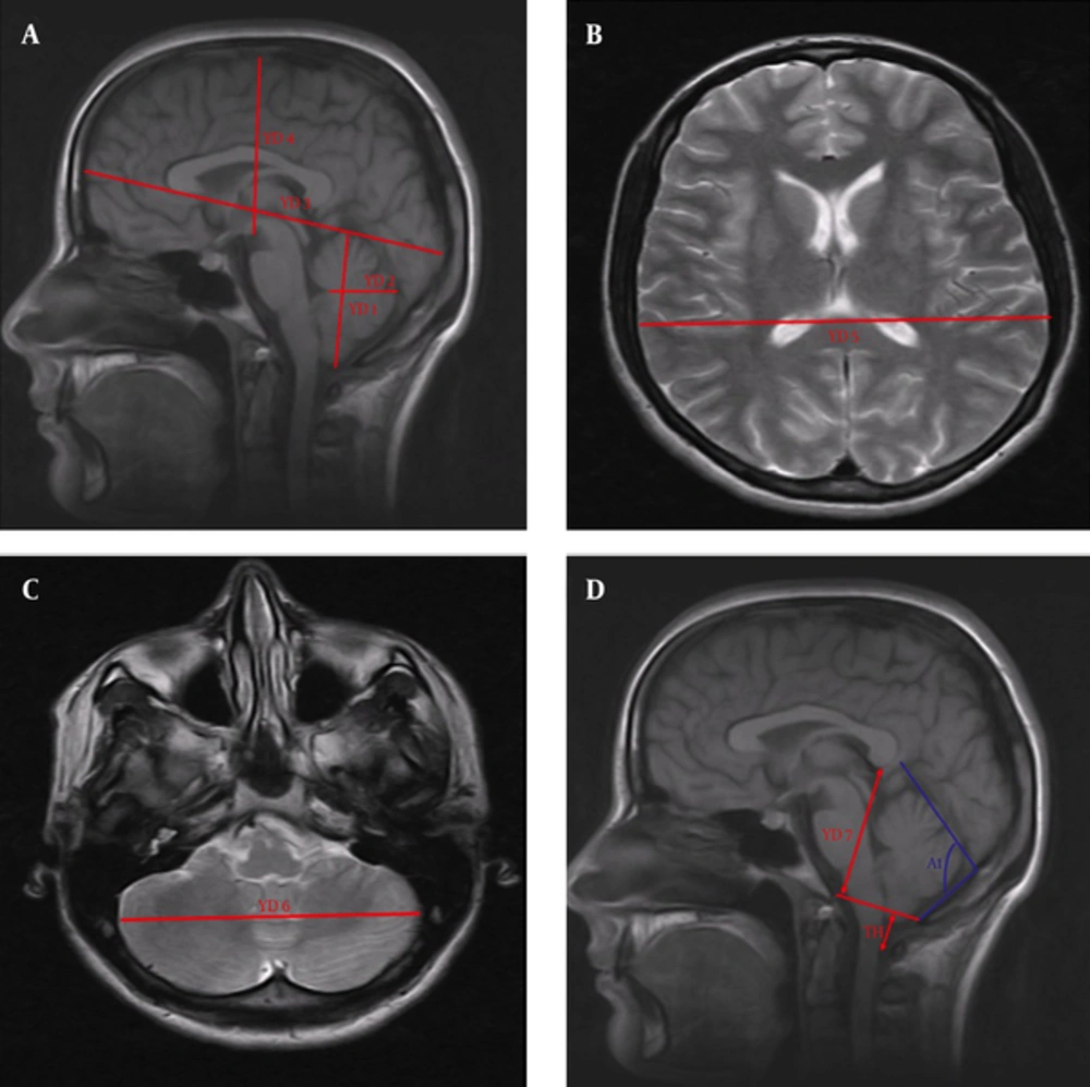
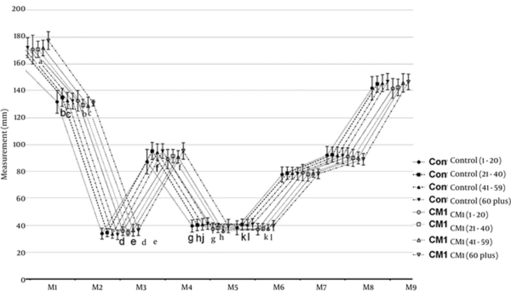
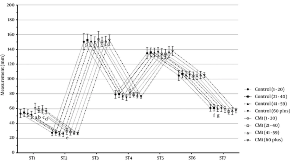
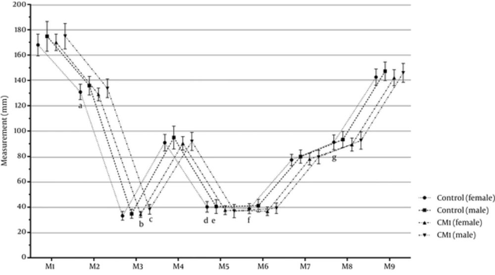
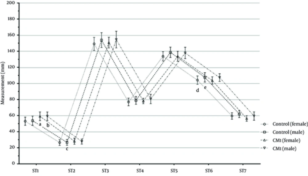
The anteroposterior diameter of the foramen magnum was higher in the people who developed tonsillar herniation than in the healthy people. On the other hand, maximum cranial height, supraocciput length, and clivus length were lower in the patients with tonsillar herniation.
In numerous existing studies, the anteroposterior diameter of the foramen magnum was defined by measuring the distance between the basion and the opisthion (
3,
20-
22,
26,
27).
Dağtekin et al. reported that the foramen magnum diameter was found to be 40.1 mm in the CMI group and 32.4 mm in the controls, and that the difference was statistically significant (
26).
Aydın et al. (
21) found that the foramen magnum diameter was 31.7 ± 6.1 mm in the CMI group and 25.2 ± 3.8 mm in the controls, and that the difference was statistically significant. Based on their results, they proposed that an underdeveloped bony structure in the intrauterine phase causes the contents of the posterior fossa to become a downward hernia when the caudal metencephalon is normally developed. They also reported that this may lead to expansion of the anteroposterior diameter of the foramen magnum.
In contrast, Hwang et al. (
27) found the diameter of the foramen to be 24.78 mm in the CMI patients and 29.54 mm in the controls. The authors reported that this difference was statistically significant.
Likewise, Leikola et al. (
28) found that the diameter of the foramen magnum was smaller in pediatric patients with nonsyndromic craniosynostosis.
On the other hand, despite a larger diameter of the foramen magnum ın the CMI group, Karagoz et al. (
20), Sekula et al. (
3), and Milhorat et al. (
29,
30) reported that the difference was not statistically significant.
In this study, we found that the anteroposterior diameter of the foramen magnum was 35.43 ± 3.34 mm in CMI patients and 33.74 ± 3.57 mm in the control group. The difference between the CMI and control groups was statistically significant (P = 0.001). Although the increase in the diameter of the foramen magnum that we found in our CMI patients showed similarities with the results of Dagtekin et al. (
26) and Aydın et al. (
21), Hwang et al. (
27) and Leikola et al. (
28) reported that the diameter of the foramen magnum was lower in the CMI patients. This difference may have resulted from the low number of patients, as noted by the authors. We also thought that this outcome may have been due to the fact that patients with craniosynostosis were included in the study by Leikola et al. (
28).
In their study, Aydın et al. (
21) referred to hypoplasia and the fact that, unlike the normal development of soft tissues, bony structures in the fossa may lead to herniation toward the cervical canal. Secondary to this, the diameter of the foramen magnum may be larger in CMI patients, as was found in our study.
One of the measurements required in order to elucidate the etiology of tonsillar herniation is the length of the supraocciput. This length was found to be smaller in CMI patients in the literature, with researchers reporting that the difference was statistically significant (
4,
8,
20,
26,
29,
30). Sekula et al. (
3), Aydın et al. (
21), Furtado et al. (
19), Hwang et al. (
27), and Heiss et al. (
31) did not report a statistically significant difference between the groups.
In this study, we found that the length of the supraocciput was 37.22 ± 4.17 mm in the CMI patients and 40.29 ± 4.79 mm in the controls. This difference between the patient and control groups was statistically significant (P = 0.001). A decrease in supraocciput length in the CMI patients might be attributed to occipital bone hypoplasia, which is a factor in the etiology of the condition.
Our other finding suggesting occipital bone hypoplasia in the CMI patients was the decrease in the length of the clivus. In our study, we found that the length of the clivus was 37.46 ± 3.75 mm in the CMI patients and 39.78 ± 4.62 mm in the controls. This difference between the patient and control groups was statistically significant (P = 0.001).
Like our outcomes, many authors reported that the length of the clivus was statistically significantly lower in the patients who developed herniation (
3,
4,
20,
21,
27,
29,
31).
On the other hand, Nishikawa et al. (
8) and Dagtekin et al. (
26) found the clivus length shorter in people with tonsillar herniation, although they reported that the difference was not statistically significant.
In the literature, the anteroposterior diameter of the cranial fossa was found to be smaller in CMI patients (
3,
20,
21).
Karagöz et al. proposed that this smaller length in CMI patients might lead to compensatory anterior growth of the tentorium (
20).
In the current study, despite the fact that the anteroposterior length of the posterior fossa was slightly bigger in the CMI patients, this difference was not statistically significant.
Researchers who examined the change of the angle between the length of the supraocciput and the tentorium cerebelli line (tentorial angle) in the people with herniation found that this angle was greater than in the healthy individuals (
3,
4,
26,
27).
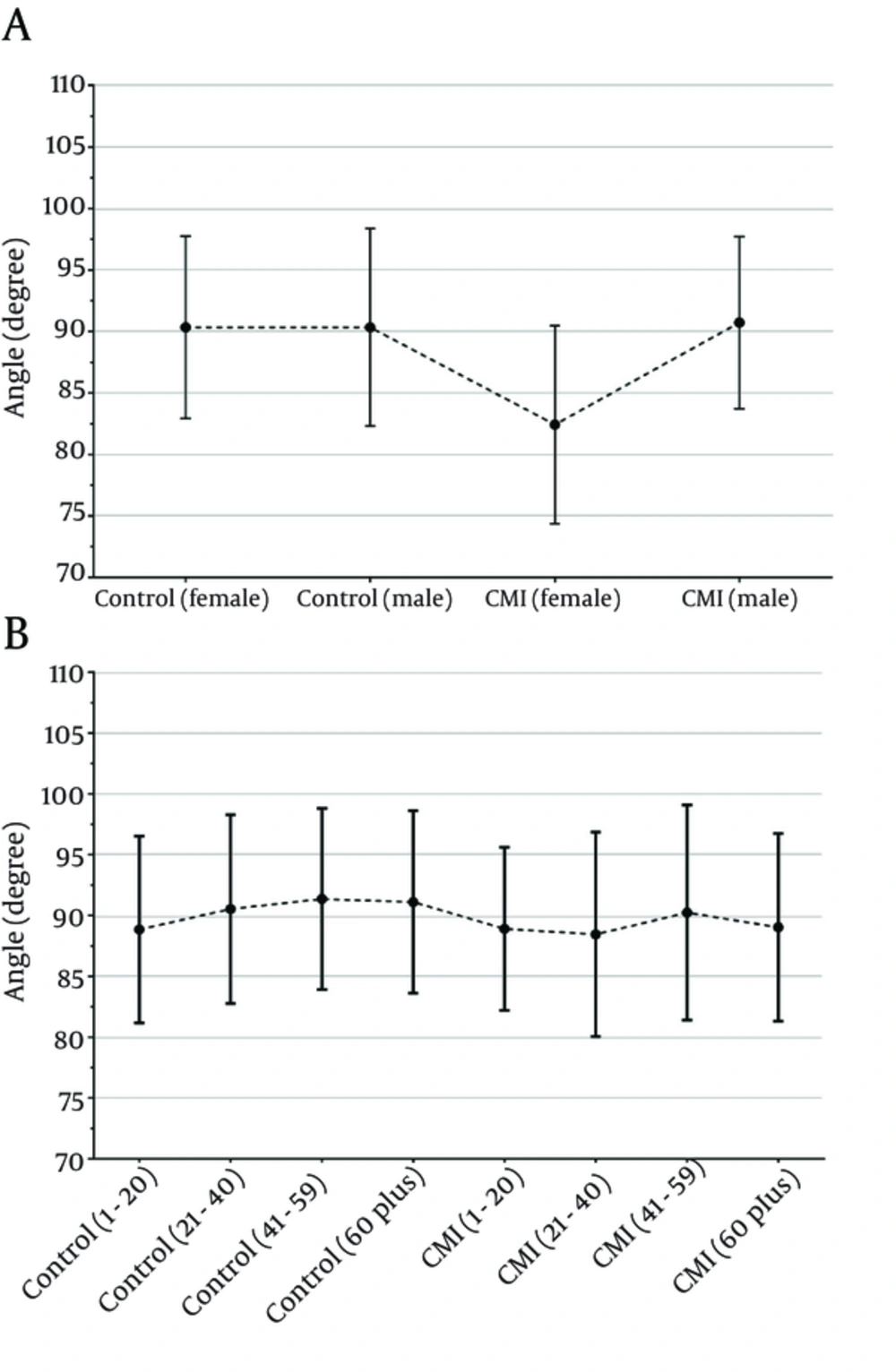
In this study, we found that the tentorial angle was 89.08 ± 7.78o in the CMI patients and 90.33 ± 7.66o in the controls. This difference between the patient and control groups was not statistically significant (P = 0.209).
Dagtekin et al. (
26) found that the mean length of the tonsillar herniation was 7.4 mm (4.9 - 12.1 mm). On the other hand, in a study by Aydın et al. (
21), the mean quantity of the tonsillar herniation was found to be 12.6 mm (5 - 38 mm). Milhorat et al. found that tonsillar herniation was 9.8 ± 5.8 mm in the CMI patients and 2.1 ± 3.7 mm in the controls (P < 0.001) (
4).
Elster and Chen (
32) found that the degree of tonsillar herniation was higher in those having the findings of spinal cord, cerebrum and cerebral trunk.
Mikulis et al. (
33) found that the mean extent of tonsillar herniation was 6 mm in the first decade, 5 mm between the second and third decades, 4 mm between the fourth and eighth decades, and 3 mm in the ninth decade. The authors suggested that there was a statistically significant decrease in the degree of the herniation with aging.
Aboulezz et al. (
34) claimed that tonsillar herniation lower than 5 mm from the level of the foramen magnum was pathologic.
Barkovich et al. (
23) reported that in the absence of syringomyelia, a herniation of 2 mm or less was clinically insignificant.
Urbizu et al. (
25) used a tonsillar herniation greater than 3 mm on MRI as a base, and this resulted in several symptoms and findings (neural compression in the craniovertebral junction, syringomyelia, and cerebellar or intracranial hypertension) for the diagnosis of CMI. Using the results of that study, the authors supported the hypothesis that variability in the genes relating to the paraxial mesoderm might affect the size of the FCP, causing CMI.
Heiss et al. (
31) found the mean value of tonsillar ectopia to be 12.3 mm (5 - 22.7 mm) before the patients were taken for the operation.
Milhorat et al. (
30) defined herniation as tonsilla cerebelli lying more than 5 - 7 mm below the level of the foramen magnum; those authors described a prolapse of 0 - 4 mm as cerebellar tonsils.
Aitken et al. (
35) described tonsillar herniation of between 2 and 4 mm as borderline herniation. In essence, 5 mm is considered to be a cut-off value for the overall majority in terms of tonsillar ectopia, although it has been reported that increasing symptoms and syringomyelia requiring surgical intervention have been seen in patients with lower degrees of herniation.
Sahuquillo et al. (
24) defined CMI tonsilla cerebelli as sloping down at least 3 mm from the foramen magnum.
In this study, the mean amount of herniation was found to be 4.85 ± 3.09 mm.
Aydın et al. (
21) found the height of the posterior fossa to be 124.7 ± 15.7 mm in the CMI group and 141.2 ± 6.8 mm in the control group; they also reported that this difference was not statistically significant.
In our study, we found the mean height of the posterior fossa to be 57.31 ± 4.21 mm in the CMI patients and 60.5 4 ± 4.30 mm in the controls. Additionally, we found the height of the posterior fossa statistically significantly smaller in the CMI patients compared to the controls. The reduction we found in the height of the FCP was similar to that found in the study by Aydın et al. (
21). However, the difference between the means, despite the measurements being taken from the same points, was remarkable. We believe that the small height of the posterior fossa in the CMI patients might indicate bone hypoplasia and cause the posterior fossa organs to slope down.
Sekula et al. (
3), Nishikawa et al. (
8), and Hwang et al. (
27) found the length of the superior and inferior cerebellar hemisphere to be higher in the CMI patients. However, only Hwang et al. (
27) highlighted that the difference was statistically significant.
In our study, we found that the maximum height of the cerebellum was 59.13 ± 5.33 mm in the CMI group and 53.32 ± 5.37 mm in the controls. This difference between the CMI and control groups was statistically significant (P = 0.001).
Hwang et al. (
27) found that the axial length of the cerebellar hemisphere (the most remote lateral distance) was 86.93 mm in the CMI group and 98.83 mm in the control group; they reported that the difference was not statistically significant.
In this study, we found the above parameter to be 104.65 ± 4.93 mm in the CMI group and 105.51 ± 5.75 mm in the control group. This difference between the patients and the controls was statistically insignificant (P = 0.249).
The results for this parameter showed similarities with the results of the study by Hwang et al. (
27).
In our study, we found that the axial length of the cerebellar hemisphere on the midsagittal section was statistically greater in the CMI patients.
On the other hand, in the measurements they carried out on the transverse section, Hwang et al. (
27) found that the axial length of the cerebellar hemisphere on the midsagittal section was statistically less in the CMI patients.
Unlike the other studies, we measured other parameters in this study such as maximum cranial height, occipital cord, cranial base length, and maximum cranial width in the CMI patients. We found the maximum cranial height to be 130.33 ± 6.17 mm in the CMI group and 132.94 ± 7.09 mm in the controls. This distance was statistically shorter in the CMI group (P = 0.004), which suggests that the total size of the cranium might be smaller in the CMI patients.
No statistical significance was found in the differences between the CMI and control groups in terms of the anteroposterior diameter of the cerebrum, maximum height of the cerebrum, and maximum cerebrum weight that we measured to ascertain whether the size of the cerebrum might affect CMI.
In conclusion, the size of the posterior cranial fossi was smaller and the size of the cerebellum was greater in the CMI patients. Thus, the risk of tonsillar herniation may be higher than the normal population in patients with a small posterior cranial fossa and a large cerebellum.