Based on current advances in basic medical research and the development of translational medicine (
14), the journey towards pharmacological intervention for hydrocephalus is arduous (
15). This is due to the fact that hydrocephalus is not merely a single, well-defined etiological disease but rather a syndrome encompassing multiple etiologies with common clinical manifestations (
16). Identifying drug targets relies heavily on continuous in-depth research by scientists (
17). Therefore, the most effective treatment modality currently is surgical intervention (
18). Surgical interventions can be broadly categorized into four main types, each serving different purposes: Relieving intraventricular obstruction, reducing cerebrospinal fluid secretion, establishing new intracranial circulation, and establishing extracranial shunting. Each of these categories has its own detailed subdivisions and specific operative techniques. The selection of a particular procedure depends on the patient's physical condition and the surgeon's proficiency in the technique. However, the contraindications for these surgeries often overlap and include conditions such as unresolved intracranial hemorrhage, uncontrolled intracranial infection, preterm neonates, and other systemic illnesses. These contraindications fall within the scope of the complex definition of hydrocephalus presented in this paper. Therefore, comprehensive preoperative assessment of the patient's condition, early identification, and prediction of the possible occurrence of complex hydrocephalus, and taking corresponding measures before surgery are crucial. This preoperative preparation should not be perceived as a single surgical failure but rather as a preparatory step for formal surgical treatment, serving as the cornerstone for a successful single surgical intervention. It avoids the prolonged suffering and adverse effects on the patient's physical and mental health, as well as their economic well-being, associated with repeated shunting procedures, fenestration, and cauterization.
The Ommaya reservoir, invented and first applied by Ayub Khan Ommaya in 1963 (
19), has gradually gained widespread use in surgical procedures (
20). This device offers unique advantages due to its small size, minimizing the impact of foreign bodies implanted in the patient's body and thus reducing iatrogenic damage. Importantly, compared to direct implantation of shunting tubes, the Ommaya reservoir minimizes the risks of blockage and infection, which are common causes of postoperative complications leading to complex hydrocephalus and significantly impacting patients. Moreover, the Ommaya reservoir allows for repeated drainage of cerebrospinal fluid without the need for multiple ventricular or lumbar punctures. Additionally, it facilitates the convenient intraventricular administration of medications to control intracranial infections and promote hematoma absorption.
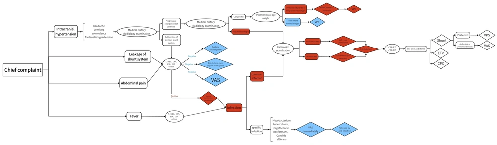
This article categorizes the common chief complaints of patients into four main groups. The first category comprises manifestations of intracranial hypertension, including headaches, vomiting, somnolence, and fontanelle hypertension. The second category involves leakage of the shunt system, which had been placed in the first operation, either at the scalp or anal region. The third category includes abdominal pain, and the last one is fever. Following the identification of these four major categories of chief complaints, further investigations are conducted to confirm the underlying causes. This process involves the utilization of medical history, radiology examination, serological analyses, and microbiological examinations. The objective of all these auxiliary investigations is to ascertain the underlying etiology, thereby determining the presence of high-risk factors for complex hydrocephalus and assessing the likelihood of its occurrence. Subsequently, a two-stage surgical plan is tailored based on the patient's individual circumstances, such as overall health status and personal preferences, significantly improving the prognosis for complex hydrocephalus.
If there are manifestations of intracranial hypertension, it may indicate progressive enlargement of the ventricular system primarily or inadequate drainage by the shunt system used in previous shunt surgery, which can be discriminated by medical history and radiology examination. Primary hydrocephalus can be confirmed through radiology examinations, distinguishing between congenital hydrocephalus (commonly due to developmental abnormalities of the ventricular system) and secondary hydrocephalus resulting from intracranial hemorrhage. In cases of congenital hydrocephalus where the etiology is clear and the condition is straightforward, if the patient is a term infant with normal birth weight, symptoms can often be effectively alleviated through standard shunt surgery, leading to gradual normalization of the ventricular system over time (
21). However, if the patient is a preterm infant with very low birth weight, it will become complex hydrocephalus, which requires EVD with an Ommaya reservoir first, followed by VPS (
22). Meanwhile, hydrocephalus secondary to intracranial hemorrhage is not only a contraindication for routine shunt surgery but also represents a form of complex hydrocephalus. Additionally, the presence of intraventricular blood can lead to the formation of fibrous septa within the ventricular system, further complicating the condition into multiloculated hydrocephalus (
23).
If malfunction of the previous shunt system is identified, indicating initial surgical failure and meeting the definition of complex hydrocephalus, it is crucial to assess for any signs of infection through temperature measurement, white blood cell count, C-reactive protein, erythrocyte sedimentation rate, and CSF culture. The above tests are also required in the conditions mentioned previously, where the second category involves leakage of the shunt system and the third indicates abdominal pain. If there is no infection, it will be considered simple hydrocephalus that can be resolved by replacing the shunt system, placing the original shunt system following disinfection, or altering to a VAS. However, in cases of intracranial infection, it will be classified as complex hydrocephalus, necessitating the immediate removal of the shunt, followed by appropriate management of the infection.
If a patient presents with fever along with changes in the characteristics of CSF, broad-spectrum antimicrobial therapy should be initiated alongside serological and microbiological examinations to identify the pathogen. Once the pathogen is identified, targeted antimicrobial therapy should be administered. It is essential to differentiate between different pathogens to determine the sequence of treatment modalities. For pathogens such as
Mycobacterium tuberculosis (
24),
Cryptococcus neoformans (
25), or
Candida albicans (
26), which are considered specific types of intracranial infections, the treatment approach differs. The significant goal in these cases is to reduce intracranial pressure through shunt surgery, followed by anti-infection treatment. If the infection is caused by common gram-positive or gram-negative bacteria, it is considered a standard infection. Infection is a contraindication for routine shunt surgery and represents a form of complex hydrocephalus. Similarly, due to the action of microorganisms within the skull, an immune response is triggered (
27), leading to the proliferation of substances like collagen and the formation of septa within the ventricular system, resulting in multiloculated hydrocephalus. Existing studies mostly regard infection as a postoperative complication of hydrocephalus, but less attention is paid to infection as a major factor of complex hydrocephalus (
28). In this paper, it is believed that for hydrocephalus post-infection, the most important step is to control the infection until the cerebrospinal fluid is clear and free of pathogenic microorganisms, after which surgery can be carried out.
In cases where intracranial hemorrhage or infection is suspected, radiology examinations are crucial to assess the presence of septations within the ventricular system and the circulation of cerebrospinal fluid, aiding in the diagnosis of complex multiloculated hydrocephalus (
29). If multiloculated hydrocephalus is confirmed, the initial preparatory step involves endoscopic fenestration of the cysts to establish communication between them, converting multiloculated hydrocephalus into uniloculated hydrocephalus. This reduces the number of required shunt catheters and minimizes the need for multiple shunting procedures. Subsequently, EVD is used to temporarily drain CSF. Existing research mostly focuses on comparing the advantages and disadvantages of craniotomy and endoscopic surgery, which is of certain significance (
30). Through such research, medical institutions can learn and master relevant surgical skills accordingly, and also guide doctors' choice of surgical methods to a certain extent. However, this study holds that compared with the choice of surgical form, for multiloculated hydrocephalus, the most important step is to overcome the obstacles and establish communication first, rather than adhering strictly to a particular surgical form.
For patients with uniloculated hydrocephalus, the first stage involves EVD and placement of an Ommaya reservoir. This allows intermittent drainage of CSF to promote hematoma resolution and facilitates intraventricular administration of antimicrobial agents (
31). After the first stage of surgery, anti-infection treatment is continued until CSF routine examination and biochemical tests confirm that the CSF is clear and sterile (
32), indicating successful control of intracranial hemorrhage and infection. The patient can then proceed to the second stage of formal surgery.
At this point, the patient often no longer has contraindications to shunt surgery, and the likelihood of developing complex hydrocephalus is significantly reduced. Based on the patient's current overall health status, VPS should be the preferred choice if there are no abdominal or peritoneal diseases; otherwise, VAS is preferred. Patients typically experience symptom relief shortly after formal surgery, and regular follow-up examinations post-discharge will show gradual normalization of the ventricular system.
In cases of severe and refractory hydrocephalus with special circumstances, which match the first definition of complex hydrocephalus mentioned above, it is necessary to consider the procedure of surgical management for complex hydrocephalus again. Alternative surgical approaches should be considered because VPS or VAS may be ineffective for that patient. These may include ETV to establish new intracranial circulation or CPC to reduce cerebrospinal fluid secretion.
In addition to the scenarios mentioned above, there is another significant category of complex hydrocephalus cases involving preterm newborns. Preterm newborns often have very low birth weights and poor overall health, making them intolerant to surgery. Even if surgery is attempted, the outcomes are usually poor, leading to prolonged non-recovery, repeated surgeries, and significant physical, psychological, and financial burdens for the patients. Additionally, they are often associated with intracranial hemorrhage and are more susceptible to intracranial infections due to their weakened immune systems, representing two or more types of complex hydrocephalus. Thus, whether the infant has intracranial hemorrhage and infections or is diagnosed with congenital hydrocephalus, it is undeniable that preterm newborns should be managed according to the complex hydrocephalus protocol.
To address this, we typically perform a two-stage approach: In the first stage, EVD with Ommaya reservoir implantation is carried out. This approach allows us to control the existing intracranial hemorrhage and infection while delaying the need for further interventions. During this time, the infant can continue to develop in a relatively stable environment. Once the infant reaches a postmenstrual age greater than 37 weeks and a weight exceeding 2.5 kilograms, their overall health status becomes more tolerant of surgery. At this point, the second stage involves VPS. For these infants, this treatment approach yields more significant therapeutic effects and a better prognosis.
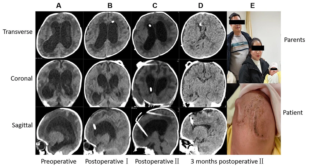
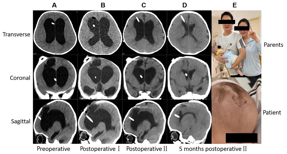
In clinical practice, clinicians should carefully understand the process, starting from the initial diagnostic symptoms, and use various auxiliary examination methods to clarify the real situation of patients. They should assess whether there are factors that may lead to complex hydrocephalus, prevent problems before they occur, and prepare in advance for diagnosis and treatment according to the two-stage surgical procedure. Based on the analysis of typical cases in this study, it is evident that all three patients presented with two or more high-risk factors for complex hydrocephalus during their initial consultations. It can be almost conclusively determined that these patients did not have simple hydrocephalus, and the likelihood of successful cure through a single surgery was minimal.
For the first two cases, we adopted the procedure of surgical management for complex hydrocephalus proposed in our new summary: EVD with Ommaya reservoir implantation to control existing intracranial hemorrhage and infection in the first stage. After confirming that the cerebrospinal fluid is clear, the hematoma is absorbed, and the infection is controlled, the second stage involved VPS placement. Symptoms were alleviated immediately postoperatively, and subsequent imaging results showed normalization of the ventricular system.
Since complex hydrocephalus is a chronic condition, a discussion on long-term complications will be beneficial. From our results, patients who underwent the two different procedures of surgical management showed almost no difference in subsequent symptom relief, normal imaging ventricles, and survival rate. This also indicates that for complex hydrocephalus, unless individual patients have special circumstances, there is generally no difference in the cure of the disease. However, inappropriate surgical procedures will greatly increase the number of operations undergone by patients, especially for children, who are in a critical period of growth and development. The impact of surgery can affect their subsequent cognition, development, and function, which is also reflected in the results of this article. In addition to these injuries, continuous surgery affects the quality of life and economic situation of patients, bringing both physical and psychological distress.
Moreover, long-term complications are a major factor affecting patients' lives. Slit ventricle syndrome is mainly due to the siphoning effect of the shunt tube, which leads to excessive shunting of cerebrospinal fluid. Eventually, the ventricle becomes narrow and slit-like, requiring reoperation to replace the shunt device (
33). Shunt dependency syndrome often arises due to the long-term placement of the shunt, decreased brain tissue compliance, and increased intracranial pressure from a small amount of cerebrospinal fluid, causing headaches. It is often difficult to remove the shunt and switch to ETV (
34). Sylvian Aqueduct Syndrome often compresses the brain tissue due to the reversal of supratentorial and infratentorial pressure after shunt operation, resulting in ocular symptoms, and ETV is needed again to balance the pressure (
35).
Of course, we also acknowledge that this study has its limitations. The patients selected in this paper are all from Xiangya Hospital, making it a single-center study. The temporal and regional characteristics of the sample sources have not been further analyzed, and they do not have a wider range of universality. Additionally, it is a retrospective study, so it is not random in patient selection, and some selection bias may occur. To conduct better research, we plan to collaborate with several affiliated units to collect samples for analysis in future work. Simultaneously, randomized controlled trials will be gradually carried out after careful design of the research protocol.
In summary, through meticulous review of the literature and consolidation of clinical cases within our institution, we have pioneered a relatively comprehensive and systematic procedure of surgical management for pediatric complex hydrocephalus. This protocol encompasses early identification and diagnosis, followed by personalized therapeutic interventions tailored to the etiology and individual patient circumstances, implemented in two stages. By intervening early and effectively, we have succeeded in curtailing the progression of complex hydrocephalus, thereby enhancing the therapeutic experience and quality of life for our patients.
5.1. Conclusions
(1) Complex hydrocephalus is defined as hydrocephalus requiring multiple surgeries for cure, caused by hemorrhage and/or infection, multiloculated hydrocephalus, and preterm infants with very low birth weight.
(2) The treatment principle lies in early identification for prompt diagnosis and timely staged treatment in both early and late phases. Early comprehensive treatment aims to prevent progression to complex hydrocephalus, with personalized interventions tailored accordingly.
(3) In the early first stage, EVD with Ommaya reservoir implantation is performed. This aims to control pre-existing intracranial hemorrhage and intracranial infection while allowing preterm infants to gain developmental time. Endoscopic cyst fenestration converts multiloculated hydrocephalus into uniloculated hydrocephalus.
(4) There is no absolute preference among various surgical techniques in the subsequent second stage. The choice depends on the patient's condition, sometimes necessitating sequential usage to manage extremely refractory hydrocephalus.
(5) Postoperative follow-up is essential, with regular imaging examinations to assess the restoration of the ventricular system. Timely identification and implementation of new interventions are crucial in managing any evolving complications.