1. Background
Coccydynia, or coccygodynia, is defined as pain in or around the region of the coccyx without significant radiation to other sites. The pain is usually triggered by direct compression of the area or by changing the position from sitting to standing (1). The condition may be idiopathic or may be due to some underlying pathologies or conditions, and it is five times more common in women (2-4). Trauma is a known precipitant in many patients, but the precise etiology of the condition is uncertain; infection and tumors are only rare causes (5). Abnormal coccygeal mobility associated with changes in posture may account for the pain in some cases (2). In others, pain may be generated by coccygeal intervertebral disc pathology (6), pericoccygeal soft tissue inflammation (6), sacrococcygeal cornual junction pathology (7), or coccygeal nerve entrapment (4, 7). The condition is associated with severe pain that causes daily activities to be limited. Thus, early diagnosis and treatment is necessary to restore the functional outcome (1, 4, 8-10). Several nonoperative and operative strategies have been introduced and tested for treatment of chronic coccydynia, with controversial results. The available nonoperative modalities include using donut pillows, physical therapy and massage, radiofrequency ablation of the coccyx, injection of glucocorticoids into the coccyx, and different coccygeal manipulations (4, 8, 11-16). For those who are resistant to medical and physical therapy, surgical resection of the mobile portion of the coccyx and coccygectomy is recommended (5, 17, 18).
Extracorporeal shock wave therapy (ECSWT) is a method of propagating shock waves into musculoskeletal tissues in order to maintain function and to limit pain and disability. Currently, ECSWT is being applied in many musculoskeletal disorders such as plantar fasciitis, lateral epicondylitis of the elbow, calcific tendinopathies of the shoulder, nonunion of long bone fractures, avascular necrosis of the femoral head, jumper’s knee, and Achilles tendinopathy (19, 20). Although the mechanism of action of ECSWT in relieving the pain of musculoskeletal conditions is not clearly understood, it is believed that the neovascularization and the increase in blood supply resulting from this therapy initiate healing and repair (21, 22). A recent study has shown that application of ECSWT resulted in relieved pain in two patients with chronic coccydynia; however these results are limited (23).
2. Objectives
The aim of the current study was to determine the effects of ECSWT on the pain scores of patients with chronic coccydynia.
3. Patients and Methods
3.1. Study Population
This was a quasi-experimental study performed in outpatient rehabilitation clinics of Kashani hospital, a tertiary healthcare center affiliated with Isfahan University of Medical Sciences, during a one-year period from February 2014 to March 2015. We included all patients with chronic coccydynia referred to our center for physical therapy and rehabilitation. We included adult patients (> 18 years) who had at least a 24-month history of pain in the coccygeal area, triggered by changing position and without radiation. We excluded those patients with acute coccygeal fractures, tumors, or osteomyelitis, those with pilonidal abscess, those with genitourinary incontinency, those with a neurologic deficit, and those with opium addiction. Those with old fractures without current dislocation were included in the study. All patients had received conservative therapies for at least one year before inclusion in the study. Thus they were considered to suffer from refractory chronic coccydynia. The study protocol was approved by the institutional review board and medical ethics committee of Isfahan University of Medical Sciences. All patients provided their informed written consent before inclusion in the study. The study is registered with the Iranian registry for clinical trials (IRCT2015041021673N1; www.irct.ir).
3.2. Study Protocol
All patients were evaluated meticulously regarding their past medical history and were examined by a physical medicine resident on presentation. Those eligible for the study were further investigated by radiography for coccygeal fractures. Patients received ECSWT with a radial probe delivering 3,000 shock waves per session of 2 bar at 21 Hz frequency directed to the coccyx. The probe was placed in direct contact with the coccyx in the sagittal plane in the intergluteal cleft. The coccyx was investigated first by finger examination, and the probe was directed toward it. Each patient received four sessions of ECSWT at one-week intervals. The patients’ pain severity was measured and recorded according to a 10-point visual analogue scale (VAS) before intervention. The patients were visited at one, two, three, and four weeks after initiation of therapy. They were also visited at one and six months after the cessation of therapy (two and seven months after initiation of treatment), and the VAS score was recorded on each visit.
3.3. Statistical Analysis
All statistical analysis was performed by statistical package for social sciences (SPSS Inc., Illinois, USA) version 19.0. Data are presented as mean ± SD and proportions as appropriate. As the data did not have normal distribution, we used the Wilcoxon signed ranks test to compare the VAS scores before and after the intervention. A two-sided P value of less than 0.01 was considered to be statistically significant.
4. Results
Overall, we included 10 patients with chronic coccydynia who were found to be eligible for the study and who finished the study. Most of the participants were women (90.0%), and the participants’ mean age was 39.1 ± 9.1 (ranging from 28 to 52) years. Most of the patients (80.0%) had a previous history of trauma to the coccygeal area, while only 20.0% had signs of radiological fractures without dislocations. The baseline characteristics of the patients are summarized in Table 1.
| Characteristics | Valuea |
|---|---|
| Age, y, mean ± SD | 39.1 ± 9.1 |
| Gender | |
| Male | 1 (10.0) |
| Female | 9 (90.0) |
| Pain duration, y, mean ± SD | 5.6 ± 3.4 |
| History of coccygeal trauma | 8 (80.0) |
| Radiological finding | |
| Normal | 8 (80.0) |
| Fracture without dislocation | 2 (20.0) |
aValues are expressed as No. (%) unless otherwise indicated.
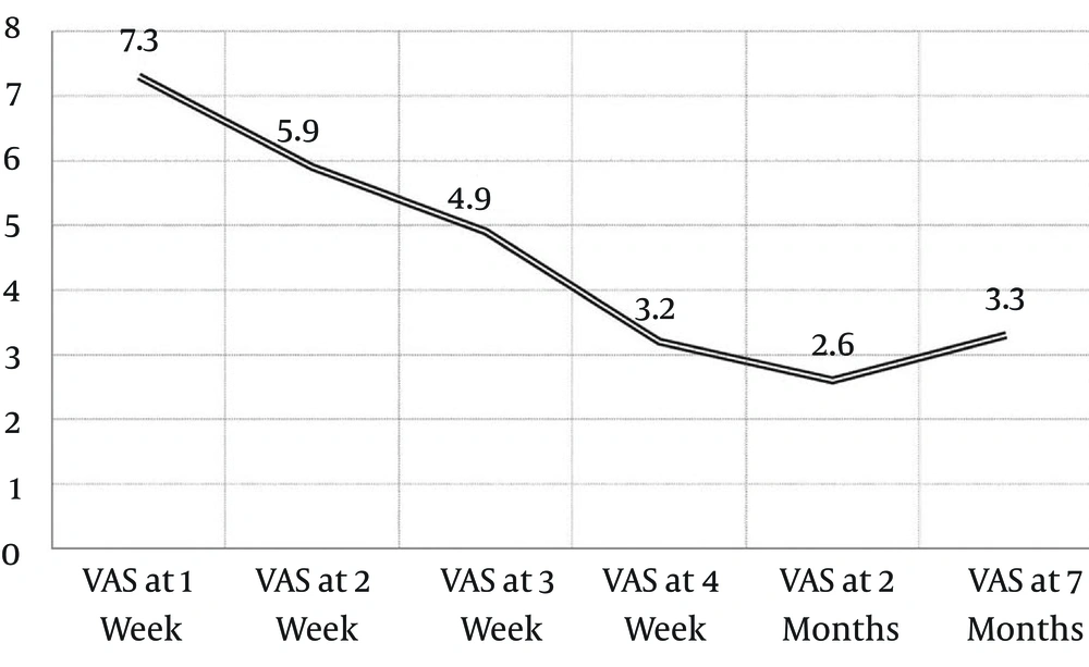
The VAS score was compared to baseline seven months after therapy (3.3 ± 3.6 vs. 7.3 ± 2.1; P = 0.011). However, the VAS score at two months (2.6 ± 2.9 vs. 7.3 ± 2.1; P = 0.007) and at four weeks (3.2 ± 2.8 vs. 7.3 ± 2.1; P = 0.007) was significantly decreased when compared to baseline. The VAS score at three weeks (4.9 ± 3.2 vs. 7.3 ± 2.1; P = 0.020) and at two weeks (5.9 ± 2.5 vs. 7.3 ± 2.1; P = 0.024) was not statistically different from baseline. In other words, the VAS score decreased after four weeks of therapy, and the decrease was persistent two months after therapy; however, the pain increased seven months after therapy. The trends in changes of VAS scores during the seven months of the study are demonstrated in Figure 1. All the patients’ information is summarized in Table 2.
| Patient | Age, y | Sex | Pain Duration, y | Trauma History | VAS 1 Week | VAS 2 Weeks | VAS 3 Weeks | VAS 4 Weeks | VAS 2 Months | VAS 7 Months |
|---|---|---|---|---|---|---|---|---|---|---|
| 1 | 52 | Woman | 10 | Yes | 10 | 2 | 1 | 0 | 0 | 1 |
| 2 | 28 | Woman | 9 | Yes | 9 | 9 | 8 | 6 | 3 | 9 |
| 3 | 33 | Woman | 10 | No | 5 | 4 | 0 | 0 | 0 | 0 |
| 4 | 40 | Woman | 3 | Yes | 4 | 3 | 2 | 1 | 1 | 1 |
| 5 | 42 | Woman | 5 | Yes | 8 | 8 | 9 | 4 | 6 | 7 |
| 6 | 52 | Woman | 2 | Yes | 9 | 9 | 9 | 9 | 9 | 9 |
| 7 | 29 | Man | 2 | No | 5 | 5 | 5 | 2 | 1 | 0 |
| 8 | 38 | Woman | 3 | Yes | 9 | 8 | 6 | 2 | 0 | 0 |
| 9 | 30 | Woman | 9 | Yes | 7 | 6 | 5 | 5 | 3 | 3 |
| 10 | 47 | Woman | 3 | Yes | 7 | 5 | 4 | 3 | 3 | 3 |
5. Discussion
ECSWT has been used successfully to treat several musculoskeletal disorders, especially those involving the tendons and cartilages (19, 20). Currently, ECSWT is commonly used to treat many conditions such as plantar fasciitis, lateral epicondylitis of the elbow, calcific tendinopathies of the shoulder, nonunion of long bone fractures, avascular necrosis of the femoral head, jumper’s knee, and Achilles tendinopathy (19, 20). In this quasi-experimental study, we demonstrated that ECSWT resulted in decreased pain measured by VAS score in patients with chronic coccydynia in the early phases. The significant effect of ECSWT on pain intensity was observed after four weeks of therapy and was persistent for two months after the therapy. The interesting finding of the current study is that the favorable outcome was observed after four weeks of treatment. However, the decline trend in VAS score was observed even two months after the cessation of the therapy. The pain intensity increased seven months after the therapy, which was comparable with baseline. In other words, patients received four weeks of treatment in which the pain severity decreased significantly. However, the effect remained constant and the VAS score decreased until one month after the cessation of treatment, and then the pain intensity increased six months after therapy cessation. This shows that ECSWT provided a constant effect on the inflammation of the coccyx, resulting in decreased pain in the early phase.
Currently, data is scarce regarding the efficacy of ECSWT in patients with chronic coccydynia. A recent study by Marwan et al. (23) investigated this issue in two cases. The researchers included two patients with chronic coccydynia who failed to respond completely to other conservative management. A numerical pain scale (NPS) and VAS were used to assess the pain. Before starting ECSWT, Patient 1 reported a pain intensity of 6/10 and 5.1/10 on NPS and VAS, respectively, whereas the intensity of pain in patient 2 was 7/10 and 6.9/10 on NPS and VAS, respectively. Four weeks after ECSWT, patient 1 reported a complete relief of pain on NPS and VAS, whereas patient 2 reported a pain intensity of 1/10 and 0.8/10 on NPS and VAS, respectively. The same intensity of pain was reported by both patients after 12 months of follow-up (23). In a larger study, we showed that the pain intensity had decreased significantly seven months after therapy. The significant effects of ECSWT on the pain intensity of patients with chronic coccydynia appeared after four weeks of therapy. The interesting finding of our study is that the pain duration of the patients was 5.6 ± 3.4 years, and they had tested many conservative managements of chronic coccydynia without improvement. The conservative managements tested by the included patients involved physical therapy, donut pillow, electrostimulation, ultrasonic rehabilitation, exercise, and steroid injections. But the pain intensity decreased significantly after four weeks of ECSWT and reached its minimum at seven months.
Inflammation in the coccyx and its joint with the sacral vertebra is the proposed mechanism of pain in patients with chronic coccydynia (11). Several etiologies could lead to chronic inflammatory changes of the coccyx, such as trauma, instability, pregnancy and delivery, and hypermobility (4, 8, 10). In our series, 80.0% of patients had a previous history of trauma to the coccyx, and 20.0% had radiologic evidence of coccygeal trauma. Thus, inflammation is the most important etiology of pain in chronic coccydynia. The mechanism of action for ECSWT has yet to be well identified. The most important physical parameters of shock wave therapy for the treatment of musculoskeletal disorders include pressure distribution, energy flux density, and total acoustic energy. In contrast to lithotripsy, in which shock waves disintegrate renal stones, musculoskeletal shock waves are not being used to disintegrate tissue but rather to microscopically cause interstitial and extracellular responses, leading to tissue regeneration (20). It is believed that shock wave therapy alleviates pain by the induction of neovascularization and improvement of blood supply to the tissue and by initiating repairs to the chronically inflamed tissues by tissue regeneration (21). The experimental findings confirm that ECSWT decreases the expression of high levels of inflammatory mediators (matrix metalloproteinases and interleukins). Therefore, ECSWT produces a regenerative and tissue-repairing effect in musculoskeletal tissues, not merely a mechanical disintegrative effect, as was previously generally assumed (24).
We note some limitations to our study. First, the study population was limited, because of the low incidence of the condition. This may affect the power of the study in a negative fashion. Second, we used a quasi-experimental study design. This means that we did not include a control or placebo group. Thus, we cannot exclude the placebo effect of the procedure. Third, we followed the patients for seven months. As we obtained favorable results, we did not continue the study. Longer follow-up periods are required to determine the long-term results and outcome. The other limitation is that we only used VAS for clinical evaluation, which has its own shortcomings. Other clinical indices should be used in future studies. However, this is the first study to investigate this issue using a standard methodological approach.
In conclusion, extracorporeal shock wave therapy is an effective modality in relieving the pain intensity in the early phase in patients with refractory chronic coccydynia. The application of ECSWT for coccydynia could effectively reduce the pain intensity, especially in those resistant to other conservative therapies. However, the issue should be addressed in tests in larger placebo-controlled clinical trials before being applied in medical practice.
