1. Introduction
Neurofibromatosis type 1 (NF-1) or von Recklinghausen’s disease is an autosomal dominant disease, which affects about 1 out of 3000 individuals and has different clinical features involving bones, skin, eyes, nervous, and vascular systems (1). Due to delayed or a missed diagnosis of vascular involvement, they may rapidly develop hypovolemic shock and could potentially cause early death in NF-1 patients. Here we report a case on a NF-1 patient who was diagnosed after developing cervical hematoma following a jaw thrust maneuver.
2. Case Presentation
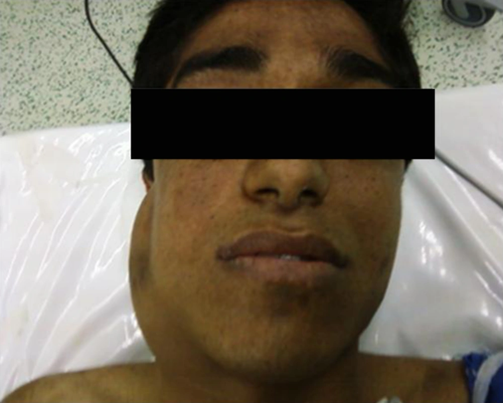
A 25-year-old man is presented with dull pain during activity in the posterior right thigh for 7 months. A 5 × 3 cm mass was detected in deep palpation of the mid-posterior right thigh. The borders of the mass and its depth were not clearly specified on examination. On physical examination, small size freckles less than 5 mm were noted on his face without involvement of other parts of the body. Routine laboratory data was normal and magnetic resonance imaging (MRI) of the patient’s right thigh confirmed a 37 × 65 × 38 mm oval lesion in close proximity to the neurovascular structures. Excision of the mass was planned as an elective surgery. General anesthesia was chosen based on the patient’s preference. On the day of the operation, hemodynamic parameters were completely normal. His weight was around 70 kg and was pre-medicated by an intravenous injection of 2 mg midazolam and 150 µg fentanyl followed by induction of anesthesia using 350 mg thiopental and 35 mg atracurium. After 3 minutes of oxygenation by the flow of 7 lit/min of 100% Oxygen, the patient was intubated using a cuffed endotracheal tube (n = 8) that was fixed in the appropriate position so that 1 finger could pass freely under the fixation band. Maintenance of anesthesia was based on 100µg/kg/min propofol plus N2O: O2 equaled to 2:2 liters. We packed a soaked band into the patient’s mouth and turned him to prone position. Every 10 minutes the position of the patient’s head and neck was checked. An operation was done by an excision of the mass during 1.5 hours without any major issues. Estimated blood loss was 250 mL. After returning the patient to the supine position and the reversal of paralytics by 3.5 mg neostigmine and 1.5 mg atropine, he was extubated. The patient was completely awake afterwards with 97% O2 saturation (Spo2) on room air by a pulse oximeter and was transferred to the recovery room. A face mask with a flow of 8 lit/min of 100% O2 was applied. After 5 minutes in the recovery room the saturation of oxygen decreased to 90% while the patient became drowsy and less irritable. Due to a lack of neuromuscular monitoring, hypoxia might occur. Nonetheless, Spo2 further declined to 84%, so we attempted a jaw thrust maneuver to maintain oxygenation. After 30 seconds of jaw thrust, the patient’s oxygen saturation increased and reached 98% by face mask and he regained wakefulness. During the next 10 minutes after the maneuver, a small hematoma was formed in the right side of the patient’s neck under the mandibular angle. Compression by ice bag did not help and a mass increased in size and extended toward the right ear (Figure 1). We decided to transfer the patient to the operation room (OR) to explore the cervical region. In the OR, the patients’ heart rate was 121 beats/min and blood pressure was 135/86 mmHg. The patient had no respiratory distress. Following an injection of 100 µg fentanyl, 20 mg etomidate and 100 mg succinylcholine intubation with a cuffed endotracheal tube (n = 7.5) was done without difficulty. By a classic cervical incision and proximal vascular control, tearing of the facial artery and vein as well as a traumatic lesion to the parotid tail were found. The vessels were legated and after achievement of homeostasis, a cervical drainage tube was left in place. During the operation, propofol was infused at 100 - 150 µg/kg/min and 30 mg atracurium was also injected. The proportion of N2O to Oxygen was 2:2. Duration of the second surgery was about 40 minutes. We did not encounter any hemodynamic instability during the second surgery and the estimated blood loss was 350 mL, including the formed clot. During both operations, if the heart rate and systolic blood pressure increased more than 20% of baseline, by considering the sufficient depth of anesthesia and appropriate level of muscle relaxation, 50 µg fentanyl was injected intravenously as an analgesic agent. Moreover, the patient received N2O as an analgesic substance. In the first surgery, all induction agents were acceptable and thiopental was chosen by the anesthesiologist. However, in the second surgery, with the aim of establishment of better hemodynamics in a patient with growing hematoma and active bleeding, etomidate was used. The preference of expert anesthesiologists in our medical center for maintenance of anesthesia is Total IV Anesthetic (TIVA) method, therefore propofol was administered in this regard.
The patient was extubated successfully after injection of 3 mg neostigmine and 1.5 mg atropine and was transferred to the recovery room followed by uneventful ICU admission. The patient was discharged from the hospital 2 days later. Pathological evaluation of the lesion revealed a low grade malignant peripheral nerve sheath tumor. Further evaluations confirmed the diagnosis of NF-1.
3. Discussion
Hypoxia in the postoperative period could result from various reasons. Since neuromuscular monitoring was not done, postoperative residual curarization (PORC) could be considered as one of the factors for the hypoxia in this patient. PORC is defined as a residual paresis early after completion of general anesthesia using neuromuscular blocking agents. PORC could potentially increase morbidity and may prolong the postoperative recovery period. Continued monitoring at the recovery room and assessing the patients by acceleromyography would result in a better outcome (2).
Opioid induced depression was also a rationale behind this situation that should be kept in mind. Evaluating the patients by checking the opioid toxidrome may be helpful. However, since the opioids used during general anesthesia are commonly short-acting ones, it is a bit unlikely (3).
Whatever happened, the evaluation led to diagnosis of NF-1 in this patient. Intrinsic lesions of vascular walls in NF-1 are of considerable problems, which its pathogenesis have not been completely defined. Occlusion, stenosis, aneurysm, arterial rupture, fistulas, and ectasia were reported as different vascular manifestations of NF-1 (4). Therefore it is apparent that such disorders can involve large to small vessels and some lesions may be clinically silent. It is important for emergency physicians and anesthesiologists to consider the probable risk of arising unexpected hematoma. Hematoma formation is unusual in NF-1 especially in the maxillofacial area where it has rarely been reported (5). It is thought that the walls of large vessels in NF-1 patients may have some pathologic alterations (6). It is still debatable whether our NF-1 case was only prone to hematoma formation by aggressive force or trivial forces might induce such a huge hematoma. Our case did not have any sign of vascular lesion in the jaw before the jaw thrust maneuver. Therefore, it might actually be the loosen wall of arteries that may be ruptured with any trivial trauma. However we could not exclude the presence of the preexisted vascular lesion that was triggered with the jaw thrust maneuver. The vessels, which are injured, are not always directly associated with NF-1 and the pathologic mechanism of lesions may be due to intimal thickening of the vascular wall that creates obstructive lesions (7). Hematoma formation is not common in NF-1, however there are some reports regarding traumatic or spontaneous vascular ruptures in NF patients (4, 8-12). Moreover, it is important to perform a complete preoperative evaluation, to consider related imaging and to be aware of the probable anesthetic complications associated with the disease (13).
3.1. Conclusion
NF-1 patients should be closely observed for any sign of vascular abnormality and danger of a vascular rupture. It seems that in these patients who need a jaw thrust maneuver, after general anesthesia or in emergency cases, vigilant considerations regarding their ventilation and meticulous jaw thrust maneuver are reasonable to avoid the probable risk of vascular lesions.
