1. Background
Leishmania is an obligatory intracellular protozoan that has two stages during its life cycle: promastigote that develops in the midgut of sand fly (as a vector) and culture medium, and amastigote that multiplies in the host macrophage (1, 2). Leishmania is transmitted by the bite of the female sandfly. Different clinical manifestations of leishmaniasis include Cutaneous Leishmaniasis (CL), Mucocutaneous Leishmaniasis (MCL), and Visceral Leishmaniasis (VL) (1, 3). Cutaneous leishmaniasis is endemic in different parts of the world, particularly in the Middle East (Afghanistan, Algeria, Iraq, Yemen, Saudi Arabia, and Iran) (1, 3-5). The topical treatment of CL includes the use of pentavalent antimony compounds such as sodium stibogluconate and meglumine antimoniate; but unfortunately, these drugs are not effective for the treatment of CL (4). Meanwhile, they are toxic, expensive, painful, and associated with adverse side effects, drug resistance, relapse after treatment, and without satisfactory results (1, 3, 5).
Regarding the lack of effective drugs, it is necessary to search for a new alternative drug for the treatment of CL. Compounds with an origin in natural sources including plants have been paid more attention (1, 6). Propolis is one of the herbal remedies with longstanding and brilliant properties in the healing of different infectious diseases. Propolis is a resinous mixture produced by honey bees from plants, buds, and exudates (7). It is the bee glue and protects the hive against external invaders by its waxy nature and mechanical properties. Propolis is lipophilic and sticky (when heated). It has a pleasant aromatic smell and different colors depending on its origin and age. Raw propolis is composed of 50% resin, 30% waxes, 10% essential oils, 5% pollen, and 5% of various organic compounds that have various biological activities based on its place, time of collection, and geographical and climatic conditions. These compounds belong to the following groups: polyphenols; benzoic acids and derivatives; cinnamic alcohol and cinnamic acid and its derivatives; sesquiterpene and triterpene hydrocarbons; benzaldehyde derivatives; other acids and respective derivatives; alcohols, ketones, and heteroaromatic compounds; terpene and sesquiterpene alcohols and their derivatives; aliphatic hydrocarbons; minerals; sterols and steroid hydrocarbons; sugars and amino acids, flavonoids, aliphatic acids, esters, chalcones, and lignans. Some compounds are common in all propolis samples and determine their characteristics (4, 7). In folk medicine, it is used as an antiseptic and oxidant to treat several diseases, such as acne, viral infection, microbial infection, burns, and wounds.
2. Objectives
There are a few reports of the anti-cutaneous leishmaniasis properties of propolis in Iran. Thus, the aim of the present study was to evaluate the propolis extract against Leishmania major via in vivo and in vitro models.
3. Methods
3.1. Preparation Plant
Propolis was collected from the Zagros mountain, Western Iran, and broke into pieces. Then, it was dissolved in ethanol 96%. The extracts were filtered via Whatman filter paper (No. 3), and then concentrated by a rotary evaporator. For the in vivo model, the ointment was synthesized in two different percentages by mixing the Ethanolic Extract of Propolis (EEP) and vaseline-oserin: Ointment 1% [EEP (1 g)+ vaseline-oserin (99 g)] and ointment 4% [EEP (g)+ vaseline-oserin (96 g)]. For the in vitro model, different concentrations were prepared including 9.375 μg/mL, 18.75 μg/mL, 37.5 μg/mL, 75 μg/mL, 150 μg/mL, and 300 μg/mL.
3.2. Parasites
Leishmania major strain (MRHO/IR/75/ER) promastigotes were provided from the Department of Parasitology, Faculty of Health, Tehran University of Medical Sciences, Tehran, Iran. It was cultivated in Novy-MacNeal-Nicolle (NNN) medium and then in Roswell Park Memorial Institute (RPMI) medium supplemented with 10% Fetal Bovine Serum (FBS) and 2 Mm L-glutamine. The stationary phase of parasites was obtained.
3.3. Animals
Twenty female BALB/c mice (3-4-weeks-old) were purchased from the Pasture Institute, Tehran, Iran. The animals were maintained under standard conditions (temperature 22 ± 2ºC, lights on at 6:0 to 18:00) with free access to food and water. After one week of the acclimatization period in the Faculty of Veterinary, Urmia University, Urmia, Iran, the experiments started. All of the procedures were carried out in accordance with the Animal Care Guidelines of the Ministry of Health and Education of Iran.
3.4. In Vitro Model
The effect of EEP on the standard number of parasites was evaluated. First, EEP in different concentrations was added to the culture containing promastigotes (n = 2 × 10⁶), and then incubated at 22ºC. The number of promastigotes was counted 24, 48, and 72 h after adding EEP. Besides, the proportion (quantity) of alive promastigotes (viability) was assessed via the colorimetric cell viability MTT assay.
3.5. In Vivo Model
Mice were inoculated subcutaneously in a shaved and disinfected area at the base of the tail with approximately 2 × 10⁶ the stationary-phased Leishmania major (MRHO/IR/75/ER) promastigotes; then, they were randomly divided into four groups (n = 5 in each group) including a negative control group (treated with Vaseline Eucerin), a positive control group (treated with standard drug, Glucantime), and two experimental groups (treated with 1% EEP ointment and 4% EEP ointment).
After the ulcers were observed in the local inoculation, the treatment started daily for four weeks. Two diameters [small (d) and big (D)] of ulcers were measured by a caliper at the beginning and weekly for four weeks. Then, their sizes were computed using the formula D+d/2.
3.6. Statistical Analysis
All the statistical analyses were performed using SPSS (SPSS Inc, Chicago, Illinois, USA) version 22. The data were analyzed by one-way ANOVA and Tukey post hoc test for in vivo and in vitro models, respectively. The P values of < 0.05 were considered statistically significant. All measures were expressed as means (Mean ± S.E.M.).
4. Results
4.1. Antipromastigote Effect
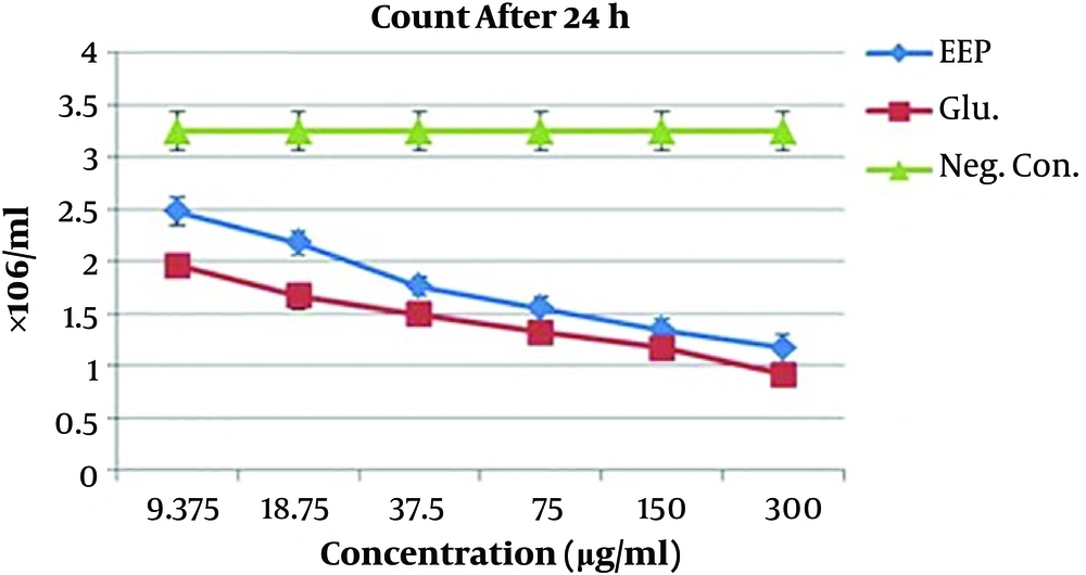
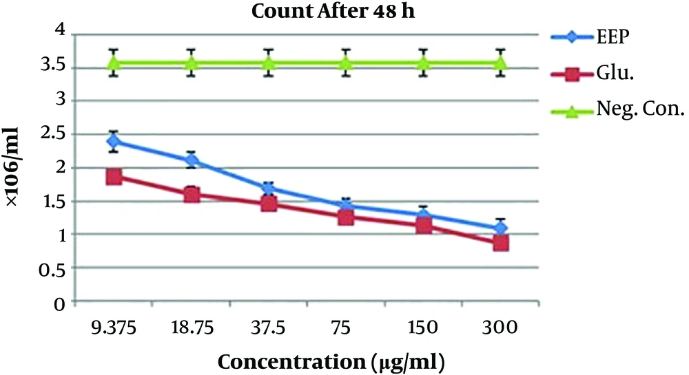
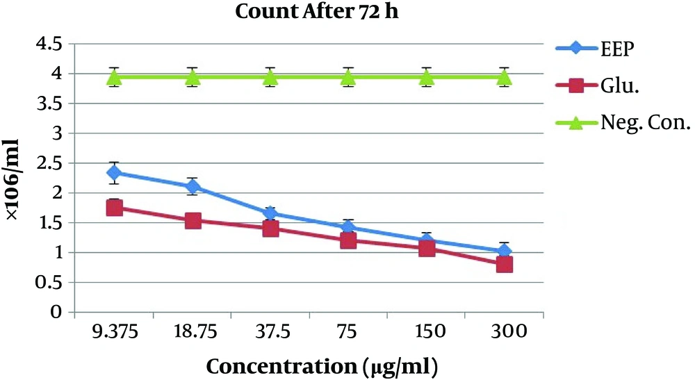
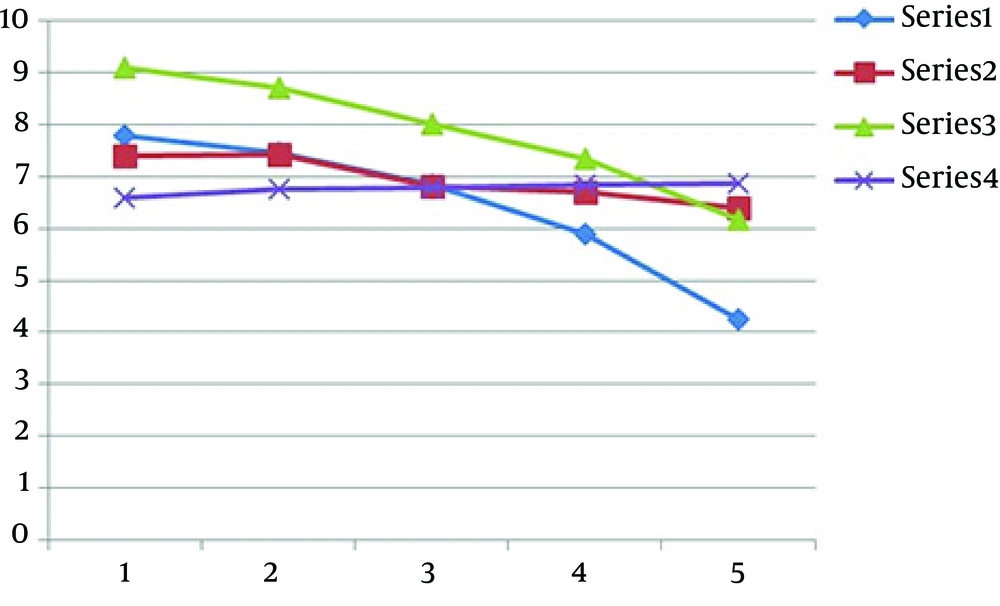
Figures 1-3 show the mean numbers of promastigotes after 24, 48, and 72 h of exposure to different concentrations of EEP and Glucantime (positive control), besides negative control counts. The counts were 1.03 and 0.82 at 300 μg/mL concentration of EEP and Glucantime, respectively, after 72 h (Figure 1). The t test showed significant differences between the Glucantime and EEP groups at 9.375 and 18.75 µg/mL concentrations but there were no significant differences between the Glucantime and EEP groups at 37.5, 75, 150, and 300 µg/ml concentrations. Besides, both EEP and Glucantime groups were significantly different from the negative control group (Figures 1-3).
4.2. MMT Test
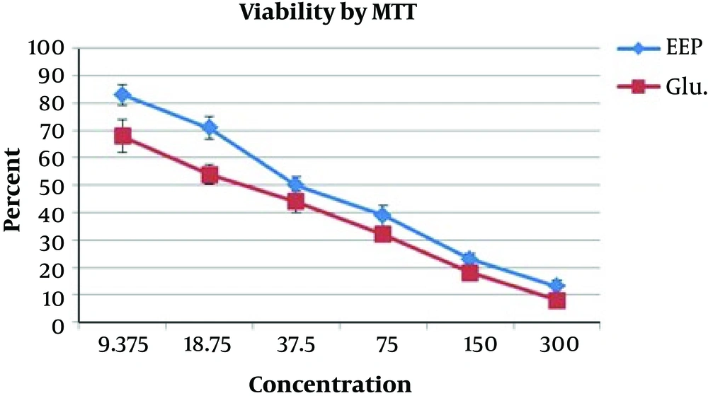
The mean ± SD values of the viability of parasites exposed to EEP and Glucantime were 13 and 8, respectively, at 300 μg/mL after 72 h (Figure 4). According to the t test, the differences of groups were not significant at 37.5, 75, 150, and 300 µg/mL concentrations (Figure 4).
4.3. In Vivo Model
The mean sizes of ulcers in different groups at different times are shown in Figure 5. By using the one-way-ANOVA, the treatment of ulcers was similar with EEP 4% (experimental group) and Glucantime (positive control group) and there was no significant difference between these two groups; but these drugs both were significantly more effective than EEP 1% in the experimental group and Vaseline Eucerin in the negative control group. However, EEP 1% decreased the size of ulcers more effectively than Vaseline Eucerin did in the negative control group but not significantly (Figure 5).
5. Discussion
Based on the lack of reliable standard drugs in the treatment of CL, the current study was designed to assess the effect of EEP against Leishmania major in vitro and in vivo. The results of the in vitro model showed the decreased number of promastigotes by EEP that was similar to Glucantime (as a standard drug) at the concentration of 300 µg/mL. This is confirmed by other studies (8-10). Machado et al. (9) demonstrated that the antileishmanial effects of propolis were associated with flavonoids and derivatives of caffeic acid compounds; amyrins (phenolic compounds) were also probably involved in the leishmanicidal activity (8, 9, 11), which is consistent with a study by Ayres et al. (8). Besides, it was reported that Korean propolis induced macrophages by producing interleukin-1, tumor necrosis factor-α, and nitric oxide; macrophages are able to control Leishmania amazonensis inside itself (8, 9). The study by Reboucas-Silva et al. demonstrated that propolis induced-inflammatory imbalance involving cytokines and oxidative response which are the characteristic symptoms of leishmaniasis (10).
The in vivo model showed the decreased size of ulcers by both EEP4% and Glucantime more significantly than the negative control and EEP1% (P < 0.05). It has been confirmed by other studies (4, 10). Similar to the present study, a study by Nilforoushzadeh et al. (4) found that propolis had high efficacy against leishmania in vivo but there were some differences between these studies, including the lack of negative control, the method of analysis, the propolis concentration, the different durations of using the standard drug and propolis, and the lack of in vitro model. Moreover, unlike the current study, Nilforoushzadeh et al. demonstrated the effectiveness of Glucantime as a standard drug (4).
On the contrary, a study by Ayres et al. (12) showed that the Brazilian red propolis gel (propane) and Glucantime alone were not effective against L. amazonensis but red propolis in combination with the Glucantime showed the potential value for treatment of cutaneous leishmaniasis; the reasons for this property were not known and further studies were encouraged. It is probable that the different species of Leishmania and propolis types may lead to different results.
The essential principle compounds responsible for biological activities of propolis are polyphenols, aromatic acids, diterpenic acids, flavonoids, and phenolic esters that play important roles against Leishmania (4, 7, 13). Leishmania causes high inflammatory and propolis has antioxidant and anti-inflammatory properties that may lead to healing wounds such as cutaneous leishmaniasis (7, 13-15). Indeed, different activities of propolis including enhancing the expression of collagens Type I and III (16, 17) and IFNγ (16), influencing the elimination or reduction of fibrosis development (17), decreasing the release of fibronectin (18), forming biocellulose membranes (10), stimulating natural killer cells, and influencing the release of nitric acid and tumor necrosis factors from macrophages (4) have been reported to help the tissue repair or heal cutaneous leishmaniasis.
5.1. Conclusion
Propolis as an herbal drug could decrease the number of promastigotes of Leishmania major in vitro and the size of scars in cutaneous leishmaniasis in vivo model similar to the standard drug. We suggest using it for complementary treatment.

