Alteration of Serum Acetylcholinesterase Activity in Patients with Gastroesophageal Reflux

authors:

avatar Mohammad-Reza Mirzaii-Dizgah ORCID 1 , avatar Iraj Mirzaii-Dizgah ORCID 2 , * , avatar Marjan Mokhtare ORCID 3

Student Research Committee, School of Medicine, Iran University of Medical Sciences, Tehran, Iran
Department of Physiology, School of Medicine, AJA University of Medical Sciences, Tehran, Iran
Department of Internal Medicine, Colorectal Research Center, Hazrat-e Rasool General Hospital, School of Medicine, Iran University of Medical Sciences, Tehran, Iran

how to cite: Mirzaii-Dizgah M, Mirzaii-Dizgah I, Mokhtare M. Alteration of Serum Acetylcholinesterase Activity in Patients with Gastroesophageal Reflux. Ann Mil Health Sci Res. 2022;20(2):e130616. https://doi.org/10.5812/amh-130616.

Abstract

Background:

Gastroesophageal reflux disease (GERD) is one of the most common gastrointestinal diseases. One of the factors that are known to play a role in reducing inflammation is acetylcholine (Ach), which decreases cholinesterase to increase Ach. The present study investigated cholinesterase activity in the serum of patients with GERD.

Methods:

In this cross-sectional study, 30 patients with GERD were referred to Imam Reza Hospital, and 30 healthy individuals were included in the study. Serum was collected in the morning and the fasting state. Cholinesterase activity was measured photometrically and analyzed by unpaired student t-test and receiver operating characteristic (ROC) curve analysis.

Results:

The mean serum cholinesterase activity was significantly lower in the GERD group (5,445 ± 411 IU/L) than in healthy individuals (6,728 ± 305 IU/L). The cut-off for the detection of patients suffering from GERD by the serum activity of cholinesterase was 5,637 IU/L.

Conclusions:

It seems that the cholinesterase activity reduces in the serum of patients with GERD.

1. Background

Gastroesophageal reflux disease (GERD) is one of the most common diseases of the digestive system, which is caused by the dysfunction of the lower esophageal sphincter and also by causing a disturbance in the clearing of stomach acid (1, 2). This disease is especially common in developed countries. Its prevalence is 18 - 27% in North America, 8 - 25% in Europe, 23% in South America, 11% in Australia, and 7 - 2% in East Asia (3).

Compared to other Asian countries, it seems that GERD is more common in Iran, and its prevalence is similar to Western countries. The prevalence of 21.2% in a study in Tehran is the best estimate for reflux in Iran (1). The estimate of 21.5% in one study, and 25.5% in one study in Shahrekord, can indicate the high prevalence of the disease in Iran. The annual prevalence rate in one of the studies is estimated to be more than 85% (4). This high prevalence affects not only the quality of life but also the economy. The financial burden of GERD in the United States of America may directly reach 9 - 10 billion dollars annually. This amount is apart from the indirect costs that are imposed on society due to the reduction of productivity in daily work (3).

Gastroesophageal reflux disease is a disorder in which the reflex passage of stomach contents into the esophagus leads to signs such as stomachache, chest pain, hoarseness, asthma, painful throat, globular feeling, and tooth decay (5). The frequent presence of gastric contents with pH < 1 in the mouth may create a persistent acidic situation that can harm the mouth's solid and soft tissues. A sufficient amount and quality of salivary secretions are necessary to protect the teeth and mucous membranes of the mouth, pharynx, and esophagus (5).

In the pathophysiology of GERD, dysfunction of various mechanisms known as the "anti-reflux barrier" occurs, one of the most obvious of which is the dysfunction of the autonomic nervous system, especially the parasympathetic nerves, so that parasympathetic activity is impaired in all patients with GERD (6, 7). Inflammation motivates releasing of acetylcholine (Ach) from the parasympathetic end. Ach can suppress inflammation in both the central nervous system (CNS) and peripheral tissues. Cholinesterases include a group of enzymes that hydrolyze acetylcholine and other choline esters. Cholinesterase activity is reduced in various chronic and severe inflammatory diseases (8-12).

Today, although the ambulatory pH monitoring method is the diagnostic gold standard, its sensitivity for esophageal reflux is lower than for non-esophageal reflux. Studies have shown that there are day-to-day changes in pH meter results, with one patient having a regular test on one day and an abnormal test on another day. Finally, none of the above diagnostic methods have been proven as a reliable tool for GERD, and the search for more accurate diagnostic methods is necessary (13, 14).

2. Methods

In a cross-sectional study, 30 patients with GERD referred to the gastroenterology clinic of Imam Reza Hospital who had a return of stomach contents to the mouth and pharynx and grade B or higher esophagitis based on the Los Angeles classification and 30 healthy individuals selected from the people who visited the hospital for annual monitoring and were healthy were included in the study. Two milliliters of venous blood were taken from all the participants and poured into the gel clot tube. Clotted blood was centrifuged at 5,000 rpm for ten minutes. The samples were stored at -70°C. The cholinesterase activity kit was purchased from Pars Azmoun (Karaj, Iran). Photometric measurement was done according to the manufacturer’s instructions. Samples were assayed at least twice. The data were analyzed in SPSS software version 25 with the unpaired student t-test and receiver operating characteristic (ROC) curve analysis.

3. Results

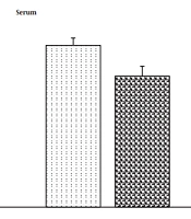
The average serum cholinesterase activity in the GERD group was lesser than that of healthy subjects (P = 0.001) (Figure 1).

Mean serum cholinesterase activity in patients with gastroesophageal reflux disease (GERD) and healthy subjects. Data are stated as mean ± SEM. *: Indicates P < 0.05.

The results of ROC curve analysis showed that the evaluation of cholinesterase activity in serum could be used to distinguish patients with GERD from healthy individuals. The serum cholinesterase activity cut-off point for distinguishing patients with GERD from healthy people was 5,637 IU/L with 61% sensitivity and 54% specificity (P = 0.027).

4. Discussion

Gastroesophageal reflux disease is one of the most public digestive diseases, and its prevalence is relatively high in Iran. Its economic burden is high, so it is estimated that Americans spend more than 10 billion dollars per year on proton pump inhibitors (PPIs) for its treatment (15). The reduction of the quality of life and the occurrence of many problems such as absenteeism, reduction of leisure time programs, and increase of problems related to doing household chores are among the problems of patients with GERD. However, most cases of GERD (79.2%) are non-complicated (16).

Today, there are various methods for diagnosing this disease, some of which do not have sufficient sensitivity and specificity. Therefore there is still no gold standard diagnostic test for GERD, and the diagnosis is based on a combination of PPI tests, ambulatory pH monitoring, and esophagogastroduodenoscopy (EGD). Other methods, such as barium esophagogram, high-resolution esophageal manometry, and gastric emptying studies can help rule out other diseases that may contribute to or mimic the signs of GERD (17). Most of these diagnostic methods are expensive. Therefore, finding a solution to diagnose this cost-effective disease seems necessary. In the pathophysiology of GERD, dysfunction of various mechanisms known as the "anti-reflux barrier" occurs, one of the most obvious of which is the dysfunction of the autonomic nervous system, especially the parasympathetic nerves, so that parasympathetic activity is impaired in all patients (6, 7). Inflammation motivates the “cholinergic anti-inflammatory pathway,” which motivates the release of Ach (18). Ach can suppress inflammation in both CNS and peripheral tissues (19). Acetylcholine affects macrophages through the α7 nicotinic receptor, which results in stopping the NF-κB nuclear transmission signaling pathway, which leads to a decrease in the making of pro-inflammatory cytokines. In this way, the cholinergic system with a reflex mechanism can quickly recover inflammation by getting evidence from various body parts (11, 18).

Cholinesterase activity is reduced in various chronic and severe inflammatory diseases (9-13). Since cholinesterases are made in the liver, their amount decreases in liver patients and malnutrition (19-21). Also, cholinesterase activity in acquired immunodeficiency syndrome (AIDS) (22), burns (20), in patients with gingivitis and periodontitis (23), cancer (24), brain damage (25), ischemic stroke (26), anorexia nervosa (27), Alzheimer’s disease (28) and multiple sclerosis (MS) (29) is reduced. Regulating the systemic level of Ach requires continuous control of the balance between the production of Ach by the vagus nerve and its hydrolysis by acetylcholinesterase and butyrylcholinesterase. Since the serum levels of cholinesterase reflect fluctuations in the body’s total capacity to hydrolyze Ach; therefore, the reduction in the serum level of cholinesterase is related to the downregulation of cholinesterase activity as a compensatory reaction of the body to increase the anti-inflammatory activity of acetylcholine (18).

The acid and pepsin contents of the stomach can cause damage to the esophagus and mouth and then lead to an increase in inflammatory factors and the occurrence of inflammation. Since the level of cholinesterase activity decreases in inflammatory diseases, it can justify the decrease in the serum level of cholinesterase activity in patients with GERD. The results showed that the cut-off point of cholinesterase activity in serum was significant for distinguishing GERD. Therefore, assessing cholinesterase activity may be helpful in the diagnosis of GERD.

4.1. Conclusions

It seems that the amount of cholinesterase activity is reduced in the serum of patients with GERD.

References

  • 1.

    Delavari A, Moradi G, Elahi E, Moradi-Lakeh M. Gastroesophageal reflux disease burden in Iran. Arch Iran Med. 2015;18(2):85-8. [PubMed ID: 25644795].

  • 2.

    Yandrapu H, Marcinkiewicz M, Poplawski C, Han K, Zbroch T, Goldin G, et al. Role of saliva in esophageal defense: implications in patients with nonerosive reflux disease. Am J Med Sci. 2015;349(5):385-91. [PubMed ID: 25789686]. [PubMed Central ID: PMC4418785]. https://doi.org/10.1097/MAJ.0000000000000443.

  • 3.

    Menezes MA, Herbella FAM. Pathophysiology of Gastroesophageal Reflux Disease. World J Surg. 2017;41(7):1666-71. [PubMed ID: 28258452]. https://doi.org/10.1007/s00268-017-3952-4.

  • 4.

    Delavari A, Moradi G, Birjandi F, Elahi E, Saberifiroozi M. The Prevalence of Gastroesophageal Reflux Disease (GERD) in the Islamic Republic of Iran: A Systematic Review. Middle East J Dig Dis. 2012;4(1):5-15. [PubMed ID: 24829629]. [PubMed Central ID: PMC4017699].

  • 5.

    Sujatha S, Jalihal U, Devi Y, Rakesh N, Chauhan P, Sharma S. Oral pH in gastroesophageal reflux disease. Indian J Gastroenterol. 2016;35(3):186-9. [PubMed ID: 27211893]. https://doi.org/10.1007/s12664-016-0659-7.

  • 6.

    Blaut U, Dobrek L, Laskiewicz J, Thor PJ. [Disturbances of the autonomic nervous system in gastroesophageal reflux disease]. Folia Med Cracov. 2001;42(1-2):63-73. Polish. [PubMed ID: 11712327].

  • 7.

    Dobrek L, Nowakowski M, Mazur M, Herman RM, Thor PJ. Disturbances of the parasympathetic branch of the autonomic nervous system in patients with gastroesophageal reflux disease (GERD) estimated by short-term heart rate variability recordings. J Physiol Pharmacol. 2004;55 Suppl 2:77-90. [PubMed ID: 15608363].

  • 8.

    Jin QH, He XJ, Li TL, Chen HH. Predictive value of serum cholinesterase for the prognosis of aged patients with systemic inflammatory response syndrome. Chin Med J (Engl). 2011;124(17):2692-5. [PubMed ID: 22040426].

  • 9.

    Lampón N, Hermida-Cadahia EF, Riveiro A, Tutor J. Association between butyrylcholinesterase activity and low-grade systemic inflammation. Ann Hepatol. 2012;11(3):356-63. https://doi.org/10.1016/s1665-2681(19)30932-9.

  • 10.

    Bahloul M, Baccouch N, Chtara K, Turki M, Turki O, Hamida CB, et al. Value of Serum Cholinesterase Activity in the Diagnosis of Septic Shock Due to Bacterial Infections. J Intensive Care Med. 2017;32(5):346-52. [PubMed ID: 26951579]. https://doi.org/10.1177/0885066616636549.

  • 11.

    Zivkovic AR, Tourelle KM, Brenner T, Weigand MA, Hofer S, Schmidt K. Reduced serum cholinesterase activity indicates splenic modulation of the sterile inflammation. J Surg Res. 2017;220:275-83. [PubMed ID: 29180192]. https://doi.org/10.1016/j.jss.2017.07.024.

  • 12.

    Peng ZL, Huang LW, Yin J, Zhang KN, Xiao K, Qing GZ. Association between early serum cholinesterase activity and 30-day mortality in sepsis-3 patients: A retrospective cohort study. PLoS One. 2018;13(8). e0203128. [PubMed ID: 30161257]. [PubMed Central ID: PMC6117034]. https://doi.org/10.1371/journal.pone.0203128.

  • 13.

    Hayat JO, Gabieta-Somnez S, Yazaki E, Kang JY, Woodcock A, Dettmar P, et al. Pepsin in saliva for the diagnosis of gastro-oesophageal reflux disease. Gut. 2015;64(3):373-80. [PubMed ID: 24812000]. https://doi.org/10.1136/gutjnl-2014-307049.

  • 14.

    Du X, Wang F, Hu Z, Wu J, Wang Z, Yan C, et al. The diagnostic value of pepsin detection in saliva for gastro-esophageal reflux disease: a preliminary study from China. BMC Gastroenterol. 2017;17(1):107. [PubMed ID: 29041918]. [PubMed Central ID: PMC5645897]. https://doi.org/10.1186/s12876-017-0667-9.

  • 15.

    Shaheen NJ, Hansen RA, Morgan DR, Gangarosa LM, Ringel Y, Thiny MT, et al. The burden of gastrointestinal and liver diseases, 2006. Am J Gastroenterol. 2006;101(9):2128-38. [PubMed ID: 16848807]. https://doi.org/10.1111/j.1572-0241.2006.00723.x.

  • 16.

    Gong EJ, Choi KD, Jung HK, Youn YH, Min BH, Song KH, et al. Quality of life, patient satisfaction, and disease burden in patients with gastroesophageal reflux disease with or without laryngopharyngeal reflux symptoms. J Gastroenterol Hepatol. 2017;32(7):1336-40. [PubMed ID: 28052406]. https://doi.org/10.1111/jgh.13716.

  • 17.

    Chatila AT, Nguyen MTT, Krill T, Roark R, Bilal M, Reep G. Natural history, pathophysiology and evaluation of gastroesophageal reflux disease. Dis Mon. 2020;66(1):100848. [PubMed ID: 30803725]. https://doi.org/10.1016/j.disamonth.2019.02.001.

  • 18.

    Shao X, Yang L, Hu K, Shen R, Ye Q, Yuan X, et al. Serum Cholinesterases, a Novel Marker of Clinical Activity in Inflammatory Bowel Disease: A Retrospective Case-Control Study. Mediators Inflamm. 2020;2020:4694090. [PubMed ID: 32733165]. [PubMed Central ID: PMC7376425]. https://doi.org/10.1155/2020/4694090.

  • 19.

    Pavlov VA, Ochani M, Gallowitsch-Puerta M, Ochani K, Huston JM, Czura CJ, et al. Central muscarinic cholinergic regulation of the systemic inflammatory response during endotoxemia. Proc Natl Acad Sci U S A. 2006;103(13):5219-23. [PubMed ID: 16549778]. [PubMed Central ID: PMC1405626]. https://doi.org/10.1073/pnas.0600506103.

  • 20.

    Santarpia L, Grandone I, Contaldo F, Pasanisi F. Butyrylcholinesterase as a prognostic marker: a review of the literature. J Cachexia Sarcopenia Muscle. 2013;4(1):31-9. [PubMed ID: 22956442]. [PubMed Central ID: PMC3581611]. https://doi.org/10.1007/s13539-012-0083-5.

  • 21.

    Ogunkeye OO, Roluga AI. Serum cholinesterase activity helps to distinguish between liver disease and non-liver disease aberration in liver function tests. Pathophysiology. 2006;13(2):91-3. [PubMed ID: 16530396]. https://doi.org/10.1016/j.pathophys.2006.01.002.

  • 22.

    Ott M, Wegner A, Caspary WF, Lembcke B. Intestinal absorption and malnutrition in patients with the acquired immunodeficiency syndrome (AIDS). Z Gastroenterol. 1993;31(11):661-5. [PubMed ID: 8291278].

  • 23.

    Apatzidou DA, Iskas A, Konstantinidis A, Alghamdi AM, Tumelty M, Lappin DF, et al. Clinical associations between acetylcholine levels and cholinesterase activity in saliva and gingival crevicular fluid and periodontal diseases. J Clin Periodontol. 2018;45(10):1173-83. [PubMed ID: 30022504]. https://doi.org/10.1111/jcpe.12989.

  • 24.

    Mitsunaga S, Kinoshita T, Hasebe T, Nakagohri T, Konishi M, Takahashi S, et al. Low serum level of cholinesterase at recurrence of pancreatic cancer is a poor prognostic factor and relates to systemic disorder and nerve plexus invasion. Pancreas. 2008;36(3):241-8. [PubMed ID: 18362836]. https://doi.org/10.1097/MPA.0b013e31815b6b2b.

  • 25.

    Zhang QH, Li AM, He SL, Yao XD, Zhu J, Zhang ZW, et al. Serum Total Cholinesterase Activity on Admission Is Associated with Disease Severity and Outcome in Patients with Traumatic Brain Injury. PLoS One. 2015;10(6). e0129082. [PubMed ID: 26107885]. [PubMed Central ID: PMC4479571]. https://doi.org/10.1371/journal.pone.0129082.

  • 26.

    Ben Assayag E, Shenhar-Tsarfaty S, Ofek K, Soreq L, Bova I, Shopin L, et al. Serum cholinesterase activities distinguish between stroke patients and controls and predict 12-month mortality. Mol Med. 2010;16(7-8):278-86. [PubMed ID: 20464061]. [PubMed Central ID: PMC2896466]. https://doi.org/10.2119/molmed.2010.00015.

  • 27.

    De Caprio C, Alfano A, Senatore I, Zarrella L, Pasanisi F, Contaldo F. Severe acute liver damage in anorexia nervosa: two case reports. Nutrition. 2006;22(5):572-5. [PubMed ID: 16600819]. https://doi.org/10.1016/j.nut.2006.01.003.

  • 28.

    Hosoi M, Hori K, Konishi K, Tani M, Tomioka H, Kitajima Y, et al. Plasma Cholinesterase Activity in Alzheimer's Disease. Neurodegener Dis. 2015;15(3):188-90. [PubMed ID: 26138498]. https://doi.org/10.1159/000381532.

  • 29.

    Polachini CR, Spanevello RM, Casali EA, Zanini D, Pereira LB, Martins CC, et al. Alterations in the cholinesterase and adenosine deaminase activities and inflammation biomarker levels in patients with multiple sclerosis. Neuroscience. 2014;266:266-74. [PubMed ID: 24508813]. https://doi.org/10.1016/j.neuroscience.2014.01.048.