


1. Background
Identification means recognizing human personality and individuality, in other words, recognizing the sum of signs and effects that distinguish one person from others (1). Gender determination in the adult skeleton is usually the first step in the identification process, as subsequent methods for determining age and height depend on gender estimation. Confidence in correctly determining gender depends on the completeness of the remains and the degree of intrinsic gender difference in a population (2). The skull and pelvis are widely used as the most accurate remains to determine gender (3). Previous studies have focused on using different human skeleton parts, including the humerus, radius, ulna, femur, tibia, talus, ribs, and metatarsals (4). After development, the morphological changes of the scapula bone are usually insignificant during life (5, 6). Also, the processes of fossilization, decomposition, and feeding of carnivores cause damage to bones such as the pelvis and skull, but usually, the scapula is less damaged (7). However, the scapula, part of the short and wide bones, is not as important as other bones in determining gender (4).
2. Objectives
Based on mentioned issues, various studies have been performed on the dimorphic characteristics of scapula bone in different populations. At the time of writing, only one study had been conducted on this bone in the Iranian population (8). Therefore, this study evaluated the accuracy of scapula metric indices to determine gender in the Iranian population. This study aimed to investigate and locate the anthropometric indices of scapula bone in the Iranian population to determine gender.
3. Methods
This study was based on reconstructed CT scan images of 259 Iranian adults aged 18 to 90 years referred to the Imaging Center of Besat Hospital in Tehran between January 2020 and January 2021 for chest imaging for any reason, including lung infection. Exclusion criteria were any trauma, congenital or acquired defect(s), and burn(s) in the thorax and back.
Four anthropometric indices were examined in the right scapula, including the mean maximum length of the scapula (MLS) (maximum distance between the highest point of the scapula and its lowest point), mean maximum width of the scapula (MWS), mean maximum length of the glenoid cavity (MLG) and mean maximum width of the glenoid cavity (MWG) (4, 7).
Imaging was performed with a GE-64 slice CT scanner (General Electric Company, United States), and images were evaluated in PACS software (version 3.7.3.7). To prevent measurement errors, they were performed by two persons independently. Data were analyzed using SPSS 22 software. The Kolmogorov-Smirnov test was used to determine the normality of quantitative variables. Parametric tests were used for variables with a normal distribution, and non-parametric tests were used for variables without a normal distribution. The ROC curve was also used to assess the differentiating ability of the four scapula indices in gender differentiation in the studied population. Using the ROC curve and the data results, the cutoff point was determined, based on which the sensitivity and specificity of each indicator in determining gender were clarified. Youden's J statistics were used to determine the optimal cutoff and evaluate the accuracy of the obtained differentiation points, which are obtained through the below formulation:
J = sensitivity + specificity - 1
Based on this index, the optimal shear point will be whenever the sum of its sensitivity and specificity is the highest (data that has the highest "sensitivity" and the lowest value for "characteristic-1").
In all analyses, a significant level of less than 0.05 was considered.
4. Results
In this study, 259 individuals were studied by available methods, including 126 men (48.6%) and 133 women (51.4%) (Table 1).
| Variables and Categories | Frequency (%) |
|---|---|
| Gender | |
| Female | 133 (51.4) |
| Male | 126 (48.6) |
| Age | |
| 30 > | 64 (24.7) |
| 30 - 50 | 129 (49.8) |
| 50 < | 66 (25.5) |
Demographic Characteristics of Study Subjects
The mean age of the subjects was 41.21± 15.4, with a minimum of 18 and a maximum of 90 years.
The mean measured values of MLS and MWS indices were 145.00 and 93.41 in men and 129.51 and 82.17 in women, respectively. Also, the average MLG and MWG indices were reported to be 32.23 and 24.71 in men and 30.03 and 22.70 in women, respectively.
The differences between max length glenoid, max width glenoid, max width scapula, and max length scapula in two groups of men and women are presented in Table 2.
| Index and Gender | Mean ± SD | Max | Min | P-Value |
|---|---|---|---|---|
| Max width glenoid | ≤ 0.001 | |||
| Male | 24.71 ± 3.1 | 32.6 | 19.0 | |
| Female | 22.71 ± 1.9 | 31.5 | 19.2 | |
| Max length glenoid | ≤ 0.001 | |||
| Male | 32.23 ± 3.9 | 40.5 | 20.9 | |
| Female | 30.04 ± 3.7 | 40.6 | 19.5 | |
| Max width scapula | ≤ 0.001 | |||
| Male | 93.41 ± 8.7 | 116.8 | 68.0 | |
| Female | 82.17 ± 8.0 | 103.9 | 66.6 | |
| Max length scapula | ≤ 0.001 | |||
| Male | 145.01 ± 8.9 | 163.7 | 124.8 | |
| Female | 129.51 ± 9.0 | 156.5 | 103.9 |
Comparison of Structural Features of Scapula in Men and Women
The ROC curve was used to evaluate the differentiation power of four scapula indices in gender differentiation in the studied population.
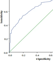
The area under the curve for the max length of scapula index in the whole population for gender differentiation was 88.3% with a confidence interval of 84.3% to 92.3%. (P ≤ 0.001) (Figure 1).

ROC curve for max length of scapula index
The area under the curve for the max width of the scapula index in the whole population for gender differentiation was 82.7% with a confidence interval of 77.7 to 87.6%. (P ≤ 0.001) (Figure 2).

ROC curve for max width of scapula index
The area under the curve for the max length of glenoid index in the whole population for gender differentiation was 66.3%, with a confidence interval of 59.7 to 72.9%. (P ≤ 0.001) (Figure 3).

ROC curve for the max length of glenoid index
The area under the curve for the max width of the glenoid index in the whole population for gender differentiation was 69.8% with a confidence interval of 63.3 to 76.3%. (P ≤ 0.001) (Figure 4).

ROC curve for the max width of glenoid index
To obtain the sensitivity and specificity of the four indicators for differentiation between two genders, the demarcation point (DP) was determined using the ROC curve, and based on this, the following results were obtained (Table 3):
| Index | Demarking Point (mm) | Sensitivity (%) | Specificity (%) | Accuracy (%) | Youden's J Statistics |
|---|---|---|---|---|---|
| MLS | 139.60 | 71.4 | 90.2 | 80.8 | 0.616 |
| MWS | 89.35 | 71.4 | 78.9 | 75.1 | 0.503 |
| MLG | 31.25 | 65.1 | 61.7 | 63.4 | 0.268 |
| MWG | 24.05 | 54 | 80.5 | 67.2 | 0.345 |
Points of Distinction and Their Sensitivity and Specificity
5. Discussion
Among the most important pieces of information that can be obtained from bones is determining gender. Although non-metric skeletal measurements were used to determine gender initially in the literature, scientific metric measurements were later used. Metric measurements are preferred because of their easy reproducibility, high accuracy, and lack of special skills (9). Previous studies have shown that populations differ in size and proportion, which can affect gender assessment (10). Studies in the Chinese population show that using scapula bone for sex determination is reliable (11), while the results of a study in the Egyptian population were not (12). According to conventional morphometric studies of the scapula, the human scapula exhibits sexual dimorphism concerning its size (13). Although it is possible to determine sex accurately and quickly through the skeleton, it is not always possible to find all skeletal bones at one time in forensic medicine; Hence it is possible to obtain maximum information from each bone and extract information such as age and gender of the owner at the time of analysis.
The results of the present study showed that the mean size of scapula anthropometric indices, including MLS, MWS, MLG, and MWG, is significantly higher in men than women, which is in complete accordance with other similar studies in other populations. This result was similar to the result obtained in the study of Tripathi (14-16).
In this study, the highest accuracy in determining gender is for the MLS index, with 88.9%, and the lowest accuracy is related to the MLG, with 66.3%. In Ozer et al. (17), Torimitsu et al. (4), and Papaioannou et al.'s (1) study, the highest accuracy in determining gender was related to MLS and MWS. In our study, the highest accuracy in determining gender was equal to 88.3% and 82.7%, respectively, to mentioned indicators. In these studies, the accuracy of the MWG index is about 88%; however, in our study, it had a different result, as much as 69.8%. Another point is about the MLG index, which in the study of Papaioannou et al. (1) and Ozer et al. (17) had a high accuracy of about 88% in determining gender; but in our study and Torimitsu et al., the lowest accuracy was related to this index (4). This indicates, to some extent, that this index is not a good criterion for gender differentiation in our studied population and is less accurate than other indicators.
The mean sizes obtained in this study are different from previous studies and, in almost all cases, are less than them, indicating structural differences in the scapula of different populations. Comparison of the results of our study with other studies conducted among residents of different regions showed that there is a statistically significant difference in the values of the four indicators studied between different ethnicities (different countries), which can be due to various reasons such as racial differences, genetics, and environment, job stress, and growth hormone levels, etc. (11). However, the value of each of these indicators in different studies conducted in other countries is higher in the male population than females, and in this respect, there is a special similarity between all these studies; in fact, there was a statistically significant difference in the measured values between men and women of all races, which in our study is similar to previous studies.
The results of this study were gained and collected by comparing anthropometric indices of the scapula in reconstructed CT scan images, so it is recommended for comparative comparison, simultaneous research on radiographic stereotypes, and direct measurement of the mentioned indices on bone. Since this research has been done on adults, in order to compare it and its values in gender differentiation, it is recommended to do studies before puberty. Considering that this research has been conducted on different Iranians living in Tehran, it seems that its results can be generalized to other parts of the country. However, it is recommended that similar studies should be conducted in other parts of the country. Similar to other studies on scapula anthropometric indices, all these values are higher in Iranian men than women, and due to the significant difference between genders with acceptable sensitivity and specificity, it helps to identify genders as well. Therefore, if mutilated remains or incomplete bone fragments, including the scapula, are present due to natural and unnatural events, it is possible to predict a gender by preparing a CT scan graph and measuring its anthropometric indices with a high percentage. Because the scapula bone usually does not undergo significant morphological changes during a person's lifetime after evolution, and according to this study conducted on graphs, if there is a need to specifically identify an individual, it will be possible to match graphs before death and graphs taken from skeletal remains, which are simple and accessible to do, and the result is largely reliable. Finally, based on the results obtained in this study, the accuracy of MLS and MWS indices to determine gender is high and is very helpful in determining gender. However, the two indices, MLG and MWG, have lower accuracy in determining gender in the Iranian population, and dimorphic characteristics of the scapula bone are shown to a lesser extent.
