1. Introduction
Lipoid proteinosis (LP), also known as Urbach-Wiethe disease, is a rare inherited metabolic disorder that follows an autosomal recessive pattern of inheritance. Lipoid proteinosis hallmark feature is the deposition of glycoprotein, forming an amorphous hyaline-like substance in various tissues, such as the skin, mucous membranes, salivary glands, and internal organs (1, 2). Characteristic histopathological findings include melanin incontinence and hyperkeratosis in the basal layer. Additionally, periodic acid‐Schiff (PAS)-positive hyaline deposits are typically found in the upper dermis, often localized around blood vessels and sweat glands (3, 4).
In many affected individuals, the first clinical manifestation is a hoarse or completely voiceless condition, which can become apparent either at birth or later in life (2, 5). Clinical manifestations typically commence during childhood and progress with age. Skin manifestations encompass acne, inflammation, scarring, and ulcers. The presence of amorphous hyaline deposits in these ulcerated areas results in a thick, waxy skin texture. Furthermore, the ulcers tend to leave atrophic scars resembling chickenpox marks (6). Lipoid proteinosis predominantly affects the oral cavity (7), with nearly all oral tissues exhibiting infiltrated white plaques and chickpea-sized ulcers. These ulcers often appear before adolescence and gradually increase in size (8).
Macroglossia, characterized by a firm, wood-like consistency of the tongue tissue and difficulty protruding the tongue, is a common feature of LP (8). Approximately half of LP patients display rows of yellowish papules around all four eyelids (9). Despite LP being a sporadic disease, it occurs globally, with approximately four hundred reported cases (6). Lipoid proteinosis is a slowly progressing disorder, and although it follows a benign course, extensive skin lesions, dry mouth, and hoarseness can lead to severe psychosocial complications. This study presents a case report detailing the oral and skin manifestations of this syndrome in a 36-year-old female, along with a brief review.
2. Case Presentation
A 36-year-old female case who had been diagnosed with LP and confirmed through genetic testing sought evaluation at the Department of Oral and Maxillofacial Medicine, Faculty of Dentistry, Tehran University of Medical Sciences, Tehran, Iran, on October 31, 2023. The case presented with complaints of dry mouth and difficulty in speaking and swallowing. She expressed the need to drink water frequently to moisten her mouth. Her xerostomia had started approximately 10 years ago and had progressively worsened, significantly impacting her ability to communicate and sleep. She reported that her xerostomia was particularly severe at night, often disrupting her sleep. In her medical history, it was noted that she had been taking vitamin D, calcium, and the medication “Prolia” (denosumab). The patient also reported that her teeth had severely decayed around 1.5 years ago, ultimately leading to the extraction of all her teeth.
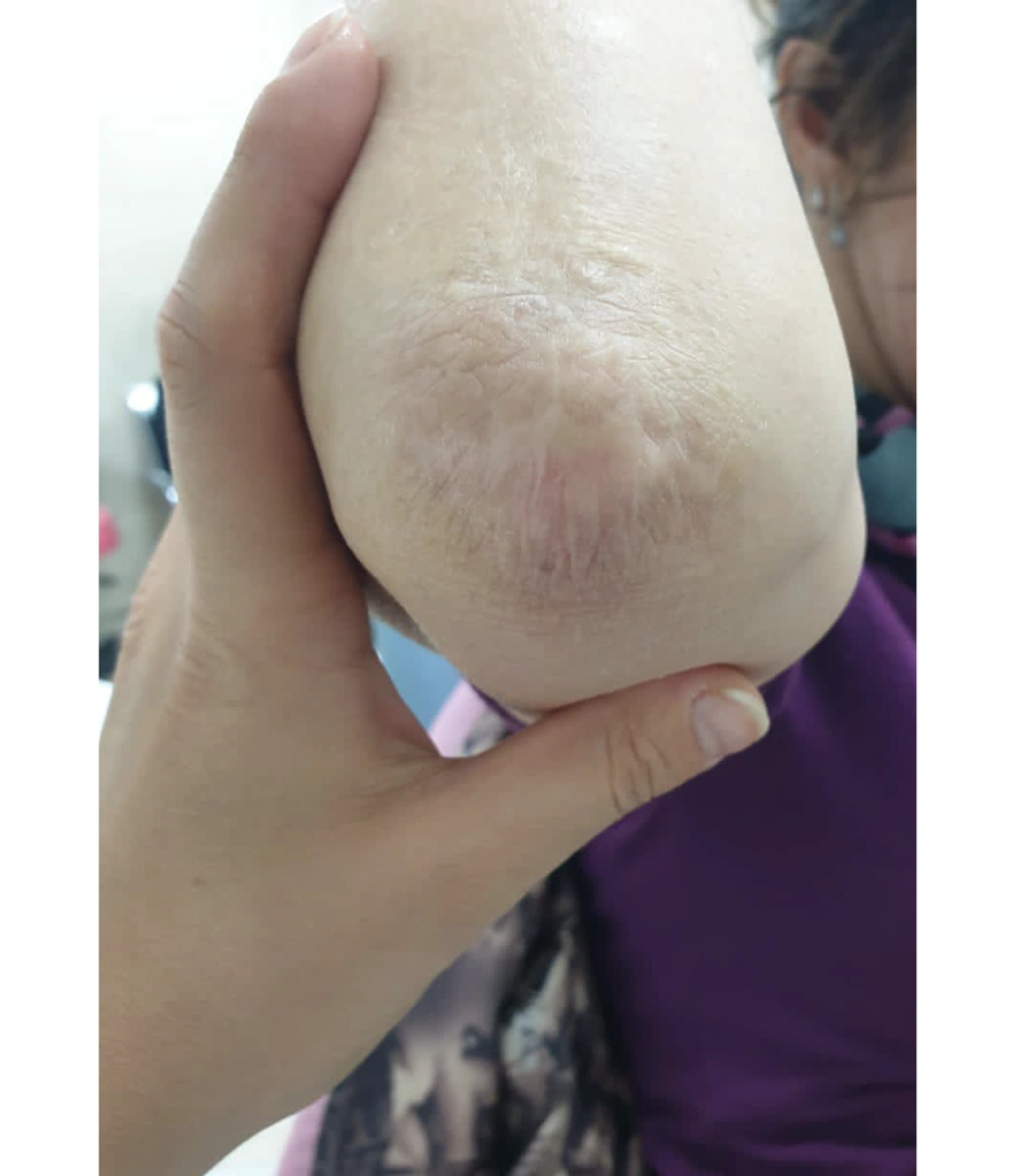
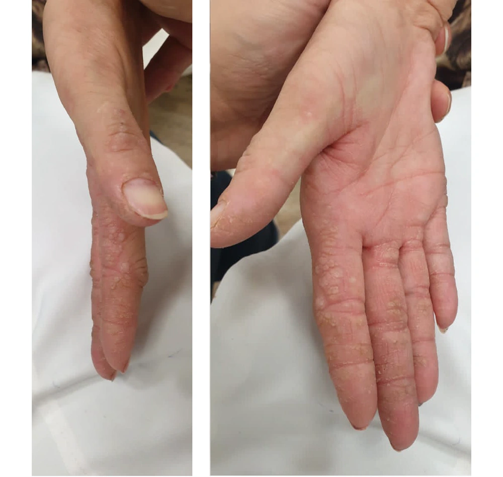
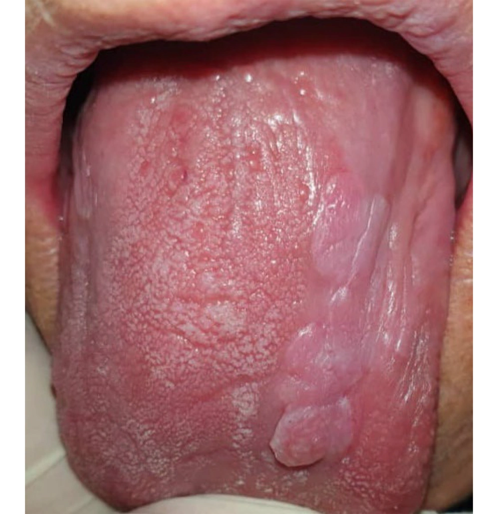
Upon general examination, several findings were observed, including voice hoarseness, hyperkeratosis on her elbows and hands (Figure 1), and multiple papules on her fingers (Figure 2). The oral and maxillofacial examination revealed limitations in mouth opening (with a maximal incisal mouth opening of 30 mm), along with firm, fibrotic, dry mucosa and multiple papules on the tongue. Her tongue appeared desiccated and depapillated (Figure 3).
The left parotid duct orifice could not be located, and dry mouth and hyposalivation were evident. Saliva collected from the patient appeared frothy and hazy, and the tongue blade test yielded a positive result. The patient did not exhibit any significant abnormalities in her eyelids or face. Furthermore, there were no signs or symptoms of temporomandibular joint disease, such as muscular tenderness, joint sounds, or pain, suggesting that reduced skin elasticity was the primary cause of her restricted mouth opening. There was no history of seizures, visual disturbances, or disorientation.
Regarding her family history, there were no reports of consanguineous marriages. The case hailed from a middle economic and social class and had no siblings. None of her family members displayed similar symptoms. To assess salivary flow rate, unstimulated whole saliva was collected, with the patient instructed to collect her saliva and spit it out every minute for a total of 5 minutes, yielding a flow rate of 0.2 mL/min. The patient was educated on the importance of sipping a mixture of water, one tablespoon of fresh lemon drops, and one teaspoon of olive oil throughout the day. She was also advised to use a room humidifier at night to stimulate her parotid and submandibular glands 4 - 5 times daily and to rinse her mouth with olive oil before bedtime. The patient was cautioned against consuming alcohol, sugar, or strongly flavored substances that could irritate her dry and sensitive mucosa. On a follow-up contact one month later, the patient reported an improvement in her symptoms.
We searched MEDLINE, Scopus, Web of Science, and Cochrane databases with the following keywords. ((((lipoid proteinosis[Title]) OR (lipoid proteinosis syndrome[Title])) OR (Urbach-wiethe syndrome[Title])) OR (Urbach-wiethe disease[Title])) OR (Hyalinosis cutis et mucosae[Title]))) AND (((oral manifestations[Title]) OR (oral[Title])) OR (oral manifestation[Title])). Table 1 shows a summary of LP oral manifestation reports.
| Authors (y) | Oral Manifestations | Case |
|---|---|---|
| Jahanimoghadam and Hasheminejad (2022) (3) | Restricted mouth opening | 10-year-old male |
| Teeth hypoplasia or aplasia | ||
| Lee et al. (2018) (10) | Pallor and fibrotic texture with induration of the lingual and buccal mucosa | 10-year-old Asian female |
| Maximal inter-incisal opening 20 mm | ||
| Loss of the dorsal tongue papillae | ||
| A thick sub-lingual frenum | ||
| Multiple atrophic papules extending from the soft and hard palates to the crypts of the tonsillar | ||
| Microdontia of the maxillary permanent premolars with congenitally missing teeth | ||
| Ravi Prakash et al. (2012) (11) | Firm, thickened, and pale tongue with large crenations with cobble stoning with yellow to white papules | 32-year-old male |
| Nodular and thickened mucosa with hyperkeratosis of the buccal, labial, and palatal mucosa | ||
| Aroni et al. (1998) (12) | White to yellow patches with micro-papules on the labial, dorsum of the lingual, faucal, and uvula mucosa | 37-year-old female |
| Thickened oral mucosa on palpation with inflamed and flabby appearance | ||
| Shortened and indurated lingual frenulum | ||
| The patient could not protrude her tongue | ||
| Ivory-colored depositions and bulging in the region of the sublingual gland | ||
| Enlarged gingiva | ||
| Xanthinaki et al. (2008) (13) | The oral mucosa appeared nodular, diffusely enlarged, and thickened due to infiltration with waxy-yellowish-white plaques and nodules. | 14-year-old male |
| Thickened, furrowed appearance of the skin with several skin scars, eyelid nodules, loss of eyelashes, and voice hoarseness | ||
| Hofer (1975) (14) | Lingual ulceration | 27-year-old female |
| Lingual stiffness with reduced mobility and depapillation | ||
| Partially everted lips and fissures at the corner of the mouth | ||
| Xerostomia | ||
| Agenesis of teeth 12 and 22 | ||
| Sabater-Abad et al. (2019) (15) | Well-defined ulcer with fibrin coating on the dorsum of the tongue | 50-year-old female |
| No dental anomalies | ||
| Difficulty when protruding the tongue | ||
| Tongue and lips were thickened and indurated | ||
| Finkelstein et al. (1982) (16) | Leathery, fibrotic, and thickened gingiva, lips, and tongue | 5-year-old white female |
| Kabre et al. (2015) (7) | Scrapable whitish patches on the bilateral buccal, floor of the mouth, and palate mucosa | 19-year-old male |
| Missing 4 second premolars | ||
| A super-numerary tooth between 16 and 14 | ||
| Kabre, V., et al. (2015) (7) | Lingual ulcerations | 11-year-old male |
| Voice Hoarseness started in infancy | ||
| Buccal mucosa ulcerations | ||
| Recurrent throat pain and dysphagia for the past 2 to 3 years | ||
| Anil et al. (1993) (17) | Inability to protrude the tongue, crenated borders | 18-year-old female |
| Irregular nodules on lower lips | ||
| Lips were thickened, firm, and sclerotic | ||
| Ulcerations on gingiva | ||
| Gingival hypertrophy | ||
| Deshpande et al. (2015) (18) | Crenations of an enlarged tongue | 28-year-old male |
| Sublingual frenum stiffness and reduced lingual movement | ||
| Yellow waxy papules in all of the mouth | ||
| Mahalingam et al.(2015) (19) | Crenations of an enlarged tongue | 61-year-old male |
| Sublingual frenum stiffness and reduced lingual movement | ||
| Yellow waxy papules on lips | ||
| Mittal et al. (2016) (20) | Sublingual frenum stiffness and reduced lingual movement | 6-year-old male |
| Vesicules and ulcers of the thickened lips mucosa | ||
| Jansen et al. (2016) (21) | Xerostomia | 22-year-old female |
| Destroyed dentition | ||
| Kartal et al. (2016) (22) | Sublingual frenum stiffness and reduced lingual movement | 42-year-old female |
| Burning sensation during mastication | ||
| Yellow and waxy papules on the lips |
3. Discussion
Lipoid proteinosis typically follows a slow and benign course and exhibits varying prevalence rates across different regions, with a higher incidence reported in Sweden and South Africa (12). The oral symptoms of this rare syndrome tend to manifest earlier than other skin-related signs (10). Therefore, raising awareness among dentists about the oral indicators of this syndrome can lead to early identification and effective mitigation of its impact. One of the initial telltale signs of this condition is the hoarse voice and weak crying of a child immediately after birth (14, 23, 24). Additional symptoms encompass the presence of papules on the skin, eyelids, elbows, and hands, along with hyperkeratosis and scarring (12, 25). Prominent oral signs include tongue firmness and the inability to protrude the tongue due to hyaline deposition in the tongue and its frenum. This results in a thick and rough tongue texture, along with the formation of palatine and infratemporal papules.
In contrast to the current case, no signs of tongue thickness, inability to protrude the tongue, or ankyloglossia were observed. Xerostomia, tooth agenesis, and excessive gingival growth are infrequent oral manifestations of this disease (26). Xerostomia has been previously reported in LP cases, as evidenced in the case described here. Notably, non-specific salivary gland inflammation has been observed in LP patients, emphasizing the importance of ruling out conditions such as sialadenitis and Sjögren’s syndrome. In the present case, the right parotid papilla could not be located, and the salivary gland flow rate was less than 1 mL in 5 minutes.
Similarly, previous reports have documented hyperkeratosis in specific body regions that are subject to continuous trauma, such as the hands (22, 27). Recently, Frenkel et al. conducted an extensive review of the oral manifestations in LP patients, analyzing 133 cases within 1948 and 2016. Frenkel et al. observed that the most commonly affected oral areas in this condition were the lingual (68%), the floor of the mouth (56%), the labial (43%), the buccal mucosa (40%), the palate (25%), and the gingival (6%) (27). However, in the present case, only the buccal mucosa was affected, presenting as firm and papillomatous.
Furthermore, there was no observed tongue involvement or any reports of such symptoms by the patient. Certain articles have reported findings, such as microdontia of permanent molars and congenital absence of lateral incisors and second premolars in LP cases (28). However, in this instance, the patient had already lost all her teeth during the examination, making it impossible to assess dental agenesis. It is crucial to recognize that LP can manifest differently in each case, with variations in clinical presentations and oral involvement. Therefore, a comprehensive examination by healthcare professionals plays a pivotal role in accurately diagnosing and managing the condition.
Lipoid proteinosis is a noteworthy and rare mucocutaneous disease primarily observed in infancy and early childhood. Given that oral manifestations of LP often precede skin and neurological symptoms, general dentists and dental specialists are ideally positioned to diagnose LP early, initiate appropriate referrals, and manage the condition effectively, thereby reducing its complications and morbidity.


