Stimulated and Unstimulated Saliva Glucose Levels in Children Suffer Insulin-Dependent Diabetes Mellitus

authors:

avatar Mina Khayamzdeh 1 , avatar Iraj Mirzaii-Dizgah ORCID 2 , * , avatar Parastoo Rostami 3 , avatar Farimah Izadi 1 , avatar Marjan Khayamzadeh 4 , avatar Mohammad-Javad Kharazifard 1

Department of Oral Medicine, International Campus School of Dentistry, Tehran University of Medical Sciences, Tehran, Iran
Department of Physiology, AJA University of Medical Sciences, Tehran, Iran
Department of Pediatrics, Tehran University of Medical Sciences, Tehran, Iran
Endocrine Research Center, Shahid Beheshti University of Medical Sciences, Tehran, Iran

how to cite: Khayamzdeh M, Mirzaii-Dizgah I, Rostami P, Izadi F, Khayamzadeh M, et al. Stimulated and Unstimulated Saliva Glucose Levels in Children Suffer Insulin-Dependent Diabetes Mellitus. Ann Mil Health Sci Res. 2017;15(2):e64373. https://doi.org/10.5812/amh.64373.

Abstract

Objectives:

Occurrence insulin dependent diabetes mellitus (IDDM) or Type 1 DM is growing worldwide. Checking serum glucose is necessary for management of DM. Serum glucose assessment involves needle puncture or venipuncture. The aim of this study was to assess the salivary glucose level in monitoring glycaemia in children with IDDM.

Methods:

Serum as well as stimulated and unstimulated saliva were collected from 34 IDDM and 34 non-diabetic subjects. Serum and saliva glucose levels were measured by GOD-POP. For the statistical analysis of student’s t-test, Pearson correlation test and Receiver operating characteristic (ROC) analysis was used.

Results:

Stimulated and unstimulated salivary levels of glucose were significantly higher in children with IDDM compared to control subjects. Serum glucose concentration correlates with stimulated (r = 0.407; P = 0.005), but not with unstimulated salivary concentration of glucose (r = 0.189; P = 0.145). Serum HbA1c correlates with unstimulated (r = 0.404; P = 0.001), but not with stimulated salivary concentration of HbA1c (r = 0.0.95; P = 0.526). The cut-off value of stimulated and unstimulated salivary glucose for the diagnosis of IDDM were 2.15 mg/dL and 1.05 mg/dL, respectively.

Conclusions:

It seems that saliva glucose is higher in Type 1 diabetic mellitus subjects and checking of glucose in saliva may be applied as an index of DM.

1. Background

Diabetes mellitus (DM) is a carbohydrate metabolism disorder specified by increasing glucose in plasma and urine as well as demonstrating an alteration in the glucose balance between consumption and release. The lack, demolition, or other loss of the β cells of the islets outcomes in Type 1 diabetes (insulin-dependent diabetes mellitus (IDDM)). IDDM is a long-lasting autoimmune disease in which there is T cell-mediated demolition of the pancreatic β cells (1).

The occurrence of diabetes was estimated to be 2.8% in 2000 and 4.4% in 2030 (2). Totally, 8.4% of all-cause deaths were ascribable to DM in adults aged 20 - 79 years (3). If DM cannot be well qualified, the tasks and metabolism of some organs will be disturbed, which eventuates in debility, poor immunity, and complicacies. These complicacies can take enormous pain to subjects and even endanger their existences. Diabetes can be well qualified by such proper means as adjusting their diet if it is diagnosed in time. Otherwise, when it reaches an advanced phase, it can lead to severe diseases, such as heart diseases, renal diseases, blindness, and paraplegia (4).

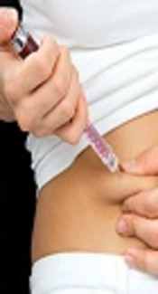
Frequent checking of glucose in plasma of individuals with DM aids well-timed recognition of hyper-glycaemia and is vital to limiting the adverse effects that can be controlled by poor control of the disease. A common method of blood-based research is to provide invasive methods for collecting blood samples from patients. This can cause needle anxiety or the risk of blood-borne infections or both. Studies have revealed that the occurrence of needle anxiety in children is 27% and 22% of all ages (5).

As glucose is one of the plasma combinations that are exchangeable through the salivary gland, epithelium and saliva is the biologic watery that is simple to accumulate. The goal of the current study was to evaluate the glucose levels in saliva and serum of children with diabetes to specify the usefulness of saliva as an analytic tool.

2. Methods

2.1. Study Design and Population

The procedure was permitted by the ethics committee of Tehran University of Medical Sciences, Iran, and all participants gave informed consent before contribution in the procedure. This procedure was planned as a case-control survey in the children’s hospital Medical center of Tehran University of Medical Sciences. A total of 34 children who suffered from Type 1 diabetes mellitus (DM) (male/female: 12/22) and 34 non-diabetic children (male/female: 14/20) were recruited to the study. Children who were hospitalized for side effects of diabetes mellitus were considered as the case group. Non-diabetic children who were hospitalized in the orthopedic section, without any other pre-existing systemic diseases, were considered as the control group. Participants with wound (s) in their mouths were left out from the procedure.

2.2. Sample Collection

Venous blood and saliva were gathered concurrently from each participant between 8 - 9 a.m. Subjects expectorated about 5 mL of their resting whole saliva in a tube. Stimulated whole saliva was stimulated by a piece of natural gum. Two CC of venous blood was drawn following saliva sampling. The specimens were centrifuged at 3800 g for 10 minutes. The serum and saliva supernatants were isolated, which were kept in -70°C for later analysis of glucose.

2.3. Laboratory Measurements

Serum and salivary glucose concentrations were assessed by an enzymatic colorimetric GOD-PAP assay, using commercial kits purchased from the man diagnostics company (Tehran, Iran). Serum Hemoglobin A1c (HbA1c) was assessed by turbidimetrically at 552 nm (Roche Diagnostics GmbH).

2.4. Statistics

The data are offered as a mean ± SEM. The Unpaired 2-tailed student’s t-test, Pearson correlation, and receiver operating characteristic (ROC) analysis was used; P < 0.05 was considered as significant.

3. Results

As predictable, the mean of serum level of glucose was greater in children with DM than that of the non-diabetic (Table 1). Stimulated and unstimulated salivary levels of glucose were significantly greater in children with IDDM compared to control subjects (P = 0.049 and P = 0.047, in that order) (Table 1). Serum level of HbA1c was higher in the patients with IDDM than that of the controls (P = 0.0001). Serum glucose concentration correlates with stimulated (r = 0.407; P = 0.005) but not with unstimulated salivary concentration of glucose (r = 0.189; P = 0.145). Serum HbA1c correlates with unstimulated (r = 0.404; P = 0.001) but not with stimulated salivary concentration of HbA1c (r = 0.0.95; P = 0.526).

Table 1.

Serum and Saliva Levels of Glucose and HbA1c in Insulin-Dependent Diabetes Mellitus (IDDM)a

VariablesControlIDDMP Value
Age, y; Mean ± SD10.7 ± 2.910.0 ± 2.40.273
Fasting blood sugar, mg/dL97.2 ± 2.1265.6 ± 15.90.0001b
Serum HbA1c, %4.99 ± 0.0111.23 ± 0.510.0001b
Unstimulated saliva glucose, mg/dL1.50 ± 0.336.97 ± 2.230.049b
Stimulated saliva glucose, mg/dL2.18 ± 0.216.11 ± 1.710.047b

The cut-off value of unstimulated salivary glucose for the diagnosis of IDDM was 1.05 mg/dL (ROC-area under the curve = 0.79). With this cut-off, sensitivity was 75%, and specificity was 60%. The stimulated salivary glucose cutoff value with sensitivity (71%), specificity (80%), and the area under the ROC curve (0.77) was 2.15 mg/dL.

4. Discussion

Diabetes mellitus characterizes one of the main long-lasting health complications affecting individuals worldwide nowadays, which need continuous checking of their glucose levels. Analytical tests for DM usually use plasma and urine samples. Blood as a diagnostic instrument has a benefit as its adjacent association to the homeostasis of the body. However, blood gathering is an aggressive method and it requires a trained technician; the method is frequently traumatic, particularly in children leading to anxiety and trauma. Thus a noninvasive method is necessary to screen the glycemic regulator. Saliva is the easiest sample that can be gathered noninvasively with negligible apparatus and is associated with less side effects than blood. Therefore, the requirement for an another method arises. Today attention has been growing in the usage of saliva as a diagnostic watery. Thus, the aim of the research was to assess saliva glucose as a diagnostic instrument for checking glycemic controller in IDDM. We found that salivary levels of glucose was higher in children with IDDM and serum glucose correlates with stimulated saliva. In addition, serum HbA1c correlates with unstimulated saliva level.

The logic of the research for determining glucose in saliva is that saliva is being reflected as an analytic fluid of the upcoming. Saliva is supposed to be a reflect of the body and may be recognized as a capable fluid for checking the wellbeing and sickness conditions of a person in healthcare plans. In this respect, it has been revealed that numerous biochemical molecules may be assessed in saliva of patients, such as heart diseases (6-8), xerostomia (9-12), and oral lichen planus (13).

In this research, glucose was detectable in the stimulated and unstimulated saliva of diabetic and non-diabetic individuals. This was in accord with the reports of other studies (14-18).

Mean stimulated salivary glucose levels in IDDM were significantly greater than the levels in non-diabetic individuals in our study. This was in covenant with the study shown by Karjalainen et al., in Type 1 DM (19) and Mirzaii-Dizgah et al., in Type 2 DM (20, 21). In the current research, there was a positive association between stimulated saliva and serum glucose levels, which is in agreement with the studies of Karjalainen et al., (19) and Mirzaii-Dizgah et al. (20, 21). Hence, stimulated saliva glucose seems to be an indicator of serum glucose level in DM patients.

In this research, we showed that fasting unstimulated saliva glucose was significantly higher in diabetic children than in the controls. The same results were reported by other studies regarding type 1 DM (22, 23) and Type 2 DM (24-29). In our study, there wasn’t seen a significant correlation between unstimulated salivary and serum glucose in Type 1 DM. However, some reports indicated that unstimulated salivary glucose correlates with serum glucose in Type 2 DM (24, 25, 27), which are not in agreement with the result of this study.

Unstimulated saliva glucose correlates significantly with serum HbA1c level in this study, which is in accordance with the other studies regarding Type 2 DM (24, 27).

Saliva contains more than 100 million bacteria/mL of saliva (30); thus, salivary glucose may be consumed by this microorganism. Therefore, the time between collecting and measuring saliva and how to store it can affect the amount of salivary glucose. If this problem is resolved, fasting saliva glucose may be applied as a nonaggressive diagnostic, as well as a checking instrument to measure the glycemic status of DM patients.

4.1. Conclusion

It seems that saliva glucose is higher in Type 1 diabetic mellitus subjects and checking of glucose in saliva may be applied as an index of DM.

References

  • 1.

    Lopez del Valle LM, Ocasio-Lopez C. Comparing the oral health status of diabetic and non-diabetic children from Puerto Rico: a case-control pilot study. P R Health Sci J. 2011;30(3):123-7. [PubMed ID: 21932712].

  • 2.

    Wild S, Roglic G, Green A, Sicree R, King H. Global prevalence of diabetes: estimates for the year 2000 and projections for 2030. Diabetes Care. 2004;27(5):1047-53. [PubMed ID: 15111519].

  • 3.

    I. D. F. Diabetes Atlas Group. Update of mortality attributable to diabetes for the IDF Diabetes Atlas: Estimates for the year 2013. Diabetes Res Clin Pract. 2015;109(3):461-5. [PubMed ID: 26119773]. https://doi.org/10.1016/j.diabres.2015.05.037.

  • 4.

    Wang P, Tan Y, Xie H, Shen F. A novel method for diabetes diagnosis based on electronic nose. Biosens Bioelectron. 1997;12(9-10):1031-6. [PubMed ID: 9451791].

  • 5.

    Naing C, Mak JW. Salivary glucose in monitoring glycaemia in patients with type 1 diabetes mellitus: a systematic review. J Diabetes Metab Disord. 2017;16:2. [PubMed ID: 28127542]. https://doi.org/10.1186/s40200-017-0287-5.

  • 6.

    Mirzaii-Dizgah I, Riahi E. Salivary high-sensitivity cardiac troponin T levels in patients with acute myocardial infarction. Oral Dis. 2013;19(2):180-4. [PubMed ID: 22834943]. https://doi.org/10.1111/j.1601-0825.2012.01968.x.

  • 7.

    Mirzaii-Dizgah I, Riahi E. Salivary troponin I as an indicator of myocardial infarction. Indian J Med Res. 2013;138(6):861-5. [PubMed ID: 24521627].

  • 8.

    Mirzaii-Dizgah I, Riahi E. Serum and saliva levels of cathepsin L in patients with acute coronary syndrome. J Contemp Dent Pract. 2011;12(2):114-9. [PubMed ID: 22186754]. https://doi.org/10.5005/jp-journals-10024-1019.

  • 9.

    Agha-Hosseini F, Mirzaii-Dizgah I, Mirjalili N. Relationship of stimulated whole saliva cortisol level with the severity of a feeling of dry mouth in menopausal women. Gerodontology. 2012;29(1):43-7. [PubMed ID: 20636476]. https://doi.org/10.1111/j.1741-2358.2010.00403.x.

  • 10.

    Agha-Hosseini F, Mirzaii-Dizgah I, Mirjalili N. Relationship of unstimulated saliva cortisol level with severity of oral dryness feeling in menopausal women. Aust Dent J. 2011;56(2):171-4. [PubMed ID: 21623809]. https://doi.org/10.1111/j.1834-7819.2011.01320.x.

  • 11.

    Agha-Hosseini F, Mirzaii-Dizgah I, Moosavi MS. Relationship of lumbar spine bone mineral density and oral dryness feeling in menopause. Menopause. 2011;18(6):625-8. [PubMed ID: 21358351]. https://doi.org/10.1097/gme.0b013e31820285b2.

  • 12.

    Agha-Hosseini F, Moosavi MS, Mirzaii-Dizgah I. Salivary flow, testosterone, and femur bone mineral density in menopausal women with oral dryness feeling. Oral Surg Oral Med Oral Pathol Oral Radiol. 2013;115(5):612-6. [PubMed ID: 23433570]. https://doi.org/10.1016/j.oooo.2012.11.014.

  • 13.

    Agha-Hosseini F, Mirzaii-Dizgah I. p53 as a neoplastic biomarker in patients with erosive and plaque like forms of oral lichen planus. J Contemp Dent Pract. 2013;14(1):1-3. [PubMed ID: 23579883]. https://doi.org/10.5005/jp-journals-10024-1259.

  • 14.

    Ben-Aryeh H, Cohen M, Kanter Y, Szargel R, Laufer D. Salivary composition in diabetic patients. J Diabet Complications. 1988;2(2):96-9. [PubMed ID: 2458369].

  • 15.

    Amer S, Yousuf M, Siddqiui PQ, Alam J. Salivary glucose concentrations in patients with diabetes mellitus--a minimally invasive technique for monitoring blood glucose levels. Pak J Pharm Sci. 2001;14(1):33-7. [PubMed ID: 16414850].

  • 16.

    Aydin S. A comparison of ghrelin, glucose, alpha-amylase and protein levels in saliva from diabetics. J Biochem Mol Biol. 2007;40(1):29-35. [PubMed ID: 17244479]. https://doi.org/10.5483/BMBRep.2007.40.1.029.

  • 17.

    Bernardi MJ, Reis A, Loguercio AD, Kehrig R, Leite MF, Nicolau J. Study of the buffering capacity, pH and salivary flow rate in type 2 well-controlled and poorly controlled diabetic patients. Oral Health Prev Dent. 2007;5(1):73-8. [PubMed ID: 17366764].

  • 18.

    Naik VV, Satpathy Y, Pilli GS, Mishra MN. Comparison and correlation of glucose levels in serum and saliva of patients with diabetes mellitus. Indian J Pub Health Res Dev. 2011;2(1):103-5.

  • 19.

    Karjalainen KM, Knuuttila ML, Kaar ML. Salivary factors in children and adolescents with insulin-dependent diabetes mellitus. Pediatr Dent. 1996;18(4):306-11. [PubMed ID: 8857659].

  • 20.

    Mirzaii-Dizgah I, Mirzaii-Dizgah MR, Mirzaii-Dizgah MH. Stimulated saliva glucose as a diagnostic specimen for detection of diabetes mellitus. J Arch Mil Med. 2013;1(1):24-7. https://doi.org/10.5812/jamm.14609.

  • 21.

    Mirzaii-Dizgah I, Mirzaii-Dizgah MH, Mirzaii-Dizgah MR. Oral glucose tolerance test in unstimulated saliva of healthy individuals. Eur J Gen Dent. 2016;5(1):15-8. https://doi.org/10.4103/2278-9626.172736.

  • 22.

    Belazi MA, Galli-Tsinopoulou A, Drakoulakos D, Fleva A, Papanayiotou PH. Salivary alterations in insulin-dependent diabetes mellitus. Int J Paediatr Dent. 1998;8(1):29-33. [PubMed ID: 9558543]. https://doi.org/10.1046/j.1365-263X.1998.00057.x.

  • 23.

    Soares MS, Batista-Filho MM, Pimentel MJ, Passos IA, Chimenos-Kustner E. Determination of salivary glucose in healthy adults. Med Oral Patol Oral Cir Bucal. 2009;14(10):e510-3. [PubMed ID: 19680215].

  • 24.

    Abikshyeet P, Ramesh V, Oza N. Glucose estimation in the salivary secretion of diabetes mellitus patients. Diabetes Metab Syndr Obes. 2012;5:149-54. [PubMed ID: 22923999]. https://doi.org/10.2147/DMSO.S32112.

  • 25.

    Balan P, Babu SG, Sucheta KN, Shetty SR, Rangare AL, Castelino RL, et al. Can saliva offer an advantage in monitoring of diabetes mellitus? - A case control study. J Clin Exp Dent. 2014;6(4):e335-8. [PubMed ID: 25593652]. https://doi.org/10.4317/jced.51386.

  • 26.

    Balan P, B. Gogineni S, Kumari NS, Shetty V, Lakshman Rangare A, L. Castelino R, et al. Candida Carriage Rate and Growth Characteristics of Saliva in Diabetes Mellitus Patients: A Case-Control Study. J Dent Res Dent Clin Dent Prospects. 2015;9(4):274-9. [PubMed ID: 26889366]. https://doi.org/10.15171/joddd.2015.048.

  • 27.

    Satish BN, Srikala P, Maharudrappa B, Awanti SM, Kumar P, Hugar D. Saliva: A tool in assessing glucose levels in Diabetes Mellitus. J Int Oral Health. 2014;6(2):114-7. [PubMed ID: 24876711].

  • 28.

    Kadashetti V, Baad R, Malik N, Shivakumar KM, Vibhute N, Belgaumi U, et al. Glucose Level Estimation in Diabetes Mellitus By Saliva: A Bloodless Revolution. Rom J Intern Med. 2015;53(3):248-52. [PubMed ID: 26710500]. https://doi.org/10.1515/rjim-2015-0032.

  • 29.

    Indira M, Chandrashekar P, Kattappagari KK, Chandra LP, Chitturi RT, Bv RR. Evaluation of salivary glucose, amylase, and total protein in Type 2 diabetes mellitus patients. Indian J Dent Res. 2015;26(3):271-5. [PubMed ID: 26275194]. https://doi.org/10.4103/0970-9290.162883.

  • 30.

    Curtis MA, Zenobia C, Darveau RP. The relationship of the oral microbiotia to periodontal health and disease. Cell Host Microbe. 2011;10(4):302-6. [PubMed ID: 22018230]. https://doi.org/10.1016/j.chom.2011.09.008.