Vitamin E Supplementation Reduces Oxidative Stress in the Male Wistar Rats’ Brain Against Polyvinyl Chloride Products

authors:

avatar Abbas Sadeghi 1 , avatar Laya Ghahari 1 , * , avatar Mitra Yousefpour ORCID 2

Department of Anatomy, Medical School, AJA University of Medical Sciences, Tehran, Iran
Department of Physiology, Medical School, AJA University of Medical Sciences, Tehran, Iran

how to cite: Sadeghi A, Ghahari L, Yousefpour M. Vitamin E Supplementation Reduces Oxidative Stress in the Male Wistar Rats’ Brain Against Polyvinyl Chloride Products. Ann Mil Health Sci Res. 2019;17(2):e92768. https://doi.org/10.5812/amh.92768.

Abstract

Background:

Polyvinyl chloride (PVC) exerts toxic effects via the formation of oxidative stress on the brain. Antioxidants such as Vitamin E (Vit E) can affect free radicals.

Objectives:

This study was conducted to decrease damage and oxidative stress against PVC products in rats’ brain by vitamin E.

Methods:

In the current study, we used 24 male Wistar rats in four groups (N = 6 rats per group) as the control, Vit E, PVC, PVC plus Vit E. We orally administered vitamin E and PVC for 40 days. Then the rats were anesthetized and the brain was removed and transferred to a temperature of -70ºC in order to measure lipid peroxidation.

Results:

our results revealed that oral administration of PVC decreased catalase and total antioxidant capacity levels, and increased malondialdehyde in PVC exposed rats. Vitamin E improved spatial memories by enhancing neurogenesis in the brain.

Conclusions:

Exposure to PVC can produce oxidative stress enzymes and make brain damage. Vitamin E, as an antioxidant, can decrease the toxic effects of PVC and protect the brain.

1. Background

Polyvinyl chloride (PVC) is an important raw material in the chemical industry. It is used to make plastic containers, coated wires, and in the automotive industry. It can cause harmful effects on the central nervous system in humans (1-3). The PVC is used in most devices for humans and associated with the spread of diseases (4). Since PVC is not chemically bonded with polymer, it can be separated easily (2, 3). Therefore, it can enter the human body with eating food, drinking water, transmission through the air, or even contact with medications (1, 5) and toys (6, 7), especially in children. Another application of PVC is in the medical equipment and laboratory materials (1). Medical and surgical applications such as injection devices, blood storage bags, chip tubes, and dialysis machines contain plenty of PVCs. studies have shown that people who have used a large number of blood products, the PVC substance has been detected in their blood and tissues (8). The food is the main source of PVC transmission to humans (7).

In people who have exposed to severe PVC, liver cancer has been observed 15 times more than normal humans, and in this group of people, brain cancer has been 4 times higher than others (9). The PVC is one of the most abundant poisons in the natural environment of humans. This material is one of the factors that produce oxidative stress enzymes and also causes some enzymes to impair (10). Research results have shown that PVC contributes to some neurodegenerative diseases. Vitamin E is one of the most important antioxidants that can prevent damaging effects leading to a number of diseases, such as diabetes (11-13), stroke (14, 15), Alzheimer (16, 17), and aging (18).

2. Objectives

In this study, the effect of vitamin E was investigated against PVC in rat’s oxidative stress enzymes.

3. Methods

We used 24 male Wistar rats (weighing 180 - 200g) in the following groups, the control (N = 6), Vit E (N = 6), PVC (N = 6), PVC plus Vit E (N = 6) groups. The animals hosted in standard condition. We orally administered vitamin E (150 mg/kg.bw/day) and PVC (1000 mg/kg.bw/day) for 40 days. The PVC (Merck, Germany) and vitamin E were used as described in previous studies (11, 12).

3.1. Lipid Peroxidation Assay

Rats were sacrificed after 40 days of exposure to PVC. The right hemisphere was removed, homogenized, and the level of malondialdehyde (MDA), catalase (CAT), and total antioxidant capacity (TAOC) were evaluated.

3.1.1. Measuring Brain Malondialdehyde Level

To this end, 0.2 g of the brain tissue was transferred to phosphate buffer saline (PBS) 0ºC, 0.05 M, pH = 7.4 (10% w/v), and homogenously ground by a mortar and pestle. Then the solution was centrifuged at 1000 rpm. Afterward, 150 μg of the supernatant of the centrifuged specimen was removed, 300 μg of 10% trichloroacetic acid was added, and it was centrifuged at 1000 rpm, 4ºC, for 10 min. Then 300 μL of the supernatant was transferred to the tube and incubated with 300 μL of 0.67% thiobarbituric acid at 100ºC for 25 min. After 5 minutes of cooling the solution, the color pink resulting from the reaction between MDA and thiobarbituric acid appeared and was evaluated with a spectrophotometer at 535 nm wavelength. The concentration of MDA was calculated using the MDA absorption coefficient in nmol/g tissue (13).

3.1.2. Catalase Activity Evaluation

Catalase activity was determined based on its ability to decompose H2O2 using the Aebi method. In this regard, H2O2 decomposition can be assessed by absorption reduction at 240 nm. The difference in absorption per unit of time equals catalase activity. First, 10% w/v of 0.2 g of brain tissue was washed with ice-cold PBS (pH = 6.8) and homogenously ground by a mortar and pestle. The homogenized tissue solution was centrifuged at 5000 rpm for 5 min. Next, 100 μL of the centrifuged supernatant was added to 2.8 μL of PBS. Then 100 μL of H2O2 solution was added, and the absorption was measured in the 240 nm wavelength at 0 and 30 s. The PBS was used as the blank. In the end, values were expressed as U/g tissue (14).

3.1.3. Total Antioxidant Capacity Measurement

The TAOC of brain tissue was measured using the ferric antioxidant power test (FRAP). On the day of the evaluation, tissues were homogenized in cold KCL solution. To prepare homogeneous tissue solution, 0.2 g of brain tissue was taken, 10% (w/v) of it was added to KCL solution and homogenously ground by a mortar and pestle. Then the prepared homogeneous solution was centrifuged at 1000 rpm for 5 min. Afterward, 100 μL of the supernatant was removed and transferred to a test tube, 3 μL of FRAP indicator was added, and it was incubated at 37ºC heated-bath for 7 - 10 min. The absorption of the blue complex was read using a spectrophotometer at 593 nm. Values were expressed as mmol/g tissue (15).

3.2. Statistical Analysis

Data of the groups were analyzed by SPSS software and ANOVA and Tukey’s tests were used. The significance level was considered P < 0.05.

4. Results

4.1. Biochemical Test Results

MDA concentration was increased in the PVC group, notably compared to the control and the vitamin E groups (P < 0.05). However, MDA concentrations in the PVC + Vitamin E group were similar compared to the vitamin E and control groups (P > 0.05) (Figure 1A).

Effects of PVC and vitamin E on MDA concentration (A), CAT activity (B), and TAOC (C) of the brain in studied groups. *Indicates a significant difference (P < 0.05) compared to the control group. Data are expressed as mean ± SE.

The CAT activity demonstrated a significant reduction in groups under the treatment with PVC versus the control group (P < 0.05). Nevertheless, this increase was not significant in the vitamin E group compared to the control group (P > 0.05). No significant difference was seen between groups receiving PVC plus vitamin E and the control group (P > 0.05) (Figure 1B).

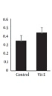
For TAOC, a considerable reduction was found in the group under the treatment with PVC compared with the control group (P < 0.05). However, no discrepancy was observed in PVC plus Vit E group (P > 0.05). Furthermore, TAOC was significantly higher in rats treated with vitamin E in comparison to the control group (P < 0.05) (Figure 1C).

5. Discussion

The present study showed that male rats exposed to PVC displayed a significant decrease in the antioxidant properties of the brain. The scientists have explained, PVC or PVC metabolite exposure could create significant neuronal degeneration and decrease the membrane’s permeability (8). Thus it seems PVC can decrease neural plasticity.

The PVC exposure may increase pathological changes in receptor activity and create toxic effects via the production of reactive oxygen species (ROS). On the other hand, PVC can reinforce the toxic effects of ROS via the reduction of antioxidant enzymes or radical scavengers (10). In the present study, PVC treatment significantly increased malondialdehyde and decreased catalase activity and total antioxidant capacity in the brain. These results confirmed that PVC is accompanied by an increased generation of ROS products (16, 17). Oxidation of the sulfhydryl is ensured from a combination of superoxide and hydrogen peroxide, which disturb glutathione synthesis via competitive antagonism of cysteine transport and provoke the genesis of free radicals through thiol oxidation (18).

The studies explained that oxidative stress had an important role in PVC-induced brain changes (19). Also, it has been shown that neurons can defend themselves and decrease excitotoxic effects of oxidative agents (18). The scientists have been interested in evaluating the antioxidant defense mechanisms of the brain (18, 20-26). On the other hand, it is evident that glial cells produce the neurotrophic factors and cytokines that decreases-induced neurotoxicity. Astrocytes have more antioxidant strength than neurons and defend neurons against oxidation and support for neuronal life (27). Induction of oxidative stress after PVC exposure and progressive increment of ROS production result in neuronal death via protein oxidation, DNA damages, and increased amount of lipid peroxidation in neural membrane (28). It has been shown that the brain produces slow endogenous levels of vitamin E (an effective antioxidant agent), and it is mainly an easy target of oxidative stress (29).

The main finding of the current research is the effects of vitamin E on oxidative stress on the brain after exposure to PVC. In recent years, it has been shown that vitamin E protects some organs against oxidative stress (30). Also, it has been explained that in the developing cerebellum, vitamin E administration reduced alcohol-induced oxidative stress and apoptosis (18). The studies established that vitamin E has similarly valuable effects on glial cells versus toxic effects of the chemical and/or metabolic agents. It can protect nerve tissues by the clearance of the free radicals and stabilizing the cell membranes (27). These data show that the administration of vitamin E may reduce lipid peroxidation and keep the neurons from PVC-related oxidative stress.

References

  • 1.

    Latini G, Ferri M, Chiellini F. Materials degradation in PVC medical devices, DEHP leaching and neonatal outcomes. Curr Med Chem. 2010;17(26):2979-89. [PubMed ID: 20858177].

  • 2.

    Talsness CE, Andrade AJ, Kuriyama SN, Taylor JA, vom Saal FS. Components of plastic: Experimental studies in animals and relevance for human health. Philos Trans R Soc Lond B Biol Sci. 2009;364(1526):2079-96. [PubMed ID: 19528057]. [PubMed Central ID: PMC2873015]. https://doi.org/10.1098/rstb.2008.0281.

  • 3.

    Hartmann S, Klaschka U. Interested consumers' awareness of harmful chemicals in everyday products. Environ Sci Eur. 2017;29(1):29. [PubMed ID: 29214119]. [PubMed Central ID: PMC5698398]. https://doi.org/10.1186/s12302-017-0127-8.

  • 4.

    Sherman M. Vinyl chloride and the liver. J Hepatol. 2009;51(6):1074-81. [PubMed ID: 19836850]. https://doi.org/10.1016/j.jhep.2009.09.012.

  • 5.

    Beko G, Weschler CJ, Langer S, Callesen M, Toftum J, Clausen G. Children's phthalate intakes and resultant cumulative exposures estimated from urine compared with estimates from dust ingestion, inhalation and dermal absorption in their homes and daycare centers. PLoS One. 2013;8(4). e62442. [PubMed ID: 23626820]. [PubMed Central ID: PMC3633888]. https://doi.org/10.1371/journal.pone.0062442.

  • 6.

    Ting KC, Gill M, Garbin O. GC/MS screening method for phthalate esters in children's toys. J AOAC Int. 2009;92(3):951-8. [PubMed ID: 19610390].

  • 7.

    Stringer R, Labunska I, Santillo D, Johnston P, Siddorn J, Stephenson A. Concentrations of phthalate esters and identification of other additives in PVC children's toys. Environ Sci Pollut Res Int. 2000;7(1):27-36. [PubMed ID: 19153836]. https://doi.org/10.1065/espr199910.007.

  • 8.

    Dhanya CR, Gayathri NS, Mithra K, Nair KV, Kurup PA. Vitamin E prevents deleterious effects of di (2-ethyl hexyl) phthalate, a plasticizer used in PVC blood storage bags. Indian J Exp Biol. 2004;42(9):871-5. [PubMed ID: 15462179].

  • 9.

    Wagoner JK. Toxicity of vinyl chloride and poly(vinyl chloride): A critical review. Environ Health Perspect. 1983;52:61-6. [PubMed ID: 6360677]. [PubMed Central ID: PMC1569348]. https://doi.org/10.1289/ehp.835261.

  • 10.

    Afolabi OK, Wusu AD, Ogunrinola OO, Abam EO, Babayemi DO, Dosumu OA, et al. Arsenic-induced dyslipidemia in male albino rats: Comparison between trivalent and pentavalent inorganic arsenic in drinking water. BMC Pharmacol Toxicol. 2015;16:15. [PubMed ID: 26044777]. [PubMed Central ID: PMC4455335]. https://doi.org/10.1186/s40360-015-0015-z.

  • 11.

    Sadeghi A, Farokhi F, Najafi G, Shalizar Jalali A. Protective effect of vitamin E against polyvinyl chloride induced damages and oxidative stress in rat testicular tissue. J Kermanshah Univ Med Sci. 2017;21(3). e69272.

  • 12.

    Sadeghi A, Ghahari L. Vitamin E as an antioxidant reduces apoptosis of cells in rat hippocampus following exposure to polyvinyl chloride. Ann Mil Health Sci Res. 2018;16(4). e85275. https://doi.org/10.5812/amh.85275.

  • 13.

    Esterbauer H, Cheeseman KH. Determination of aldehydic lipid peroxidation products: Malonaldehyde and 4-hydroxynonenal. Methods Enzymol. 1990;186:407-21. [PubMed ID: 2233308].

  • 14.

    Aebi H. [13] Catalase in vitro. Methods Enzymol. 1984;105:121-6. https://doi.org/10.1016/s0076-6879(84)05016-3.

  • 15.

    Benzie IF, Strain JJ. The ferric reducing ability of plasma (FRAP) as a measure of "antioxidant power": The FRAP assay. Anal Biochem. 1996;239(1):70-6. [PubMed ID: 8660627]. https://doi.org/10.1006/abio.1996.0292.

  • 16.

    Wang M, Su P. The role of the Fas/FasL signaling pathway in environmental toxicant-induced testicular cell apoptosis: An update. Syst Biol Reprod Med. 2018;64(2):93-102. [PubMed ID: 29299971]. https://doi.org/10.1080/19396368.2017.1422046.

  • 17.

    Mathur PP, D'Cruz SC. The effect of environmental contaminants on testicular function. Asian J Androl. 2011;13(4):585-91. [PubMed ID: 21706039]. [PubMed Central ID: PMC3739630]. https://doi.org/10.1038/aja.2011.40.

  • 18.

    Shirpoor A, Salami S, Khadem-Ansari MH, Minassian S, Yegiazarian M. Protective effect of vitamin E against ethanol-induced hyperhomocysteinemia, DNA damage, and atrophy in the developing male rat brain. Alcohol Clin Exp Res. 2009;33(7):1181-6. [PubMed ID: 19389196]. https://doi.org/10.1111/j.1530-0277.2009.00941.x.

  • 19.

    Wang X, Cai J, Zhang J, Wang C, Yu A, Chen Y, et al. Acute trimethyltin exposure induces oxidative stress response and neuronal apoptosis in Sebastiscus marmoratus. Aquat Toxicol. 2008;90(1):58-64. [PubMed ID: 18801585]. https://doi.org/10.1016/j.aquatox.2008.07.017.

  • 20.

    Silakhori S, Hosseinzadeh H, Shaebani Behbahani F, Mehri S. Neuroprotective effect of clavulanic acid on trimethyltin (TMT)-induced cytotoxicity in PC12 cells. Drug Chem Toxicol. 2019;42(2):187-93. [PubMed ID: 29764237]. https://doi.org/10.1080/01480545.2018.1468772.

  • 21.

    Saramad H, Farokhi F, Najafi G. Investigation protective effects of vitamin E against mobile radiations induced histological alterations and oxidative stress in rats’ brain. J Shahid Sadoughi Univ Med Sci. 2018;25(10):828-42.

  • 22.

    Gugliandolo A, Bramanti P, Mazzon E. Role of vitamin E in the treatment of alzheimer's disease: Evidence from animal models. Int J Mol Sci. 2017;18(12). [PubMed ID: 29168797]. [PubMed Central ID: PMC5751107]. https://doi.org/10.3390/ijms18122504.

  • 23.

    Boccardi V, Baroni M, Mangialasche F, Mecocci P. Vitamin E family: Role in the pathogenesis and treatment of Alzheimer's disease. Alzheimers Dement (N Y). 2016;2(3):182-91. [PubMed ID: 29067305]. [PubMed Central ID: PMC5651353]. https://doi.org/10.1016/j.trci.2016.08.002.

  • 24.

    Yonguc GN, Dodurga Y, Adiguzel E, Gundogdu G, Kucukatay V, Ozbal S, et al. Grape seed extract has superior beneficial effects than vitamin E on oxidative stress and apoptosis in the hippocampus of streptozotocin induced diabetic rats. Gene. 2015;555(2):119-26. [PubMed ID: 25445279]. https://doi.org/10.1016/j.gene.2014.10.052.

  • 25.

    Crouzin N, Ferreira MC, Cohen-Solal C, Barbanel G, Guiramand J, Vignes M. Neuroprotection induced by vitamin E against oxidative stress in hippocampal neurons: involvement of TRPV1 channels. Mol Nutr Food Res. 2010;54(4):496-505. [PubMed ID: 20087852]. https://doi.org/10.1002/mnfr.200900188.

  • 26.

    Babaei-Balderlou F, Zare S, Heidari R, Farrokhi F. Effects of melatonin and vitamin E on peripheral neuropathic pain in streptozotocin-induced diabetic rats. Iran J Basic Med Sci. 2010;13(2):1-8.

  • 27.

    Baydas G, Nedzvetskii VS, Tuzcu M, Yasar A, Kirichenko SV. Increase of glial fibrillary acidic protein and S-100B in hippocampus and cortex of diabetic rats: effects of vitamin E. Eur J Pharmacol. 2003;462(1-3):67-71. [PubMed ID: 12591097].

  • 28.

    Tuzcu M, Baydas G. Effect of melatonin and vitamin E on diabetes-induced learning and memory impairment in rats. Eur J Pharmacol. 2006;537(1-3):106-10. [PubMed ID: 16626697]. https://doi.org/10.1016/j.ejphar.2006.03.024.

  • 29.

    Marino MD, Aksenov MY, Kelly SJ. Vitamin E protects against alcohol-induced cell loss and oxidative stress in the neonatal rat hippocampus. Int J Dev Neurosci. 2004;22(5-6):363-77. [PubMed ID: 15380836]. https://doi.org/10.1016/j.ijdevneu.2004.04.005.

  • 30.

    Badgujar PC, Pawar NN, Chandratre GA, Telang AG, Sharma AK. Fipronil induced oxidative stress in kidney and brain of mice: Protective effect of vitamin E and vitamin C. Pestic Biochem Physiol. 2015;118:10-8. [PubMed ID: 25752424]. https://doi.org/10.1016/j.pestbp.2014.10.013.