1. Introduction
Cardiac hydatidosis (Echinococcus) is a rare manifestation, accounting for less than 0.5% of all hydatid disease cases (1). It can affect various cardiac structures, including the right ventricle, atrium, pericardium, interventricular septum, and pulmonary artery (2). Clinical presentation ranges from asymptomatic cases with small hydatid cysts or minor symptoms to severe cases involving cardiogenic shock and sudden death (3). Here, we present a case of a massive asymptomatic myocardial hydatid cyst in a male patient, diagnosed incidentally.
2. Case Presentation
A 51-year-old man weighing 63 kg presented to the Emergency Department (ED) with complaints of exertional dyspnea and recurrent palpitations. He was categorized as class III in functional capacity and had no prior hospitalizations or family history of cardiac disease. He reported sleep deprivation for approximately two months before admission due to palpitations and orthopnea, which he self-managed with oxazepam, 10 mg nightly. He also described atypical chest pain, which was neither positional nor exertion-related.
Upon admission, he was hemodynamically stable, with a blood pressure of 90/69 mmHg, a heart rate of 104 beats per minute in regular sinus rhythm, a respiratory rate of 21 breaths per minute, and afebrile. His oxygen saturation at rest, measured by pulse oximetry on the index finger, was 95%. Initial physical examination revealed a mild grade 2/6 systolic murmur along the right and left sternal borders and a diastolic murmur.
An electrocardiogram (ECG) indicated a delayed conduction pattern in the inferior leads with a sinus rhythm and normal axis deviation. A posteroanterior chest X-ray revealed an increased cardiothoracic ratio, a normal aortic arch, bulging of the main pulmonary artery, and a normal-sized right descending pulmonary artery. Sharp linear calcifications were noted along the left heart border.
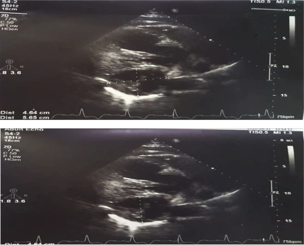
The patient was then referred for an echocardiographic assessment. Transthoracic echocardiography showed a normal left ventricular ejection fraction (LVEF) of 53%, with no regional wall motion abnormalities. There was mild diastolic dysfunction (grade 1) and no significant valvular disease. The pulmonary arterial pressure was normal (SPAP = 30 mmHg). The primary echocardiographic findings included a large (80 mm × 36 mm) cystic mass in the posterolateral wall of the left ventricle near the apical segments, with septation and spotty calcification. Another sizable septated cystic mass (46 mm × 56 mm) was detected in the basal posterior wall of the left ventricle near the mitral valve leaflets. No pericardial effusion was observed (Figure 1).
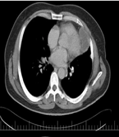
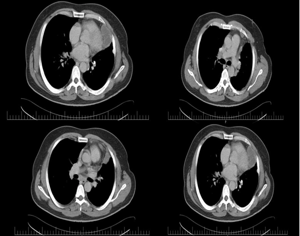
Based on the findings, the patient was referred for high-resolution chest tomography (HRCT) with contrast. The HRCT revealed multiple cystic areas with calcification in the pericardium, the largest measuring 7.50 × 5.19 cm. Additionally, a cystic area was noted on the left side of the mediastinum, measuring 5.75 × 5.42 cm (Figure 2).
Considering these findings, perimyocarditis with probable hydatid cysts was suspected, and surgery was planned for pericardiocentesis and excisional biopsy of the mass. The procedure involved general anesthesia and a median sternotomy. Following the establishment of cardiopulmonary bypass (CPB), a standard cardioplegic solution (Del Nido® solution) was infused into the aortic root to achieve complete cardiac arrest, with mild hypothermia maintained at 32°C - 35°C. To minimize the risk of fluid leakage from the hydatid cyst into surrounding tissues, two gauzes impregnated with hypertonic saline (10% NaCl) were placed around the heart.
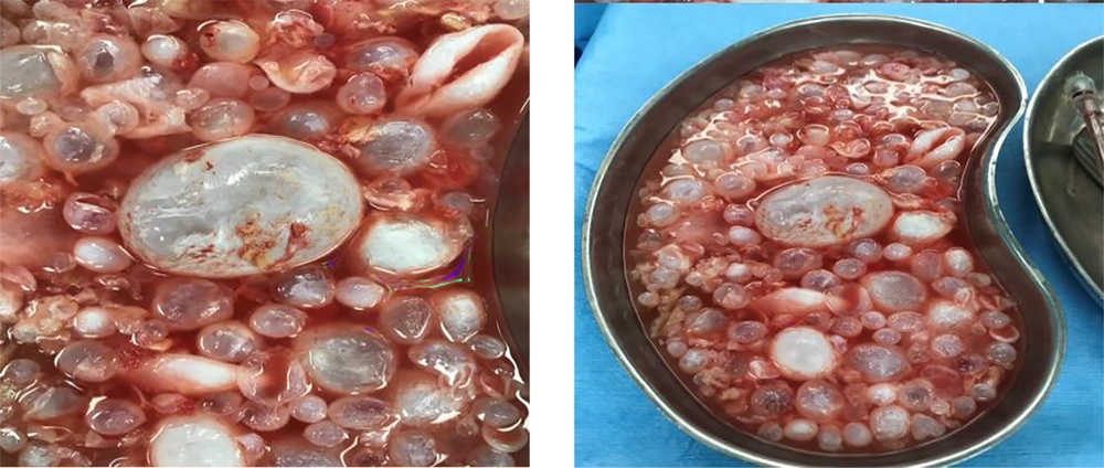
During surgery, multiple cystic structures consistent with hydatid cysts were observed within the myo-pericardium spaces of the posterior, inferior, and lateral walls. The exocyst was removed, and the cyst contents were drained, followed by total resection. Histological slides prepared from the cyst tissue confirmed the presence of a typical hydatid cyst (Figure 3). A histopathological examination provided further confirmation of the diagnosis.
After the cyst removal, the intra-myocardial cavity was irrigated with hypertonic saline. The patient was successfully weaned from CPB and transferred to the cardiac surgery ICU. He was extubated four hours after admission to the ICU. Two days later, the patient was transferred to the post-cardiac surgery ward and subsequently discharged in stable condition. Albendazole was prescribed for 21 days post-discharge. The histopathology report definitively confirmed the diagnosis of hydatidosis.
The patient was advised to visit the cardiology clinic every six months for follow-up to monitor his cardiac condition.
3. Discussion
Cardiac hydatid cysts represent a rare clinical condition, often appearing asymptomatic due to the slow growth rate of cysts. However, symptoms can vary depending on the cysts' size and location within the heart (4, 5). To our knowledge, this report includes massive, giant hydatid lesions without significant symptoms—a presentation not previously documented.
Given the life-threatening complications associated with cardiac hydatid cysts, including potential destruction of vital cardiac structures (4), compression of cardiac chambers, and even myocardial rupture or blood flow obstruction (1), surgical intervention is generally deemed essential.
The heart's contractility provides an intrinsic resistance mechanism against parasitic invasion. Nonetheless, in rare instances, this defense is inadequate, allowing the larva to infect the myocardium (6). The cyst can grow within myocardial fibers and may remain asymptomatic (1). However, some clinical manifestations may include severe dyspnea at rest, embolic events, chest pain, and even blood flow obstruction. Cardiac hydatid cysts can also disrupt the conduction system, potentially leading to dysrhythmias (7).
Despite the extensive lesions observed in our patient, his only symptom was exertional dyspnea without any classic chest pain. The cyst’s minimal impact on the cardiac conduction system, evidenced only by a delayed conduction pattern in the inferior leads on ECG, resolved post-surgery, underscoring the rarity of this case. The palpitations were likely an adverse effect of Oxazepam (8), which diminished after discontinuation.
The diagnosis of cardiac hydatid disease relies on clinical presentation and supportive paraclinical evidence, such as serologic testing and cardiac imaging (9).
Currently, advancements in cardiac imaging have improved the diagnosis of cardiac hydatid cysts. Echocardiography is recognized as a primary, sensitive tool for detecting cardiac masses, including hydatid cysts. For a more precise diagnosis, CT scans or magnetic resonance imaging (MRI) are employed to determine the exact location, size, and extent of invasion into surrounding tissues (1, 10).
In our patient’s case, a transthoracic echocardiogram was initially performed, followed by high-resolution chest CT (HRCT), which confirmed the presence of the hydatid cyst, with distinctive features such as calcification of the cyst layers that distinguished it from other masses. This imaging, complemented by cardiac MRI, may represent the gold standard for diagnosing cardiac hydatid cysts.
Surgical intervention remains the most effective treatment for cardiac echinococcosis. However, specific strategies to reduce the risk of cyst leakage and prevent involvement of surrounding tissues may vary by case (11).
In this case, we utilized hypertonic NaCl (10%) in gauze placed around the heart to minimize leakage risk. This approach, combined with careful removal of the exocysts and drainage of their contents, helped reduce the risk of contaminating adjacent tissues. Protecting the heart and other thoracic organs with this method can be a reliable approach in similar cases.
Hydatid cysts should always be considered a differential diagnosis for any cardiac mass. Successful treatment of these lesions relies on early diagnosis, a multidisciplinary approach, and meticulous surgical technique.


