1. Introduction
In recent weeks, general surgeons have been closely monitoring a concerning trend in the medical field - an increase in the diagnosis of mucocele appendix cases (1). A mucocele appendix is a condition that occurs when the appendix becomes enlarged due to a build-up of mucus in the organ. While rare, this condition can be life-threatening if left untreated (1).
Mucocele appendix is a relatively uncommon condition, but it is one that we are seeing more frequently. We believe that the increase in cases may be due to the growing use of advanced imaging techniques, which allows us to diagnose the condition more accurately (2).
Symptoms of a mucocele appendix can range from mild discomfort to severe abdominal pain. Patients may sometimes experience nausea, vomiting, diarrhea, and a fever. If left untreated, patients may develop serious complications such as peritonitis and inflammation of the lining of the abdomen (3).
While the exact cause of a mucocele appendix is not understood, general surgeons believe it is related to inflammation or obstruction of the appendix. Treatment options for a mucocele appendix can vary, depending on the severity of the case (4). Surgery from a simple appendectomy to a right hemicolectomy may be necessary, or medication may be prescribed to manage symptoms (5). In this study, we reported a rare case of mucocele of the appendix at Golestan Hospital of Ahwaz.
2. Case Presentation
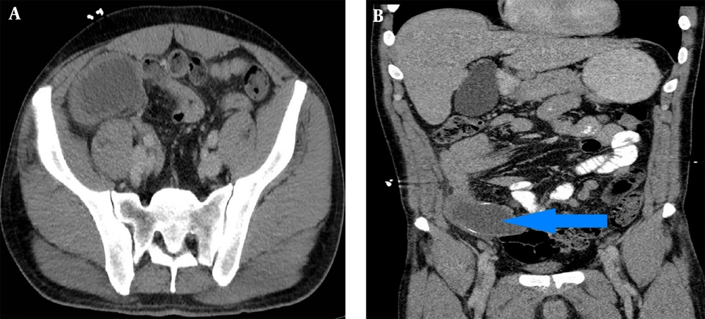
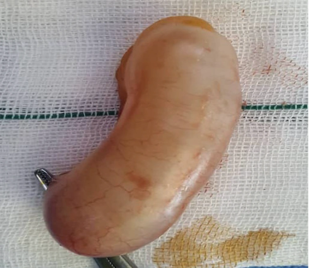
A 60-year-old female with a seven-month history of right lower quadrant pain aggravated in the last 2 weeks presented to the surgery emergency department of Golestan Hospital in Ahwaz. There was no mention of nausea, vomiting, or weight loss in the patient's history from 3 months ago, but she did mention fatigue, weakness, and decreased appetite. She was afebrile at admission. The past medical history, past surgical history, family history, drug history, and habitual history were negative. A physical exam showed tenderness and rebound tenderness in the RLQ (McBurney's point), but Rovsing's sign was negative. Other clinical examinations were normal. The patient's WBC count was 13400 (neutrophil = 82%), hemoglobin = 12.3, and had normal urine analysis. In her medical records (abdomen CT-scan with and without IV contrast), fluid signal tubular mass measured 105 * 37 mm in RLQ is noted and suggested for mucocele appendicitis (Figure 1). After admission, she became NPO (nothing by mouth). After antibiotic therapy (ceftriaxone and metronidazole IV), hydration (1/3, 2/3 1/3 - 2/3, intravenous fluid), and pack cell reservation, she is a candidate for surgical management. Appendectomy was performed through a McBurney incision, and the appendix base was free; no lymphadenopathy was seen (Figure 2).
There were no postoperative side effects. Two days later, the patient was discharged, and after follow-up, the pathology was a mucocele of the appendix with mucin-secreting mucosal hyperplasia.
3. Discussion
We were surprised to find this case in Golestan Hospital, the trauma center in the country's southwest. The appendiceal mucocele describes a mucus-filled appendix that could be neoplastic or non-neoplastic pathologies (mucosal hyperplasia, simple cysts, mucinous cystadenomas, mucinous cystadenocarcinoma) (6).
Appendiceal mucoceles are commonly seen after the fifth decade, which is similar to the result of our study.
The typical form is incidental; however, presentation with appendicitis signs and symptoms occurs in more than 30 % of cases (7). Our study case had RLQ pain, but it wasn't acute. From Alvardo criteria, our patient had RLQ tenderness (2 score), rebound tenderness (1 score), fever of more than 99.1 degrees fahrenheit (1 score), and leukocytosis > 10,000 (1 score).
On imaging findings for the neoplastic process, we expect round-encapsulated cystic mass with irregular walls and soft tissue thickening, similar to our study in which tubular mass measured 105 * 37 mm.
Careful imaging for ascites, peritoneal disease, and hepatic surface scalloping is important in the initial evaluation (8).
A reliable diagnosis is based on the combination of imaging and surgical excision. In our study, the imaging findings were adapted to surgical findings. Avoiding mucocele rupture is important during surgery because of the intraperitoneal spread of neoplastic cells and caused pseudomyxoma peritonei (9).
A certain mucocele treatment is surgery; if it has benign causes, a simple appendectomy is enough, but a right hemicolectomy is recommended for treating adenocarcinoma cysts. Colorectal, ovarian, and endometrial cancers can coexist in the setting of appendiceal mucoceles and careful examination of intra-abdominal structures.
3.1. Conclusions
An appendiceal mucocele is rare. Appendiceal tumors have a low prevalence, but due to the nature of these tumors, which can also be malignant, they should be given importance, and careful follow-up is mandatory. Patients must remain informed and proactive as the surgical community continues to learn more about this condition. The mucocele appendix can be successfully managed and treated with timely diagnosis and treatment.


