


1. Background
Given the rapid and recent development of digital technologies, there are widespread changes in all fields of dentistry with the introduction of various hardware and software (1). Therefore, various new tools can be used to train dental students in addition to the applications of digital technologies in routine dental practices (2). The use of technology in dental education can be assessed in two general categories: Training different skills and concepts and evaluating student performance (3-5).
Many researchers have significantly considered using resources and tools such as augmented reality, virtual reality, mobile applications, computer software, instructional videos, and content presented on the Internet in training, each of which can be used based on different educational purposes (6-11). Applying the mentioned facilities has attracted much attention as a substitute for routine face-to-face training due to the COVID-19 pandemic and the related quarantine and lockdowns (12-14).
Fixed prostheses are one of the main courses in dentistry, and dentists deal with them a lot in their daily practices (15). Students should be trained in dental materials, impression techniques, and crown preparation principles before entering the clinical stages (15-17). Some studies have reported the relationship between student performance in preclinical and clinical courses. Accordingly, students who outperformed in pre-clinics were more successful in the clinical courses (18, 19). However, some studies have not shown related success in clinical courses to the results obtained in the pre-clinical period (20, 21). Nevertheless, dental preparation in fixed prosthetics is irreversible, so these courses help strengthen psychomotor skills and ensure patient safety (22, 23).
Designing dental preparation depends on biological, mechanical, and aesthetic considerations. Therefore, several clinical guidelines have been mentioned in dentistry for dental preparation (24). Face-to-face and demonstration are the traditional training methods through which the basics of prosthetics are transferred to the students and their practice on their models (25). The disadvantages of the face-to-face method include student dependence on the instructor, the possibility of missing some crucial points, observing the educational process from only one side, inability to review the necessary sessions, and training several complex techniques in one session (25, 26). Substituting traditional instruction methods using multi-media-based education technologies has been considered valid by students and faculty members in crown preparation training (27).
Recently, other educational methods, such as educational videos and computer simulations, have been used, but students cannot reuse information and learn the material again.
In addition, the rubric system has been suggested to evaluate the various aspects of student’s tooth preparation systematically. Factors commonly assessed to evaluate the preparation process include occlusal reduction, presence of undercuts, taper, planar/flat reduction, line angles, smoothness, and preservation of adjacent tooth (28-30). According to the students’ opinions and results, performing occlusal reduction correctly has been considered challenging (31, 32).
Technology has also been used in student assessment to reduce the variability of inter-evaluators and intra-evaluators and enable students to self-assess (30, 33). For this purpose, applying computer-aided design (CAD) and digital image processing systems seems successful (33, 34). However, the evaluation of some preparation factors, such as smoothness and presence of undercuts, has not been examined in some of these studies or has been reported inadequately due to poor agreement with supervisors, which requires further research (30, 35).
2. Objectives
This study aimed to determine the effect of Multimedia-based teaching methods (PowerPoint, instructor demonstration, and watching educational videos) and traditional teaching methods (demonstration solely) on the quality of preparation by evaluating smoothness, occlusal reduction, and presence of undercut dental students.
3. Methods
In this interventional study, 60 third-year students completing the pre-clinical course were included and randomly allocated into groups A and B, each consisting of 30 students. Group A was trained through the multi-media-based teaching methods, including PowerPoint, instructor demonstration, and procedural videos, and group B was trained by traditional education methods, which only included instructor demonstration. The required sample size was obtained based on the formula for comparing the proportion between the two groups:
N = (z1 + z2)2 (P1(1-P1) + P2(1-P2))/d2
In which, z1 and z2 were drown from normal standard distribution considering the type of error and the power equal 0.05 and 0.80, respectively. The proportion of undercut (P) was estimated at 0.591 from the previous studies (36), and the minimum detectable difference with the test (d) was 0.4. Thus, the minimum sample size was 24 for each group. Considering the possibility of dropping samples from the study, the sample size was supposed to be 30 in each group.
Eligible participants were randomly allocated to the multimedia-based or the traditional education method groups using block randomization with block sizes of two. Randomization was not blinded to the individual participants because of the nature of the intervention. The research assistant who scored and imported data and the statistician who analyzed the results were blinded to group allocation. Participants were explicitly advised not to inform others about which group they were in and not to discuss the intervention. Participants were also advised not to give the intervention to anyone else.
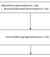
According to the exclusion criteria in evaluating tooth preparation on the second premolars, one person was excluded from group A due to their absence in the demonstration session. A total of two individuals from group B and one from group A were excluded from the study for the same reason. The number of participants in each stage of the study is given in Figure 1 based on the consort flowchart.

Number of participants in randomized control trial study
Group A was trained by Multimedia-based teaching methods, including PowerPoint, instructor demonstration, and watching videos, and group B was trained by traditional teaching methods that only included the instructor’s demonstration. Students in this group received a PowerPoint related to the session's topics before the training class, and they were encouraged to read it after receiving a guide for use. These slides included the description text, the instructor’s voice, and the pictures. The instructor presented the topics in the training session and then demonstrated how to prepare the teeth in the single crown mode. A procedural video of the instructor’s demonstration was given to each group member. The video included a step-by-step tutorial on preparing porcelain fused to metal (PFM) for second premolars and first molars teeth.
The training of group B was based only on the instructor’s theoretical teaching in the training session and the instructor’s demonstration.
After a 45-day course and students practicing and getting feedback from the supervisor, a practical exam was taken from both groups at the end of the course. The tooth preparation was performed on Typodont model teeth on the exam day, which students had previously practiced. The number of teeth on which the trial should be conducted was placed in envelopes randomly selected by the students. The students prepared the relevant teeth after determining the tooth. The students assigned each prepared tooth a code to blind the evaluator.
The desired tooth was first sprayed with a protective coating to prevent light reflection and then mounted. All prepared teeth were mounted on a dental generator and were reviewed by a Ceramill map400 optical scanner (Amann Girrbach AG, Koblach, Austria) and digitized using design software (Ceramill Mind/D-Flow; Amann Girrbach).
The expert mode of software was selected to determine the amount of undercut in the first stage to indicate the appropriate insertion path for each tooth by applying a color spectrum. Manual changes were made to choose a better path of insertion. As soon as the insertion path was fixed by clicking the "set current view as insertion axis" option, the undercut was examined from the occlusal view, and its existence or nonexistence was reported.
Students evaluated the reduction in occlusal clearance by considering 1.5 mm and 1 mm clearances for functional and non-functional cusps, respectively. Calculations were conducted using a software measurement tool, and 1.5 to 2 mm and 1 to 1.5 mm were considered sufficient for functional and non-functional cusps, respectively. Color spectra and subjective evaluation of two prosthetic faculties were used to evaluate the smoothness of the preparation.
Intra and inter-reliability for occlusal reduction, smoothness of the preparation, and undercut were calculated based on coefficient kappa. Intra reliability was 0.87, 0.90, and 0.03 for occlusal reduction, smoothness of the preparation, and undercut, respectively. In addition, the inter-reliability for these three variables was 0.82, 0.95, and 0.86, respectively. Significance levels were obtained for all kappa coefficients less than 0.001.
A chi-squared test and Fisher's exact test were used to analyze the data and compare the distributions of smoothness, undercut, and occlusal reduction between the two educational groups.
The data were analyzed using SPSS software version 18.0 (Ic., Chicago, IL, USA), and the significance level in this study was 0.05.
4. Results
First, the frequencies (%) of students who worked on teeth 5 and 6 in both groups and two genders were computed (Table 1). Results showed 59 students worked on tooth 5 in the single-crown (29 and 30 students in groups B and A, respectively). In addition, 57 students performed preparation on tooth number 6 (28 and 29 students in groups B and A, respectively).
| Tooth | Traditional | Multimedia-Based | P |
|---|---|---|---|
| Tooth 5 | |||
| Male | 13 (44.8) | 8 (26.7) | 0.145 |
| Female | 16 (55.2) | 22 (73.3) | |
| Total | 29 (100.0) | 30 (100.0) | |
| Tooth 6 | |||
| Male | 12 (42.9) | 12 (41.4) | 0.910 |
| Female | 16 (57.1) | 17 (58.6) | |
| Total | 28 (100.0) | 29 (100.0) |
The chi-squared test showed no significant difference between the frequency of gender of students who worked on teeth 5 and 6 (P = 0.145 and 0.190, respectively).
Then, the frequency distribution of occlusal reduction in teeth 5 and 6 in the single-crown state was compared between two education groups using Fisher’s exact test (Table 2).
| Tooth and Group | Sufficient | Insufficient | Too Much/Too Many | P |
|---|---|---|---|---|
| Tooth 5 | ||||
| Traditional | 12 (41.40) | 17 (58.60) | 0 (0.00) | 0.383 |
| Multimedia-Based | 22 (73.30) | 7 (23.30) | 1 (3.30) | |
| Tooth 6 | ||||
| Traditional | 11 (39.30) | 12 (42.90) | 5 (17.90) | 0.168 |
| Multimedia-Based | 16 (55.20) | 12 (41.40) | 1 (3.40) |
The results showed no significant differences between the frequency distribution of occlusal reduction in the two education groups (P = 0.383 and 0.168 for teeth 5 and 6, respectively).
The absolute and relative frequencies of smoothness in teeth 5 and 6 in the single-crown state separated by the group were presented and compared using the chi-squared test (Table 3).
| Tooth and Group | No | Yes | P |
|---|---|---|---|
| Tooth 5 | |||
| Traditional | 16 (55.2) | 13 (44.8) | 0.026 |
| Multimedia-Based | 8 (26.7) | 22 (73.3) | |
| Tooth 6 | |||
| Traditional | 15 (53.6) | 13 (46.4) | 0.022 |
| Multimedia-Based | 7 (24.1) | 22 (75.9) |
As shown in Table 3, 73.3% were smooth in group A, but 44.8% in group B for tooth 5. The chi-squared test showed a significant difference between the frequency of smoothness in the two education groups for tooth 5 (P = 0.026).
In addition, 73.3 and 44.8% were smooth in groups A and B, respectively, for tooth 5. The difference between the frequency of smoothness in the two education groups for tooth number 6 was significant (P = 0.022).
Finally, the frequencies (percent) of undercut in teeth 5 and 6 in the single crown were compared (Table 4).
| Tooth and Group | No | Yes | P |
|---|---|---|---|
| Tooth 5 | |||
| Traditional | 16 (55.2) | 13 (44.8) | 0.365 |
| Multimedia-Based | 20 (66.7) | 10 (33.3) | |
| Tooth 6 | |||
| Traditional | 15 (53.6) | 13 (46.4) | 0.078 |
| Multimedia-Based | 22 (75.9) | 7 (24.1) |
The chi-squared test showed no significant difference between the undercut in the two education groups (P = 0.365 and 0.078 for teeth 5 and 6, respectively).
5. Discussion
Besides traditional methods, information and communication technology (ICT) can be an efficient option for teaching dental students (37) as a low-cost and accessible educational tool along with students’ recreational use of videos (38).
The present study indicated that the students who used educational videos and the instructor’s demonstration had better performance on two factors of dental preparation in a single crown mode, namely smoothness and occlusal reduction. However, there was no significant difference between the study groups in evaluating the undercut.
Aragon and Zibrowski (25) indicated that the students with a personal copy of a film prepared to instruct them how to prepare teeth for full-ceramic restoration and make temporary crowns performed better in the practical test. In addition, the students’ opinions confirmed the results and 96% of the students claimed that watching the educational video helped them a lot in getting prepared for the practical exam. However, the preparation factors were not evaluated separately and objectively in their studies, and the supervisors assessed the final scores of the students. Thus, it is not clear which preparation factor was improved.
Some compelling reasons for improving the student’s performance with these educational videos include the possibility of stopping the video and taking notes, analyzing, skipping some part of the video, watching videos in groups, and discussing the various dimensions of the way of doing things, the voice of instructor becoming more apparent, the possibility of reviewing the video, improved field of view and using it at home and at the appropriate time (39).
Nevertheless, Nikzad et al. (26) concluded that the students who used procedural videos and instructed through oral presentations performed better in laboratory procedures than students who received only oral presentations. There was a slight difference between the two groups in dental preparation. The reasons for this issue can be the lack of instruction in the native language of the students in the videos, the difference between the instructors, and the different teaching methods and study design.
Another critical point is the students’ preference for using the instruction methods. Multi-media education was even more popular among students than traditional methods in studies without significant differences (40, 41). Thus, using newer methods alongside traditional ones can cover students’ preferences for learning and facilitate education.
Abitha et al. (42) found that over-reduction of the tooth surface is the most common mistake that dentists and dental students make when preparing a tooth, followed by rough surfaces as another common problem. Inadequate preparation is less common in this case. However, in the present study, the percentage of insufficient preparation was higher than over-reduction in all the study groups, which can result from the differences in the design of the two studies. In the present study, dental models and single-crown teeth were performed objectively. Whereas previous research subjectively evaluated the archives of patients for whom fixed partial denture treatment. Therefore, the students seem to be more conservative in the pre-clinic stages. Nevertheless, the present study revealed that concurrently using multimedia-based education and instructor demonstration can increase the frequency of adequate occlusal preparation and maintain smoothness in dental preparation in the preclinical stage.
Another study on the students’ teeth preparation using the rubric system and various preparation factors among the pre-clinic students claimed that the highest error rate is the presence of undercuts in the preparation (43), which was independent of the educational methods. Thus, the error has been observed in students using computer software and those trained traditionally. This issue has also been seen in the present study, so there has been no significant difference between the two groups with different educational methods.
Since objective evaluations can reduce intra and inter-examiner variability, the present study used a CAD system, which provides objective feedback (33). However, different studies have indicated contradictory results on the usefulness of applying CAD-based systems to evaluate students’ performance (30, 33, 44, 45).
According to Seet et al. (30), occlusal reduction evaluation is digitally considered a gold standard, and professors can replace it with conventional evaluations because even an evaluation by an experienced professor may not be completely objective. On the other hand, the same study claimed that evaluating the smoothness of the preparation by digital evaluations is less in agreement with the opinion of the professors. In addition, Sadid-Zadeh and Feigenbaum (46) declared that applying digital evaluation software cannot identify clinically significant undercuts and causes over-detection. Therefore, more studies are needed to determine which preparation factors can be used for digital evaluations.
This research has limitations, including an inability to control and monitor the quality of videos viewed by students. The students were also asked not to share the videos with others. However, sharing videos with the control group was possible.
Multimedia-based education and educational videos can effectively promote at least two challenging factors of teeth preparation, namely occlusal reduction and smoothness among pre-clinical students.
Technological improvements can be beneficial in student evaluation and education. Accordingly, new and multimedia-based methods for education can help promote some factors of dental preparation in dental prostheses, which at least does not decrease the quality of their training in other factors. Accordingly, using more modern and traditional methods can meet students’ preferences for learning. In addition, more efficient and accurate assessments can be provided by objective evaluations in terms of some preparation factors.
5.1. Conclusions
Based on the results, PowerPoint training, instructor’s demonstration, and watching educational videos were more successful and restraint more in creating lathes with the above characteristics compared to the instructor’s demonstration training in the parameters of occlusal tipper, undercut, smoothness, appropriate occlusal clearance, creating a finish line wing and creating a suitable finishing line.
The principles of tooth preparation are better understood by students trained with modern teaching methods, so educational videos may be more effective at teaching the principles of tooth preparation.
