Co-Incidence of Paraesophageal Hernia and Perforated Gastrointestinal Stromal Tumor: A Case Report on Rare Presentation of GIST

authors:

avatar Majid Samsami ORCID 1 , avatar Mohammad Rafiei 1 , avatar Aida Saki 1 , avatar Parham Nikraftar ORCID 1 , *

Imam Hossein Hospital, Tehran, Iran

how to cite: Samsami M, Rafiei M, Saki A, Nikraftar P. Co-Incidence of Paraesophageal Hernia and Perforated Gastrointestinal Stromal Tumor: A Case Report on Rare Presentation of GIST. Int J Cancer Manag. 2021;14(1):e111041. https://doi.org/10.5812/ijcm.111041.

Abstract

Introduction:

The most common connective tissue neoplasm of the gastrointestinal tract is gastrointestinal stromal tumors (GISTs). Its presentations are usually abdominal pain and gastrointestinal bleeding.

Case Presentation:

We report a 31-year-old man with abdominal pain presented to our hospital with 10 days history of abdominal pain. After radiologic investigations, total distended stomach paraesophageal hernia and antral deformity were seen in the CT scan. Subsequently, the patient underwent surgery and after releasing the stomach, a large sealed perforated mass was seen in the antrum near the diaphragmatic hiatus and small localized abscess that subtotal gastrectomy and hernia repair was done. Pathologic examination revealed that the perforated mass is a GIST.

Conclusions:

We report a rare complication of gist that is perforation and the first report of simultaneous paraesophageal hernia and perforated gist.

1. Introduction

The most common GI tract mesenchymal tumor is the gastrointestinal stromal tumor (GIST) (1). In the past, GIST was one of the difficult treated tumors that showed a low response to chemoradiotherapy (2, 3). Firstly, the efficacy of imatinib was shown in metastatic GIST and, then, the efficacy of imatinib was seen in the treatment of this type of tumors and used for adjuvant and neoadjuvant therapy (4-10).

GISTs are commonly presented with abdominal pain or gastrointestinal (GI) bleeding that may be acute with the presentation of upper or lower GI bleeding as melena, hematemesis, and hematochezia or chronically with symptoms of anemia such as malaise, dyspnea, and other symptoms of anemia. Other less common presentations of GIST are mass, weight loss, and sign and symptoms of obstruction as nausea, vomiting, obstipation, etc. Some rare presentations as perforation are reported in literatures (11-18).

Small size tumors may be asymptomatic and lead to delay in diagnosis. On the other hand, in other symptomatic patients, most of their symptoms are non-specific and this behavior often causes over 50% of patients diagnosed with a case of GIST to have metastasis at the first presentation and postpone diagnosis and treatment (15-19).

2. Case Presentation

A 31-year-old man presented to our hospital with 10 days history of acute onset epigastric pain that increased gradually with nausea and coffee ground vomiting. He had a history of anorexia and weight loss one month ago. When he arrived, his vital signs were normal.

Abdominal examination revealed mild tenderness in the epigastrium and other parts of the abdomen were soft and without tenderness. Laboratory data showed white blood cell count, electrolytes were normal, and renal and liver function tests were in the normal range and a hemoglobin level of 5.2 g/dL (hypochromic, microcytic). Plain abdominal radiography was unremarkable and a CT scan of thorax and abdomen was, therefore, done; large paraesophageal hernia, thickened, and enhancing mass in gastric antrum near the diaphragmatic hiatus with localized perforation and abscess around it were detected (Figure 1). After resuscitation and packed cell infusion, the patient was transferred to the operating room for urgent laparotomy.

Abdominal CT scan that shows herniation to hemithorax and antral deformity

After laparotomy, the abdomen was explored. In exploration, the total part of the stomach was herniated in right hemithorax in the paraesophageal route. The stomach was released from crosses of the diaphragm and delivered from right hemithorax to abdomen. Next, a large perforated antral mass was detected in the stomach. Another part of the abdomen was explored and no evidence of liver and peritoneal metastases and pathology of other organs was not seen. There was no contamination from the perforation in the thorax and abdomen and we did not have the pathology of this tumor; so, we planned for an oncosurgeric procedure. After releasing the stomach, hernia repair, subtotal gastrectomy, and roux-en-y antecolic gastrojejunostomy were performed (Figure 2). The patient was uneventfully recovered on the postoperative period and discharged on the fifth postoperative day.

Specimen of the gastric resection

2.1. Pathology

The incidence of GIST is 0.1% to 3% of all the gastrointestinal tumors that are mesenchymal tumors of the digestive tract, which arise from the interstitial cells of Cajal. They are mostly presented in the stomach and small intestine (20) with abdominal mass (5%-50%), hemorrhage, obstruction (5%), and rarely perforation as its symptoms (21, 22).

There are 3 morphologic types; Spindle (70%), epitheliod (20%), round cell, and admixture (23, 24).

The majority of GISTs (80%-90%) have a mutation in the C_KIT gene (80%) or platelet-derived growth factor receptor (PDGFR_alpha), which code the type III receptor tyrosine kinases; therefore, they are positive for KIT (CD117) protein staining (25).

About 95% of GISTs are positive for KIT by Immunohistochemistry. In the remaining 5%, proper morphologic findings along with DOG1 immunoreactivity can be diagnostic.

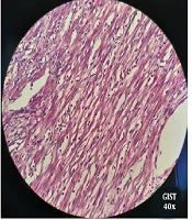
Microscopic findings revealed a spindle cell neoplasm. These cells contained eosinophilic cytoplasm and inconspicuous nucleoli.

Cut sections show a CD117 and DOG1 positive spindle cell type GIST (Figure 3).

Pathologic stain and IHC

The greatest dimension of the tumor is 8cm and the mitotic rate is > 5/5 mm2; therefore, the tumor is in the high-risk category for progressive disease on Miettinen’s criteria. Margins are clear and also 7 reactive lymph nodes are submitted with no vascular invasion.

Follow-up pathology revealed that the tumor was GIST. Tumor size was 8*5*5 cm with a clear margin and 7 dissected lymph nodes were reactive. Pathologic stage was PT3N0Mx.

He was discussed in a multidisciplinary team and further treatment with tyrosine kinase inhibitors was started.

3. Discussion

Epidemiological studies show that GIST occurs about 6.8 to 14.5 cases per million individuals every year and the mean age is 60 (26-28). The most common site for GISTs is in the stomach (51%). Other sites are the small intestine with 36% of tumors, colon with 7%, rectum with 5%, and esophagus with 1% of all GISTs (29).

The most common symptom is gastrointestinal bleeding that may present with acute melena and hematemesis with subsequent anemia. Other symptoms may present with weakness and abdominal pain and less commonly with distension, as well as discomfort due to a tumor-induced mass effect (30). Incidentally, the diagnosis of GIST occurred in 15% to 30% of patients after their death and autopsy, or patients who underwent surgery for other reasons, and this tumor was found during surgery (31).

The symptom of our patient was abdominal pain and upper GI bleeding. The operative finding was perforation of the antrum and right-sided paraesophageal hernia. Also, most previous studies such as Roy et al. (26) and Hosamani et al. (13) showed perforation of GIST on the small intestine and Meckel's diverticulum. In the patient presented here perforation was in the antrum. Skipworth et al. (Roy et al., 2012) (29) reported perforation in the stomach. The reason for the lower risk of gastric perforation is unclear but it may due to the thickness of the gastric wall compared to the large and small bowel. Perforated GISTs should undergo an operation for emergency surgical resection. The mortality rate of gastrointestinal tract perforation for all reasons is high (> 25%). Though perforation of Gastric GIST is extremely rare, it should be kept in mind while dealing with GIST, stomach, or perforation peritonitis.

3.1. Conclusions

In this study, we described one of the rarest of a gastric GIST presenting with perforation. GISTs should be in the differential diagnosis of all patients, who are diagnosed with GI tract masses and GI tract perforation. Our case shows that there is a need for more attention to new upper GI symptoms in all age groups to prevent dangerous consequences.

References

  • 1.

    Blanke C, Eisenberg BL, Heinrich M. Epidemiology of GIST. Am J Gastroenterol. 2005;100(10):2366. [PubMed ID: 16181397]. https://doi.org/10.1111/j.1572-0241.2005.50650_6.x.

  • 2.

    Chaudhry UI, DeMatteo RP. Management of resectable gastrointestinal stromal tumor. Hematol Oncol Clin North Am. 2009;23(1):79-96. viii. [PubMed ID: 19248972]. [PubMed Central ID: PMC2664970]. https://doi.org/10.1016/j.hoc.2009.01.001.

  • 3.

    Connolly EM, Gaffney E, Reynolds JV. Gastrointestinal stromal tumours. Br J Surg. 2003;90(10):1178-86. [PubMed ID: 14515284]. https://doi.org/10.1002/bjs.4352.

  • 4.

    DeMatteo RP, Ballman KV, Antonescu CR, Maki RG, Pisters PW, Demetri GD, et al. Adjuvant imatinib mesylate after resection of localised, primary gastrointestinal stromal tumour: a randomised, double-blind, placebo-controlled trial. Lancet. 2009;373(9669):1097-104. https://doi.org/10.1016/s0140-6736(09)60500-6.

  • 5.

    DeMatteo RP, Lewis JJ, Leung D, Mudan SS, Woodruff JM, Brennan MF. Two hundred gastrointestinal stromal tumors: recurrence patterns and prognostic factors for survival. Ann Surg. 2000;231(1):51-8. [PubMed ID: 10636102]. [PubMed Central ID: PMC1420965]. https://doi.org/10.1097/00000658-200001000-00008.

  • 6.

    Dudeja V, Armstrong LH, Gupta P, Ansel H, Askari S, Al-Refaie WB. Emergence of imatinib resistance associated with downregulation of c-kit expression in recurrent gastrointestinal stromal tumor (GIST): optimal timing of resection. J Gastrointest Surg. 2010;14(3):557-61. [PubMed ID: 20033343]. https://doi.org/10.1007/s11605-009-1121-2.

  • 7.

    Efremidou EI, Liratzopoulos N, Papageorgiou MS, Romanidis K, Manolas KJ, Minopoulos GJ. Perforated GIST of the small intestine as a rare cause of acute abdomen: surgical treatment and adjuvant therapy. Case report. J Gastrointestin Liver Dis. 2006;15(3):297.

  • 8.

    Fletcher CD, Berman JJ, Corless C, Gorstein F, Lasota J, Longley BJ, et al. Diagnosis of gastrointestinal stromal tumors: A consensus approach. Hum Pathol. 2002;33(5):459-65. [PubMed ID: 12094370]. https://doi.org/10.1053/hupa.2002.123545.

  • 9.

    Gold JS, Dematteo RP. Neoadjuvant therapy for gastrointestinal stromal tumor (GIST): racing against resistance. Ann Surg Oncol. 2007;14(4):1247-8. [PubMed ID: 17265116]. https://doi.org/10.1245/s10434-006-9291-6.

  • 10.

    Gold JS, Gönen M, Gutiérrez A, Broto JM, García-del-Muro X, Smyrk TC, et al. Development and validation of a prognostic nomogram for recurrence-free survival after complete surgical resection of localised primary gastrointestinal stromal tumour: a retrospective analysis. Lancet Oncol. 2009;10(11):1045-52. https://doi.org/10.1016/s1470-2045(09)70242-6.

  • 11.

    Heinrich MC, Corless CL, Duensing A, McGreevey L, Chen CJ, Joseph N, et al. PDGFRA activating mutations in gastrointestinal stromal tumors. Science. 2003;299(5607):708-10. [PubMed ID: 12522257]. https://doi.org/10.1126/science.1079666.

  • 12.

    Hirota S, Isozaki K, Moriyama Y, Hashimoto K, Nishida T, Ishiguro S, et al. Gain-of-function mutations of c-kit in human gastrointestinal stromal tumors. Science. 1998;279(5350):577-80. [PubMed ID: 9438854]. https://doi.org/10.1126/science.279.5350.577.

  • 13.

    Hosamani IR, Kodaganur S, Chaukimath SM. Perforated Gastrointestinal Stromal Tumor of the Meckel's Diverticulum: a Case Report and Review of Literature. Indian J Surg. 2016;78(5):390-5. [PubMed ID: 27994335]. [PubMed Central ID: PMC5127989]. https://doi.org/10.1007/s12262-016-1525-7.

  • 14.

    Huang CC, Yang CY, Lai IR, Chen CN, Lee PH, Lin MT. Gastrointestinal stromal tumor of the small intestine: a clinicopathologic study of 70 cases in the postimatinib era. World J Surg. 2009;33(4):828-34. [PubMed ID: 19198935]. https://doi.org/10.1007/s00268-009-9918-4.

  • 15.

    Joensuu H, Roberts PJ, Sarlomo-Rikala M, Andersson LC, Tervahartiala P, Tuveson D, et al. Effect of the tyrosine kinase inhibitor STI571 in a patient with a metastatic gastrointestinal stromal tumor. N Engl J Med. 2001;344(14):1052-6. [PubMed ID: 11287975]. https://doi.org/10.1056/NEJM200104053441404.

  • 16.

    Keung EZ, Raut CP. Management of Gastrointestinal Stromal Tumors. Surg Clin North Am. 2017;97(2):437-52. [PubMed ID: 28325196]. https://doi.org/10.1016/j.suc.2016.12.001.

  • 17.

    Kindblom LG, Remotti HE, Aldenborg F, Meis-Kindblom JM. Gastrointestinal pacemaker cell tumor (GIPACT): gastrointestinal stromal tumors show phenotypic characteristics of the interstitial cells of Cajal. Am J Pathol. 1998;152(5):1259-69. [PubMed ID: 9588894]. [PubMed Central ID: PMC1858579].

  • 18.

    Liegl-Atzwanger B, Fletcher JA, Fletcher CD. Gastrointestinal stromal tumors. Virchows Arch. 2010;456(2):111-27. [PubMed ID: 20165865]. https://doi.org/10.1007/s00428-010-0891-y.

  • 19.

    Miettinen M, Makhlouf H, Sobin LH, Lasota J. Gastrointestinal stromal tumors of the jejunum and ileum: a clinicopathologic, immunohistochemical, and molecular genetic study of 906 cases before imatinib with long-term follow-up. Am J Surg Pathol. 2006;30(4):477-89. [PubMed ID: 16625094]. https://doi.org/10.1097/00000478-200604000-00008.

  • 20.

    Miettinen M, Sobin LH, Lasota J. Gastrointestinal stromal tumors of the stomach: a clinicopathologic, immunohistochemical, and molecular genetic study of 1765 cases with long-term follow-up. Am J Surg Pathol. 2005;29(1):52-68. [PubMed ID: 15613856]. https://doi.org/10.1097/01.pas.0000146010.92933.de.

  • 21.

    Miettinen M, Wang ZF, Lasota J. DOG1 antibody in the differential diagnosis of gastrointestinal stromal tumors: a study of 1840 cases. Am J Surg Pathol. 2009;33(9):1401-8. [PubMed ID: 19606013]. https://doi.org/10.1097/PAS.0b013e3181a90e1a.

  • 22.

    Nilsson B, Andersson A, Ahlman H. Adjuvant and down-staging treatment with imatinib in gastrointestinal stromal tumors. J Surg Oncol. 2008;98(3):145-6. [PubMed ID: 18452216]. https://doi.org/10.1002/jso.21043.

  • 23.

    Nilsson B, Nilsson O, Ahlman H. Treatment of gastrointestinal stromal tumours: imatinib, sunitinib -- and then? Expert Opin Investig Drugs. 2009;18(4):457-68. [PubMed ID: 19335275]. https://doi.org/10.1517/13543780902806400.

  • 24.

    Nishida T, Blay JY, Hirota S, Kitagawa Y, Kang YK. The standard diagnosis, treatment, and follow-up of gastrointestinal stromal tumors based on guidelines. Gastric Cancer. 2016;19(1):3-14. [PubMed ID: 26276366]. [PubMed Central ID: PMC4688306]. https://doi.org/10.1007/s10120-015-0526-8.

  • 25.

    Oida Y, Motojuku M, Morikawa G, Mukai M, Shimizu K, Imaizumi T, et al. Laparoscopic-assisted resection of gastrointestinal stromal tumor in small intestine. Hepatogastroenterology. 2008;55(81):146-9. [PubMed ID: 18507095].

  • 26.

    Roy SD, Khan D, De KK, De U. Spontaneous perforation of jejunal gastrintestinal stromal tumour (gist). Case report and review of literature. World J Emerg Surg. 2012;7(1):37. [PubMed ID: 23194007]. [PubMed Central ID: PMC3537533]. https://doi.org/10.1186/1749-7922-7-37.

  • 27.

    Ruka W, Rutkowski P, Szawlowski A, Nowecki Z, Debiec-Rychter M, Grzesiakowska U, et al. Surgical resection of residual disease in initially inoperable imatinib-resistant/intolerant gastrointestinal stromal tumor treated with sunitinib. Eur J Surg Oncol. 2009;35(1):87-91. [PubMed ID: 18289826]. https://doi.org/10.1016/j.ejso.2008.01.003.

  • 28.

    Schoffel N, Groneberg DA, Kaul T, Laatsch D, Thielemann H. [Gastrointestinal stromal tumors (GIST)--literature review]. MMW Fortschr Med. 2016;158(3):60-2. [PubMed ID: 27119704]. https://doi.org/10.1007/s15006-016-7824-x.

  • 29.

    Skipworth JR, Fanshawe AE, West MJ, Al-Bahrani A. Perforation as a rare presentation of gastric gastrointestinal stromal tumours: a case report and review of the literature. Ann R Coll Surg Engl. 2014;96(1):96-100. [PubMed ID: 24417854]. [PubMed Central ID: PMC5137650]. https://doi.org/10.1308/003588414X13824511650010.

  • 30.

    Tran T, Davila JA, El-Serag HB. The epidemiology of malignant gastrointestinal stromal tumors: an analysis of 1,458 cases from 1992 to 2000. Am J Gastroenterol. 2005;100(1):162-8. [PubMed ID: 15654796]. https://doi.org/10.1111/j.1572-0241.2005.40709.x.

  • 31.

    van Oosterom AT, Judson I, Verweij J, Stroobants S, di Paola ED, Dimitrijevic S, et al. Safety and efficacy of imatinib (STI571) in metastatic gastrointestinal stromal tumours: a phase I study. Lancet. 2001;358(9291):1421-3. https://doi.org/10.1016/s0140-6736(01)06535-7.