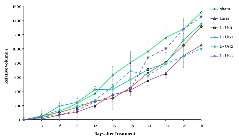
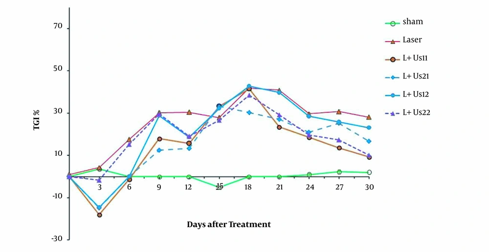
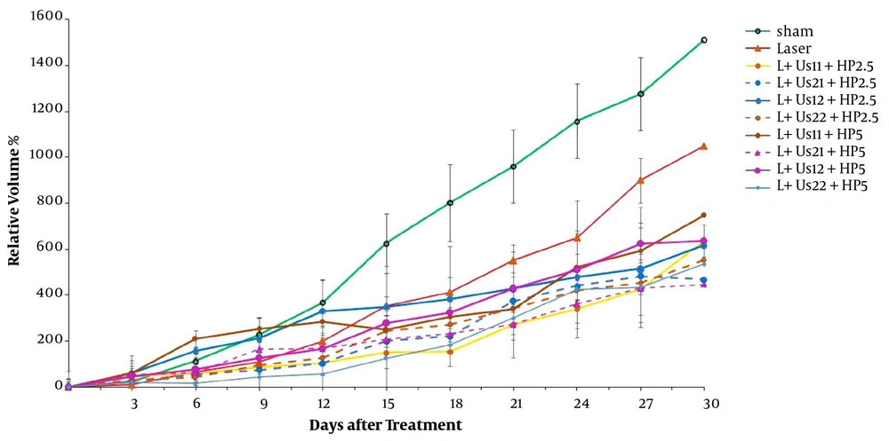
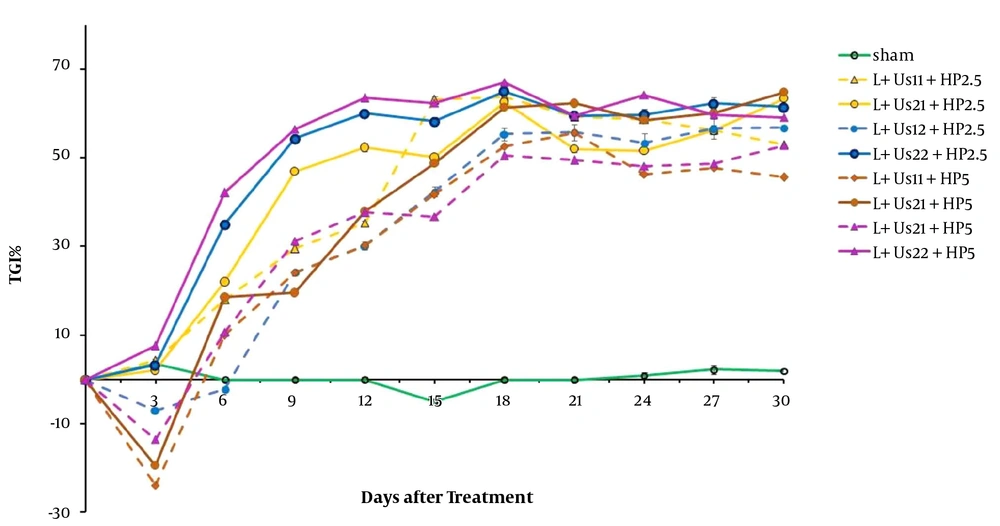
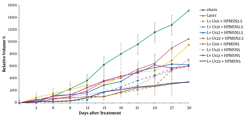
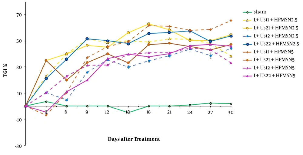
The results of the above-mentioned experiments show that the combined dual-frequency ultrasound (1 + 3 MHz) and/or laser (650 nm) radiation caused a transient inhibition effect on mice breast adenocarcinoma tumor growth. Also, HP-mediated SPDT (2.5 and 5 mg/kg) caused anti-tumor effect. Moreover, HP-MSNs-mediated SPDT (2.5 and 5 mg/kg) had inhibition effect on tumor growth. The TGI ratio increased in all experimental groups at 12 days after the initiation of radiation and persisted over 30 days of treatment. These findings are in agreement with Barati et al.'s findings that dual ultrasound (1 MHz + 150 kHz) for 30 min decreased mice breast adenocarcinoma tumor growth (
28). In addition, Guan and Xu showed that high-intensity focused ultrasound (1.6 MHz) could destroy proliferating tumor cells in human breast cancer (
29). In agreement with our results, the findings of Banerjee et al. confirm a potential role for PDT with verteporfin and laser (690 nm) in the management of early breast cancer in 12 female patients (
1). Miyoshi et al. suggested that combination therapy of SDT (titanium oxide)/ PDT (aminolevulinic acid) can help to get a reasonable anti-tumor effect on mice squamous cell carcinoma because the ultrasound (1 MHz) deeper penetration into the cancer tissue was compared with the laser light (635 nm) (
5). Moreover, the findings demonstrated that chlorin e6-mediated SPDT enhanced the antitumor efficacy on 4T1 mammary cancer cells compared with SDT (1 MHz) and PDT (laser 650 nm) alone (
30). The combination of PDT (665 nm) and SDT (3.3 MHz) have shown an improved glioblastoma cell in vitro and in vivo, which could be referred to as a synergetic effect. Exposing the nano-formulation HPPH with ultrasound also triggered the release of PS (
31). In An et al.'s study, a 630 nm semiconductor laser and 1 MHz ultrasound were used to perform SPDT. In An et al.'s study, SPDT with 630 nm laser and 1 MHz ultrasound + sinoporphyrin sodium inhibited glioma cell proliferation and induced cell apoptosis due to the generation of ROS and affecting protein expression (
32). Moreover, Hong et al. proposed SPDT with a nanoformulation Ce6-P/WNE in the treatment of prostate cancer cells. The results concluded that activated Ce6-P/WNEs in tumor cells by light (633 nm) and/or ultrasound (2.1 MHz) produced ROS even in a hypoxic environment (
19).
The structure of mesoporous channels would allow controllable drug release by mechanical and cavitation effects of ultrasound (
33). The collapse of cavitating bubbles can cause sonomechanical and sonochemical cytotoxic effects and the formation of cytotoxic reactive oxygen species (
34). Hence, this means that a combination of ultrasound and HP-MSNs could have a better treatment effect on mice breast adenocarcinoma. In agreement with our findings, Zhang et al. investigated that the therapeutic effect of encapsulated HP-SDT was better than HP or ultrasound alone (
35). In addition, during a study by Hasanzadeh et al. to review the effect of dual-frequency ultrasound radiation on nanomicellar containing doxorubicin, the SDT increased the ultrasound cavitations' efficiency (
13).
Cellular antioxidant capacity reduction may cause by oxidative stress. Free radical formation impairs the cell membrane fatty acids and proteins' function. As well, free radical production reacts with gene mutation and DNA damage, which provokes cancer development (
36). PDT applies its effects when the light is used to activate a photochemical reaction of a non-toxic PS, which generates reactive oxygen species (ROS). These ROS cause oxidative damage to lipids, proteins, and nucleic acids, which lead to the destruction of cancer cells through apoptosis or necrosis. The mentioned constructions have strong light absorption at wavelengths > 650 nm and are essential for good tissue penetration of light (
37). The destruction effect of PDT not only depends on the type of PS, but also is related to light exposure and fluency, oxygen level, localization of sensitizer, drug administration time, and other parameters (
38). Despite the satisfactory advantages of PDT, the clinical application of this approach has been limited. Several reasons can be explained such as the poor penetration of light and its dependence on the tumor tissue oxygen presence (
39).
Ultrasound good tissue penetration and wave energy focusing on the depth of tumor tissue can produce bio-effects (
11). It mentioned that acoustic cavitation is the main cause of destructive chemical reactions and the free radical production since ultrasound irradiation. During a study by Feng et al., the effect of mixing two-frequency and three-frequency ultrasonic waves on cavitations' efficiency was investigated. The results showed that irradiation of two or more ultrasound waves significantly increases the cavitations' efficiency compared to single-frequency irradiation (
40). The simultaneous effects of sensitizer and ultrasound comprise mechanical, chemical, and cavitational mechanisms (
28). Protoporphyrin that is used in PDT can generate active oxygen species after activation by visible light (
41). SDT followed by PDT can help to get a realistic anti-tumor effect because ultrasound has deeper penetration into the tumor tissue compared to laser light. Besides the singlet oxygen mechanism in PDT, mechanical stress, cavitational effects, and reactive oxygen species comprise SDT (
4).
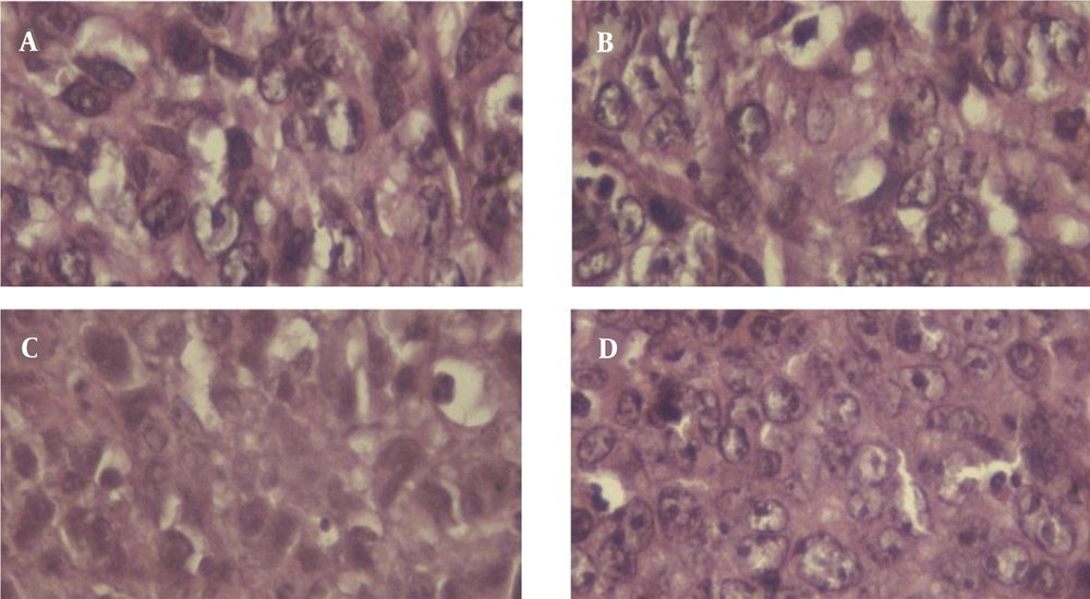
The histopathological results (Bloom-Richardson classification basis) showed that the sham and laser groups had grade III malignancy (poorly differentiated), while HP-mediated SPDT and HP-MSNs-mediated SPDT groups had grade I malignancy (well-differentiated) in the histological study of mice breast adenocarcinoma. Overall comparison test of survival equality for the different levels of groups demonstrated a significant difference between groups: Log rank (Mantel-Cox), P = 0.014. This may cause simultaneous radiation of dual-frequency ultrasound and laser radiation. On contrary, analysis of the author's previous investigation revealed that injection of HP and HP-MSNs (2.5 mg/kg) did not show any effect on T2 time and tumor relative volume, and the tumors had a poorly differentiated grading. The results of single-frequency SDT were not only determined by ultrasound wave power density, but also were related to HP-MSN injected dose (
25). Hong et al.'s study demonstrated that to overcome the limitations of each modality in the hypoxia environment, the strengths of PDT and SDT could be combined. The ultrasound deep tissue penetration combined with sensitizer activation by light will significantly enhance the therapeutic applications for prostate cancer cells (
19). In theory, the tumor cell toxicity effects of SDT and PDT are facilitated by cytotoxic drugs that are produced by sonochemical or photochemical reactions (
42).
The opinion of SPDT is based on the special buildup of sensitizer in tumors, and the improved cytotoxicity after activation by ultrasound or light. The imaginable tumor uptake could be explained as (1) selective buildup related to the tumor surrounding microenvironment, (2) lipoprotein receptors in some of the tumor cells, and (3) endocytosis mechanism to the entrance of sensitizer and low-density protein binding. In addition, because of the lower pH value in tumors, tumor-associated macrophages receive large amounts of porphyrin derivatives (
4). SPDT has been used in the treatment of many cancers with variable success, but the efficacy of breast adenocarcinoma damage induced by dual-frequency SPDT with HP-MSNs has rarely been reported. The novelty of the present SPDT study was activation of HP and HP-MSNs (2.5 and 5 mg/kg) in the presence of two intensities (1 and 2 W/cm
2) of 1 and 3 MHz ultrasound and laser radiation (650 nm) in the management of Inbred Balb/C mice grafted breast adenocarcinoma. Aside from only one laser light radiation (650 nm), another limitation of this research was once sensitizer injection with a temporary treatment effect. The fractional injection of sensitizer should be done in our future study.
5.1. Conclusions
The results of this study demonstrated that HP and HP-MSNs-mediated SPDT have an anti-tumor effect in mice breast adenocarcinoma. It can be appreciated that careful selection of the sensitizer with SPDT will play an eminent role in the success of cancer therapies. Improvement sensitizer agents used in PDT and SDT are useful drugs for SPDT and can expand cancer treatment management. However, further studies are required to optimize the sensitizer, light/laser, and ultrasound parameters to find better tumor treatment methods and explain the mechanism of SPDT.