1. Background
Skin malignancies are the most common type of malignancy and their incidence has gone through an increase of ~4 to 8% per year during the last 40 years (1). Non-melanoma skin cancer is the most common type of cancer (2) that is mainly represented by basal cell carcinoma (BCC) (80%) and squamous cell carcinomas (SCC) (20%) (3). The BCC is the most common malignancy worldwide in white people (4). Free margins are very important on large resections in a case of malignancy and therefore, for skin tumors such as BCC and SCC, occasionally need to be evaluated for the best cosmetic results (5). Traditional surgical treatment of SCC and BCC cancers include excision with subsequent assessment of margins, either with frozen sections (FSs) during surgery or after excision and closure. Mohs’ micrographic surgery (MMS) is an outpatient procedure that maximizes surgical margin assessment while reducing the amount of tissue that must be excised (6). The MMS or FS have been recommended for difficult tumors such as recurrent tumors, perineural invasion or size > 2 cm, aggressive histological subtypes and located in anatomically sensitive areas (7). The MMS is a suitable treatment (8) and FS examination is the “gold standard” of surgical margins in tumoral skins (9), but FS is a time-consuming test and expensive tool (10). On the other hand, Tzanck smear test (TST) is simple, easy to perform, inexpensive and a rapid test for cytological study to confirm or exclude malignancy. Only a few studies have investigated the value of TST in the diagnosis of BCC (11). Analysis of diagnostic accuracy between TST and FS examination for margin control in surgery for BCC is the aim of this investigation.
2. Methods
2.1. Patients
In this cross-sectional study that has been permitted by the ethics committee of Kermanshah University of Medical Sciences (Code: 93295), Kermanshah, Iran, 59 patients were known cases of BCC who previously had incisinoal biopsy and documented report of BCC on histopathology with routine Hematoxylin and Eosin (H and E) staining. The exclusion criteria were a, lack of documented report of BCC with incisional biopsy; and b, presence of additional malignant component, for example Basosquamous carcinoma. They were admitted to the surgical ward, Imam Reza hospital, Kermanshah University of Medical sciences, Kermanshah, Iran, between April 2014, and July 2015. Written consent was achieved before surgery. For complete excision of the tumor, the method of surgery was MMS by the dermatologist.
2.2. Methods
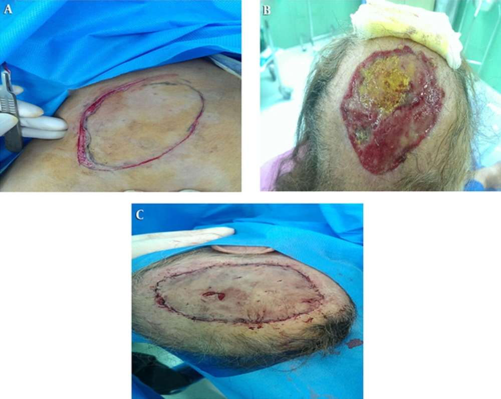
After local anesthesia (2% lidocaine and epinephrine), the tumor was excised (Figure 1) with a scalpel (no.15) and the TST was taken on a glass slide of the excised tumor for Papanicolaou staining (Pap staining) and reconfirmation of tumoral cells (BCC). Then the dermatologist took a 2 - 4 mm margin of lateral and deep border of tumoral mass for evaluation of surgical margins and divided into 0.5 cm fragments which were colored according to a map with different dyes. Marked fragments were sent to the pathology department on wet gauze with complete identification of patient for evaluation by the dermatopathologist with FS method as the gold standard (cryostat). Before dying the fragments, each margin fragment was touched with mild pressure on glass slide; immediately fixed with alcohol 96% and sent to the pathology department for Pap staining and pathology evaluation.
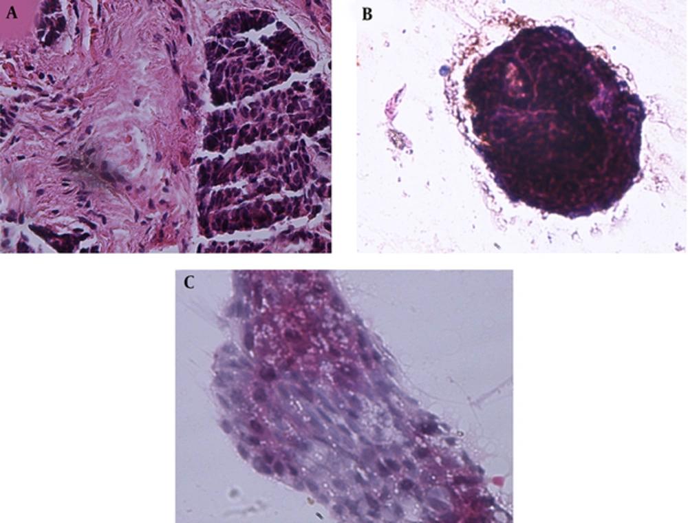
In the pathology department, each marked fragment was sectioned separately with cryostat by technicians under the supervision of the assistant to 5 µm sections and slides were provided. Then they were stained with rapid H and E method (Figure 2A) for evaluation by a dermatopathologist and assistant under two-head light microscope (Zeiss Axiostar Plus). The result of examination of each slide for the presence or absence of tumoral cells was reported to the dermatologist in the form of “involved by tumor or free of tumor” for each inked fragment immediately. If the report showed tumoral involvement, the re-excision of 2 mm was done by a dermatologist and again all the above mentioned process was repeated. This procedure was repeated until all tissue fragments were free of tumor. After that, the Pap stained slides were examined in a different session under light microscope for the presence or absence of tumoral cells. The dermatopathologist had been kept blind about the results of FS. Clusters of basaloid cells with round or oval nuclei, scanty cytoplasm and indistinct cell borders were accepted as tumoral cells and confirmed at light microscopy with magnifications of × 40, × 100 and × 400 (Figures 2B and 2C). After that, age, sex, tumor site and FS result of margins were checked in all the patients.
2.3. Diagnostic Accuracy of TST
The sensitivity, specificity, positive and negative predictive and likelihood values, Youden’s index and accuracy of TST for the evaluation of margin were calculated by comparing results of the TST and FS examinations. The kappa coefficient between the two methods was calculated. SPSS version 19.0 (SPSS, Inc., Chicago, IL) was applied for statistical analysis. P value < 0.05 was accepted as statistically significant.
3. Results
The mean age at diagnosis of 59 BCC patients was 69.9 years (range, 45 - 87 years) that 36 patients (61%) were males (Table 1).
| Variables | No. (%) | Mean | Range |
|---|---|---|---|
| Age, y | 69.9 | 45 - 87 | |
| Sex | |||
| Male | 36 (61) | ||
| Female | 23 (39) |
Out of all patients, 328 margins were obtained. The most common site of malignancies was the nose (n = 98), and then eye and forehead (each, n = 50); back (n = 37), cheek (n = 29), lip (n = 27), ear (n = 25) and scalp (n = 12) (Table 2).
| Variables | No. (%) |
|---|---|
| Tumor site | |
| Nose | 98 (29.8) |
| Eye | 50 (15.3) |
| Forehead | 50 (15.3) |
| Back | 37 (11.3) |
| Cheek | 29 (8.8) |
| Lip | 27 (8.2) |
| Ear | 25 (7.6) |
| Scalp | 12 (3.7) |
| FS result for margins | |
| Positive | 61 (18.6) |
| Negative | 267 (81.4) |
Checking blocks with FS examination showed 61 margin-positive and 267 margin-negative. In comparison between TST and FS examination, 17 true-positive and 253 true-negative results were achieved, whereas 14 false-positive and 44 false-negative. The sensitivity and specificity of TST for the evaluation of margin were 0.28 (95% CI = 0.16 - 0.42) and 0.95 (95% CI = 0.93 - 0.97), whereas positive and negative predictive values were 0.54 (95% CI = 0.36 - 0.73) and 0.85 (95% CI = 0.81 - 0.89). Also positive and negative likelihood values were 5.36 (95% CI= 2.77 - 10.19) and 0.76 (95% CI = 0.65 - 0.89), respectively. Diagnostic accuracy was 0.82 (95% CI = 0.77 - 0.87). The kappa coefficient of agreement between the two methods was 0.28 (P < 0.001). The Youden’s index was 0.23 (95% CI = 0.11 - 0.34).
4. Discussion
This study evaluates effects of TST on intraoperative clearing of tumor margin in the treatment of BCC compared with FS examination. Diagnosis of BCC is usually made clinically, which must then be confirmed microscopically. The treatment option for BCC consists of Mohs’ micrographic-controlled or en face frozen section controlled surgical excision with later routine pathology sections, cryotherapy, radiotherapy and medical treatment (12). MMS is said to be the “gold method” at some institutions (13, 14) that previous studies showed that excision of BCC with pathological evaluation of the margins, i.e. MMS and en face FS, have the lowest relapse rates and best tissue conservation rate (14). Our results demonstrate high diagnostic accuracy, but very low sensitivity and kappa coefficient for margin evaluation with TST.
The findings confirm the use of FS evaluation of margin in some patients suffering from SCC and BCC cancers of the head and neck (9, 15, 16). One study (17), reported the positive predictive value of subareolar FS is 100%, negative predictive value 83%, sensitivity 38%, and specificity 100%. The TST although is an old tool, still remains a simple, rapid, applicable, and low cost test for cutaneous lesions (11, 18). The TST may be used for assessing erosive vesiculobullous and granulomatous lesions, but more experience is needed for the evaluation of malignancies by TST (18). The diagnostic accuracy of the TST is clear, but its diagnostic reliability has been assessed only in herpetic infections and BCC (18). Durdu et al. (19) reported the diagnostic accuracy of the TST in the evaluation of pigmented skin lesions is equal to that in dermatoscopy. The TST may be a beneficial diagnostic method to dermatoscopy for identifying the melanocytic or non-melanocytic origin of certain pigmented cutaneous lesions. The results of a meta-analysis have demonstrated that this test has a very high sensitivity (97%, 95% CI 94 - 99) and specificity (86%, 95% CI 80 - 91) (20). The high accuracy of TST for margin control was encouraging to propose an applied evaluation alternative approach for well-demarcated BCC therapy (21) or other tumoral lesions (22). Compared with FS examination as “gold method for diagnosis of BCC” in study of Baba et al. (21) the sensitivity and specificity of TST for margin evaluation was 1.00 (95% CI = 1.00 - 1.00) and 0.99 (95% CI = 0.98 - 1.00), whereas positive and negative predictive values and diagnostic accuracy were 0.94 (95% CI = 0.84 - 1.05), 1.00 (95% CI = 1.00 - 1.00), and 1.00 (95% CI =0.99 - 1.00), respectively. The kappa coefficient of agreement between TST and FS examination was 0.97 (95% CI = 0.83 - 1.11), while in our study, the sensitivity and positive predictive value of TST for margin evaluation were very low (28% and 54%, respectively). Also, diagnostic accuracy was 82% and the kappa coefficient of agreement between TST and FS examination was 0.28 (P < 0.05). Also, compared with histopathology as the gold standard in the study of Dey et al. (23), the sensitivity and specificity of TST were 52.2 and 100%, respectively, and also positive predictive and negative predictive values were 100 and 21.42%, respectively. Therefore, TST may be indicated for initial evaluation to meet rapid diagnostic demand as well as in suspected recurrences. The TST for diagnosis of BCC has a number of limitations (18) and a negative cytodiagnosis should be judged with caution. Since TST does not give much information about the characteristics of the tumor and it must constantly be continued by routine pathology examination before making treatment plan (23). We can conclude that in comparing TST and Frozen section examination methods positive likelihood value and specificity of TST for margin evaluation are high and therefore, TST can be suitable in the diagnosis of BCC, but due to low sensitivity and kappa coefficient, TST alone cannot be a suitable alternative method compared to the FS examination for margin control in BCC.
In the present study BCC subtypes including nodular, micronodular, infiltrating, superficial multifocal and other less common ones are not considered in statistical analysis. We recommend further studies with considering these subtypes for better understanding of the cause of controversy in the results of researches.

