1. Background
Colorectal cancer (CRC) is a significant problem that claims many lives every year; it is the second leading cause of cancer-related death (1). The current major therapeutic approaches for CRC are chemotherapy and surgery. One of the most frequently used pharmacological treatments for cancer is 5-Fluorouracil (5-FU) with cytotoxic effects while increasing DNA damage and causing apoptosis (2). Although 5-FU is frequently applied even in patients with advanced metastatic CRC, the clinical application of 5-FU is restricted because of drug resistance development and severe side effects such as cardiotoxicity, nausea, and gastrointestinal adverse effects (3).
The current therapies for CRC are combination chemotherapy, including FOLFOX, XELOX/CAPOX, and FOLFIRI (4), as well as therapeutic antibodies against vascular endothelial growth factor (VEGF) and epidermal growth factor receptor (EGFR), which have successfully prolonged the survival of patients (5). Despite these achievements, the development of resistance, poor physicochemical properties, in vivo degradation, and side effects of current chemotherapeutic agents have made the need for urgent and essential treatment for CRC mandatory (6). In this way, identifying molecules and signaling pathways (SP) involved in CRC is imperative. According to the latest evidence, mutations lead to the abnormal expression of Wnt ligands, secreted Frizzled-related proteins (SFRP), β-catenin, and APC, which are complicated in different human cancers, especially CRC (7). The Wnt/β-catenin SP is a promising therapeutic target in CRC as more than 94% of CRC patients have shown at least 1 mutation in the proteins of this pathway (8). During stimulation of Wnt signaling, the high levels of β-catenin accumulation lead to nuclear translocation and interaction with transcription factors such as T-cell factor/lymphoid enhancer factor 1 (TCF/LEF) (9). β-catenin links to E-cadherin as the major cell-cell adhesion molecule in epithelial cells to assemble the E-cadherin/β-catenin complex. E-cadherin is a negative regulator in Wnt signaling. Meanwhile, it is believed that the loss of the E-cadherin contributes to metastasis (10).
In addition, mutations in the wnt SP may up-regulate the expression of VEGF, which plays a critical role in physiological and pathological angiogenesis (11, 12). Recently, studying anti-adhesive molecules in human milk led to the serendipitous discovery of human α-lactalbumin made lethal to tumor cells (HAMLET), a complex of oleic acid with human α-lactalbumin protein, which showed in vitro anticancer activity against different cancer cell lines notably, CRC cells, as well as in vivo antitumor activity (13). Based on the study by Puthia et al. (14), HAMLET could inhibit Wnt SP on colon cancer cells and reduce β-catenin and VEGF (15). Inspired by these results of HAMLET, complexes similar to HAMLET, such as bovine alpha-lactalbumin made lethal to tumor cells (BAMLET) (bovine lactoferrin binds oleic acid and bovine α-lactalbumin protein), were prepared to evaluate its anticancer effect (16). Tumoricidal properties were observed against several cancer cell lines, such as lung carcinoma cells (A549) and 2 colorectal adenocarcinoma cell lines (DLD1 and HT-29) similar to HAMLET. bovine alpha-lactalbumin made lethal to tumor cells could also induce cell death in vitro. It indicated a time- and dose-dependent uptake in A549, DLD1, and HT-29 cells (17). It is noteworthy that the large-scale production of HAMLET is challenging due to the limited quantities of human breast milk available (18). However, bovine α-lactalbumin has 85% homology with human α-lactalbumin and can be substituted. The effect of BAMLET complexes on Wnt SP has not yet been investigated on colon cancer.
2. Objectives
Our hypothesis was to examine how BAMLET and 5-FU are involved in targeting Wnt SP in adenocarcinoma cells to treat CRC. For this purpose, we evaluated the effect of BAMLET in comparison with 5-FU on cell viability, the expression levels of Wnt signaling-related proteins (β-catenin, E-cadherin) and genes (β-catenin and E-cadherin and VEGF), induced angiogenesis in HT-29, and HCT116 cells.
3. Methods
3.1. General Procedure for BAMLET Preparation
Following the methodology proposed by Kamijima et al., the BAMLET was prepared (Appendices) (19). Initially, α-lactalbumin was dissolved in phosphate-buffered saline (PBS) (700 µM). Then, oleic acid (120 molar equivalents) was mixed with the protein solution and kept at 50°C for 10 min, then cooling down to 25°C. The next step was centrifugation at 10 000 × g for 10 min. The excess oleic acid was removed by aspiration and dialyzed for 18 h. The complex was stored at -20°C.
3.2. Cell Culture
HCT116 cells were purchased from the National Cell Bank, Pasteur Institute of Iran. The cells were cultured in 75 cm2 falcon flasks in Roswell Park Memorial Institute media (RPMI-1640) (Bio Idea, Iran). HT-29 cells were obtained from the Bonyakhteh Institute, Iran, and cultured in Dulbecco’s Modified Eagle’s Medium high-glucose, high Glutamine (DMEM) (Bio Idea, Iran). Next, 10% fetal bovine serum (FBS) and 1% streptomycin were added to both media. A humidified atmosphere with 5% CO2 was used to keep the cells at 37°C. Then, the cells were seeded at 2000 cells/15 mL medium and subcultured weekly. Passaging was performed by washing (0.1 M PBS pH 7.2/2.6 mM EDTA) and detaching the cells by trypsinization (0.5% trypsin/2.6 mM EDTA). Two volumes of DMEM, which were mixed with 20% FBS, 100 U/mL penicillin, and 100 µg/mL streptomycin, were added to stop the enzymatic reaction. The replacement duration of the culture medium was every 48 h. To avoid passage number-related effects, the number of passages was limited to 3.
3.3. Cell Toxicity Assay
The in vitro antiproliferative activity of 5-FU and BAMLET was investigated against the human colon cancer cells HT-29 and HCT116, using the standard MTT cytotoxicity assay for 24 and 48 h. (20). The reason for selecting these cells was their differences in morphology, physiology, and expression of E-cadherin. E-cadherin expression levels are low in HCT116 cells compared to HT-29 cells. The HT-29 and HCT116 cells were cultured in DMEM and RPMI medium, respectively, as described in the previous study (21) and seeded into 96-well plates (500 cells/well) with 10% FBS and penicillin-streptomycin (500 µg/mL). They were followed by centrifugation at 37°C in a humidified atmosphere with 5% CO2 for 24 h. Freshly prepared culture growth medium containing final concentrations of 5FU ranging from 0.1 to 100 µM and BAMLET ranging from 100 to 600 µM were added to the wells for 24 and 48 h. Then, we added fresh MTT solution (5 mg/mL) to all wells, followed by incubation for 4 h at 37ºC. The medium containing unreacted MTT was removed carefully. Afterward, dissolvation of formazan crystals was performed in DMSO. The next step was determining the optical density at 570 nm, using an ELISA plate reader (BioTek, USA). Afterward, the following formula was applied to determine the cell viability:
Where, ASample, AControl, and ABlank refer to the determined absorbance for the cells treated with a tested compound, untreated cells, and blank wells, respectively. All experiments were conducted in triplicates at 5 concentrations.
3.4. Western Blot Analysis
HT-29 and HCT116 cells were treated with the compounds 5-FU (1, 0.1 µM in HT-29 and 10, 20 µM in HCT116) and BAMLET (250, 500 µM in HT-29 and 100, 200 µM in HCT116) at different concentrations according to their IC50 values for 48 h. After incubation, cells were lysed with lysis buffer (Tris buffer adjusted at pH 8.0 containing Tris-HCL, EDTA, NaCl, Sodium deoxycholate, SDS), 1 tablet protease inhibitor cocktail, and 10 µL Triton 1% and centrifuged at 10,000 rpm for 10 min at 4ºC. Using the supernatant soluble fraction, total protein was determined, using the bicinchoninic acid (BCA) Protein Assay. The Aliquots were mixed with a loading sample buffer and separated by 12% gradient SDS polyacrylamide gel (SDS-PAGE) at a constant voltage of 120 mA for 45 min. Then, the resolved proteins were transferred to the nitrocellulose membrane. The membranes were blocked for 2 h in blocking buffer (5% non-fat dry milk powder in PBS) followed by 2 h and 16 h incubation with primary and secondary antibodies, respectively (Anti-Mouse with 1: 500 concentration for primary antibody and 1: 1000 concentration for anti-mouse IgG HRP secondary antibody). Finally, after rinsing by PBS, an ECL Western Blotting analysis system (GE Healthcare, USA) was used to determine the bands (20).
3.5. Gene Expression Study
To evaluate the effects of 5-FU and BAMLET on the expression of genes that contribute to Wnt/β-catenin signaling and cancer stemness, HT-29 and HCT116 cells were treated with 5-FU (1, 0.1 µM in HT-29 and 10, 20 µM in HCT116) and BAMLET (250, 500 µM in HT-29 and 100, 200 µM in HCT116) for 48 h. The next step was harvesting cells and lysing them by scrapping directly in the Biozol reagent (1 mL for 25 cm2 flask) (Bioflux, china) and centrifuged at 12000 g for 15 min at 4°C. The RNA phase was left to precipitate at -20°C overnight. Then, it was re-suspended in 20 - 50 µL RNase-free water. Using the optical density (OD260/OD280 ratio), the purity and concentration of RNA were measured by NanoDrop 1000 spectrophotometer (Wilmington, DE, USA). The integrity of extracted RNA samples was examined by running 2 - 3 µg of RNAs on an agarose gel electrophoresis under denaturing conditions. The next step was adding DNAas and, then, synthesizing the first strand of complementary DNA (cDNA) from total RNA according to ThermoFisher Scientific cDNA Synthesis Kit instructions (USA). Finally, RT-qPCR was performed by SYBR green DNA PCR master mix (Amplicon, USA) for amplifying primers (methanation, Germany, summarized in Table 1) by the ABI real-time PCR 7500 system. The PCR reaction mixture contained 1 µL of cDNA (1 µg), 1 µL of 5 mmol/L forward and reverse primers, and 12.5 µL of SYBR green PCR master mix in a total volume of 20 µL. The RT-qPCR was performed as follows: a pre-cycling thermal activation at 95°C for 10 min, 40 cycles of denaturation at 95°C for 15 s, annealing at 60°C for 30 s, extension at 72°C for 30 s, and a final extension of 72°C for 10 min. Data analysis was administered, using the ΔΔCT technique. The experiments were performed in triplicates. GAPDH was amplified as a housekeeping gene.
| Gene | Primer Sequence |
|---|---|
| GAPDH | |
| Forward | 5'- CGACCACTTTGTCAAGCTCA -3' |
| Reverse | 5'- AGGGGTCTACATGGCAACTG -3' |
| VEGF | |
| Forward | 5 '- CCTTGCCTTGCTGCTCTACCT -3' |
| Reverse | 5' - GTGATGATTCTGCCCTCCTCCT-3' |
| β-Catenin | |
| Forward | 5′-AAAATGGCAGTGCGTTTAG-3′ |
| Reverse | 5′-TTTGAAGGCAGTCTGTCGTA-3 |
| E-Cadherin | |
| Forward | 5 '- GGATGTTGCTCAGGGTGGA -3' |
| Reverse | 5' - TAGGTAGGAGGTGAAGACGCT -3' |
Sequence of Primers
3.6. Data Analysis
The findings are described, using the mean ± standard deviation (SD) of 3 different replicates (n = 3) except flow cytometry data (n = 2). Inferential statistics were determined by Graphpad prism software v.6.0 (GraphPad Prism, RRID: SCR_002798), using one-way or two-way analysis of variance (ANOVA) and Bonferroni's posthoc test. A P-value less than 0.05 was considered statistical significance.
4. Results
4.1. In Vitro Cytotoxicity Activity of 5-FU and BAMLET on HT-29 and HCT116 Cells
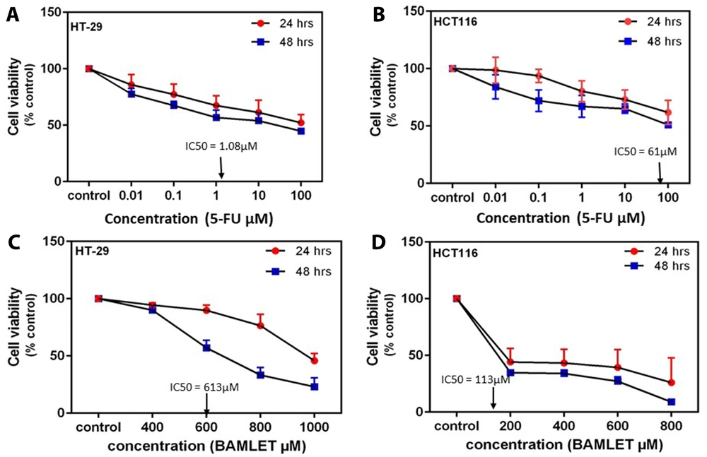
Both types of colon cancer cells responded to the cytotoxic effect of 5-FU in a dose- and time-dependent manner. For instance, treatment of HT-29 cells with 5-FU (100 µM) decreased cell viability to 49.6 and 33.4% after 24 and 48 h, respectively (Figure 1A). The results in Figure 1A and B demonstrated that 5-FU at the concentration of 100 µM reduced the percentages of cell viability in HT-29 and HCT116 cells to 33.4% and 51.02% in 48 h, respectively. Dose-response curves were constructed and the IC50 values for 5FU at 48 h treatment were obtained at about 1.08 and 61 µM in HT-29 and HCT116 cells, respectively. According to Figure 1C and D, the results of BAMLET exhibited that the cytotoxicity of BAMLET on HCT116 was not time-dependent. For example, the percentage of cell viability in HCT116 cells treated with BAMLET (600 µM) decreased to 1.6% and 24.6% in 24 and 48 h, respectively. The IC50 values for BAMLET were obtained at 113 and 613 µM in HCT116 and HT-29 cells, respectively.
Effects of different concentration of compound 5-FU; 0.01, 0.1, 1, 10, 100 µM (A and B) on the cell growth inhibition in (A) HT-29 and (B) HCT116 cells in 24 h and 48 h. Effects of different concentration of compound BAMLET; 200, 400, 600, 800, 1000 µM (C and D) on the cell growth inhibition in (C) HT-29 and (D) HCT116 cells in 24 h and 48 h. The cell viability was measured by the MTT assay as described in the experimental section. All the experiments were performed in triplicate.
4.2. Effects of 5-FU and BAMLET on the Wnt SP in HT-29 and HCT116 Cells
To understand the effect of 5FU and BAMLET on Wnt SP, the expression of β-catenin and other associated proteins like E-cadherin were investigated in HT-29 and HCT116 cells, using Western Blotting and real-time PCR analysis.
4.3. Effects of the 5FU and BAMLET on the Expression of β-Catenin
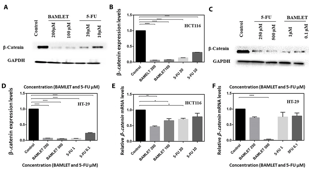
As shown in Figure 2, 5-FU and BAMLET effectively can dose-dependently suppress the expression of β-catenin in both cells (P < 0.0001). The results of the mRNA expression level of β-catenin using real-time PCR assay agree with the down-regulation of β-catenin upon exposure by 5-FU and BAMLET on HT-29 and HCT116 cells.
Effects of the 5-FU and BAMLET on the Wnt signaling pathway via expression of β-catenin in HT-29 and HCT116 cells. Cells were treated with varying concentrations of the 5-FU; 1, 0.1 µM in HT-29 (D and F) and 10, 20 µM in HCT116 (B and E) and BAMLET compounds; 250, 500 µM in HT-29 (D and F) and 100, 200 µM in HCT116 (B and E) for 48 h. The cells were harvested and lysed for detection of the expression levels of β-catenin through Western Blot analysis in (A) HCT116 and (C) HT-29 cells. Histograms display the density ratios of β-catenin to GAPDH (B) in HCT116 and (D) HT-29 cells. Effects of the 5-FU and BAMLET on relative mRNA expression for the markers associated with Wnt signaling pathway analyzed by RT-qPCR. Histograms display expression level of β-catenin in (E) HCT116 and (F) HT-29 cells. *: P < 0.05 and **: P < 0.01, ****: P < 0.0001 were considered as significant versus control.
4.4. Effects of the 5FU and BAMLET on the Expression of E-Cadherin
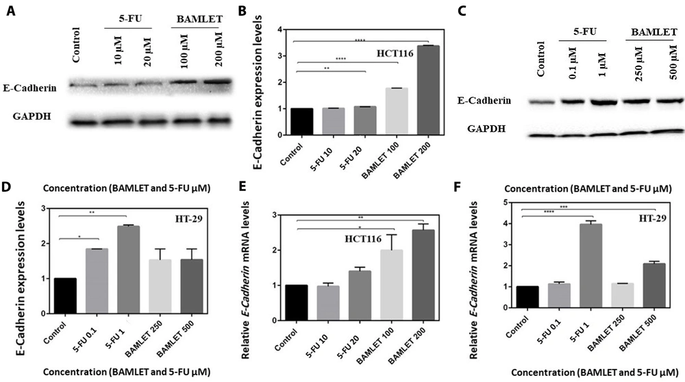
As illustrated in Figure 3, 5-FU and BAMLET led to an increase in the E-cadherin expression level proportional to 5-FU and BAMLET concentrations on both cells. Also, the validation of these findings was performed by real-time PCR analysis. The result showed that BAMLET significantly increased the E-cadherin expression level in HCT116 cells compared to the same treatment with 5-FU (P < 0.0001). However, in HT-29 cells, the most effective compound was 5-FU with an increased E-cadherin expression (P < 0.0001).
Effects of the 5-FU and BAMLET on the Wnt signaling pathway via expression of E-cadherin in HT-29 and HCT116 cells. Cells were treated with varying concentrations of 5-FU; 1, 0.1 µM in HT-29 (D and F) and 10, 20 µM in HCT116 (B and E) and BAMLET compounds; 250, 500 µM in HT-29 (D and F) and 100, 200 µM in HCT116 (B and E) for 48 hr. The cells were harvested and lysed for detection of the expression levels of Ecadherin through Western Blot analysis in (A) HCT116 and (C) HT-29 cells. Histograms display the density ratios of E-cadherin to GAPDH in (B) HCT116 and (D) HT-29 cells. Effects of 5-FU and BAMLET on relative mRNA expression for the markers associated with Wnt signaling pathway analyzed by RT-qPCR. Histograms display expression level of E-cadherin in (E) HCT116 and (F) HT-29 cells. *: P < 0.05, **: P < 0.001, ***: P < 0.001, ****: P < 0.0001 were considered as significant versus control.
4.5. Effects of the 5-FU and BAMLET on the Expression of VEGF
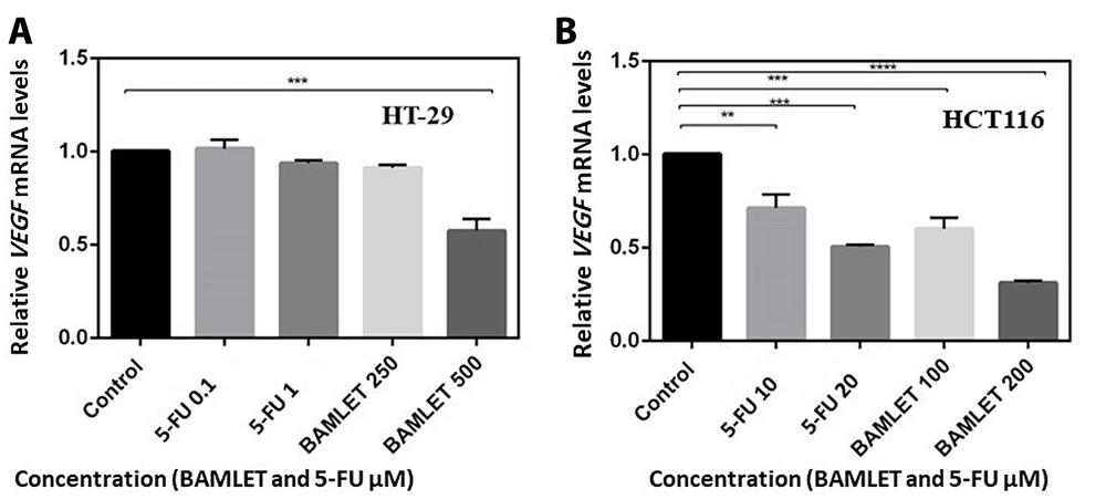
The results of 5-FU and BAMLET compounds on angiogenesis were revealed by evaluating the expression level of VEGF upon exposure to tested compounds. As shown in Figure 4, an increase in the concentration of 5-FU and BAMLET resulted in the down-regulation of VEGF expression (P < 0.0001).
Effects of different concentration of the 5-FU; 1, 0.1 µM in HT-29 (A) and 10, 20 µM in HCT116 (B) and BAMLET; 250, 500 µM in HT-29 (A) and 100, 200 µM in HCT116 (B) on the relative mRNA expression of the markers associated with angiogenesis analyzed by RT-qPCR. Histograms display expression levels of VEGF in (A) HT-29 and (B) HCT116 cells. **: P < 0.01, ***: P < 0.001, ****: P < 0.0001 were considered as significant versus control.
5. Discussion
This research demonstrated the influence of 5-FU and BAMLET on Wnt SP in HT-29 and HCT116 cells, which involves cell proliferation and angiogenesis. Previously, it was indicated that 5FU was engaged in targeting Wnt SP and inducing apoptosis in adenocarcinoma cells (22). The evaluation of cellular cytotoxicity using MTT assay showed that 5-FU is more cytotoxic than BAMLET in HT-29 and HCT116 cells with lower IC50 values in 48 h exposure. These compounds could suppress the growth of HT29 cells in a dose- and time-dependent manner. Nevertheless, the effects of BAMLET on HCT116 cells were not time-dependent (18). In a similar study, it was shown that prolonged exposure of HT-29 cells to 5FU has a significant influence on cytotoxicity as it activates autophagy in a time-dependent manner (23). To elucidate the ability of BAMLET and 5-FU to target Wnt SP in HT29 and HCT116 cells, different concentrations based on their IC50 values were tested and subjected to a series of further detection assays. Selective BAMLET concentrations were obtained based on the MTT test and determination of IC50. On the other hand, these concentrations were consistent with previous available studies. For instance, Ramer et al. indicated that BAMLET (200 mg/mL) triggers the lysosomal cell death pathway in cancer cells (24). bovine alpha-lactalbumin made lethal to tumor cells and 5-FU decreased the expression levels of β-catenin in HT-29 and HCT116 cells. β-catenin plays a significant role in regulating the β-catenin/Wnt SP, which transduces intracellular signals in transcriptional regulation (5). In canonical Wnt SP, the protein β-catenin moves from the cytoplasm to the nucleus and interacts with the transcription machinery to regulate the expression of genes with different biological effects. Therefore, the Wnt pathway regulates various processes like cell proliferation and migration, angiogenesis, and stemness, which are essential in tumorigenesis (25, 26). The absence of Wnt signaling harms the accumulation of β-catenin in the cytoplasm due to its degradation by a destruction complex including different proteins (9, 27-30). Dysregulation of the Wnt pathway contributes to the development of various cancers like CRC (31). Our findings indicated that BAMLET suppressed β-catenin expression in HT-29 and HCT116 cells more effectively than 5-FU. Consistent with our results, HAMLET, a BAMLET-like compound, reduced the β-catenin expression in APC Min /+ mice (14). Our results also showed that E-cadherin at protein and mRNA levels increased in HT-29 and HCT116 cells upon the addition of BAMLET and 5FU. In the progress of human cancers, E-cadherin, as the central cell-cell adhesion molecule in epithelial cells, plays an essential role via linking to β-catenin to assemble the E-cadherin/catenin complex (32). E-cadherin is a negative regulator of the Wnt SP. Hence, the increased cadherin expression leads to inhibition of β-catenin-dependent transcription (33). HT-29 and HCT116 cells are different cell lines from the E-cadherin status point of view (21). Although the expression level of E-cadherin is low in HCT116 cells compared to HT29 cells, a dose-dependent increase in E-cadherin expression was found in HCT116 cells exposed to 5-FU and BAMLET. Evaluation of the E-cadherin expression showed that 5FU up-regulated the expression level of E-cadherin in HT-29 cells more than BAMLET.
Accordingly, it can be suggested that BAMLET and 5-FU exert their activity partly by preventing abnormal Wnt signaling activation in HT-29 and HCT116 cells. On the other hand, loss of E-cadherin is thought to enable metastasis (10). Hence, the increase in E-cadherin expression led to a decrease in metastasis.
Recent studies have reported that Wnt SP leads to up-regulation of VEGF expression levels and phosphorylation activation of the tyrosine kinases, using VEGF regulates angiogenesis (11, 34, 35). We demonstrated that BAMLET and 5-FU suppressed the expression of VEGF in HT-29 and HCT116 cells. Therefore, it is possible to conclude that BAMLET and 5FU are involved in angiogenesis regulation and targeting the Wnt SP. In agreement with other results mentioned above, BAMLET down-regulated the VEGF expression levels effectively compared to 5-FU. The literature indicates higher secretion of endogenous VEGF by HCT116 cells and more VEGFR2 expression under hypoxia (11). However, our results suggested that the expression level of VEGF in treated HCT116 cells was lower compared to those for HT-29 cells upon exposure to BAMLET and 5-FU; hence, finding the exact mechanism for this result requires a more comprehensive investigation. To sum up, as shown experimentally in both cells, 5-FU and BAMLET may have achieved their activity via targeting Wnt SP. However, BAMLET performed more effectively than 5-FU in Wnt SP. A significant limitation of the present study is the absence of rescue experiments to compare the findings. The other is the absence of immunohistochemical investigations using clinical samples and animal experiments. Further studies are needed to extend our knowledge regarding the investigated mechanisms.
5.1. Conclusions
This research investigated the contribution of BAMLET and 5-FU in targeting Wnt SP on human colon cancer cells to find an effective therapeutic strategy for CRC. Since cell proliferation is influenced by Wnt signaling, the expression of the Wnt signaling-associated proteins was evaluated upon exposure with 5-FU and BAMLET on colon cancer cells (HT-29 and HCT116 cells). Based on the results, BAMLET targeted Wnt signaling significantly compared to 5-FU, a clinically approved anticancer drug. The positive findings of this study may pave the way for proposing an alternative treatment of CRC based on targeting Wnt SP, using BAMLET.


