1. Background
Breast cancer is the most common cancer in women worldwide with 25.1% of incidence rate and 27% among Indian women. It has been the leading cause of cancer related deaths among the women worldwide, with mortality rate of 14.7%, but 21.5% in India. (Globocan project 2012) (1). The etiology of breast cancer is been multi-factorial with various epidemiological attributes in combination with genetic factors. The role of high penetrant genes like BRCA1, BRCA2, PTEN etc. are well established in etiology of breast cancer. However, the role of low penetrant gene variations are much investigated and less established topic. Hence, our study was designed to evaluate the role of low penetrant IL-10 (-1082A/G) promoter polymorphism known for its influence on expression of the IL-10.
Evading the immune destruction is one of the eight hallmarks of cancer (2). Interleukin-10 (IL-10) is a cytokine known for its immune suppression. Normally T cells, B cells, dendritic cells and monocytes/macrophages express IL-10 during inflammation (3). IL-10 is abundantly produced by tumor associated macrophages (TAMs) which form a major component of tumor tissue (4). Hence, it is hypothesized that IL-10 might facilitate tumor cells escape immune surveillance. The immunosuppression of IL-10 is through inhibition of cytokine synthesis (TNF, IL-1, chemokine, and IL-12) by the macrophages needed for T cell activation. However, the exact role of IL-10 is controversial as there is growing evidence of anti-tumorgenic activity of IL10. It is also known that IL-10 down regulates the synthesis of VEGF, IL-1b, TNF-α, IL-6, and MMP-9 needed for angiogenesis during tumor progression, exhibiting anti-tumorigenic property (5). TAMs which produce high levels of IL-10 play an essential role in the complex process of tumor-microenvironment co-evolution and tumorigenesis (4). Previous studies have associated IL-10 promoter polymorphisms with its expression patterns, which there are of opinion that the level of expression might influence tumorigenesis (6). Further, our preliminary insilico-analysis on IL 10 promoter polymorphisms have shown an additional transcription factor binding site in IL-10 promoter region due to -1082 A/G polymorphism.
2. Objectives
The present study has intended to find the association between -1082A/G (rs1800896) promoter polymorphism in a group of breast cancer patients, and appropriate controls belonging to South India.
3. Patients and Methods
The study group of 125 patients with confirmed breast cancer who were referred to the MNJ institute of oncology and regional cancer center, Hyderabad, India. A group of 160, age and sex matched controls with no familial history of cancer were inducted as part of the study. All patients had their disease confirmed with a trucut biopsy followed by ER, PR and HER2/neu status determination.
All patients have undergone an extensive staging workup which included chest X-Rays, ultrasound, mammography, bone scans and CT scans wherever required. The study has approved by the institutional ethics committee. Epidemiological information of patients and controls was obtained through personal interview. Clinicopathological information was collected from the patient case records with oncologist help. An informed consent was taken from all the patients and controls selected for the study prior to drawing the blood samples.
3.1. Genotyping of Interleukin-10 (-1082 A/G) Polymorphism
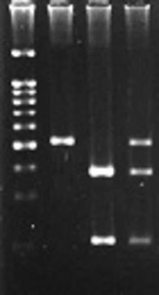
After taking informed consent, 5ml of peripheral blood was collected in EDTA vaccutainer from each subject included in the study. Genomic DNA was extracted from peripheral blood leukocytes following salting out method by Lahiri et al. (7). Genotyping was carried out by using the polymerase chain reaction with allele specific primers as described by Abdolrahim-Zadeh et al. (8). The primer sequences for genotyping is shown in Table 1. PCR was performed after standardization of the protocol and each 15 ul reaction mixture tube contained 50ng of template DNA, 0.2 μM of each primer, 120 μM of dNTP mixture, 1.5 mM of MgCl2, 10 mM of Tris hydrochloride (pH 8.3), and 0.5 U of Taq DNA polymerase. PCR conditions for amplification includes an initial melting step of 5 minutes at 95°C, followed by 25 cycles of 30 seconds at 95°C, 30 seconds at 63°C , 30 seconds at 72°C and a final elongation at 72°C for 5 minutes. The amplified products was run on 1.5% agarose gel, and then stained with ethidium bromide for visualization under ultraviolet gel documentation system.
The genotype results have reconfirmed by performing genotyping twice in randomly selected 10% of the subjects and the results were in 100% concordance.
Table 1.
Allele Specific Primers for IL-10 -1082A/G Promoter Polymorphism
| Primers | Sequences |
|---|---|
| 5’-CAGCCCTTCCATTTTACTTTC-3’ | |
| 5’-TACTAAGGCTTCTTTGGGAG-3’ | |
| 5’-CTACTAAGGCTTCTTTGGGAA-3’ |
3.2. Statistical Analysis
Statistical significance for the differences in the allele, genotype frequencies of the studied polymorphism between cases and healthy controls was determined by the Yates corrected χ2 test. The Odds ratio for genotype frequencies between the cases and the controls were also determined. All the P Values were two sided, and the level of significance was taken as P < 0.05. Further, prediction of transcription factors binding to promoter region was performed using Alibaba 2, an online bioinformatic tool.
4. Results
The genotype and allele frequency distribution of IL-10 -1082A/G promoter polymorphism in 125 breast cancer patients and 160 controls were shown in Table 2. Hardy-Weinberg equilibrium for IL-10 -1082 A/G polymorphism in breast cancer group have shown a significant deviation (χ2 = 16.92) and no significant deviation was found among controls (χ2 = 1.3). In the present study, IL-10 -1082AA genotype frequency was significantly elevated among breast cancer cases (P = 0.0001432) compared to controls under codominant model of inheritance. Further, the codominant model has revealed the existence of a statistically significant association between major genotype (AA) and breast cancer (AA vs. AG: χ2 = 14.46, P = 0.0001432, OR = 2.854, 95% CI = 1.68 - 4.849). Further dominant (AG+GG vs. AA: χ2 = 9.311, P = 0.002278, OR=2.153, 95% CI=1.336 - 3.469) and over dominant (AG vs. AA + GG: χ2 = 16.04, P = 0.00006189, OR = 2.884, 95% CI = 1.731 - 4.807) inheritance models have also supported the association of AA genotype with about 2.8 times of increased risk in breast cancer patients. It was also seen that there was a border line significant difference in allele distribution, among the patients and controls, both. (A vs. G: χ2 = 2.868, P = 0.09037, OR = 1.391, 95% CI = 0.9676 - 2.001).
Table 2.
Distribution of Genotypes and Allele Frequencies of IL-10 (A-1082 G) Promoter Polymorphism in Breast Cancer Patients and Controls
| Genotype | Disease N = 125 | Control N = 160 | χ2 | P Value | OR (95% CI) | |
|---|---|---|---|---|---|---|
| AA | 76 (60.8) | 67 (41.875) | ref | |||
| AG | 31 (24.8) | 78 (48.75) | 14.46 | 0.00014 | 2.854 (1.68 - 4.849) | |
| GG | 18 (14.4) | 15 (9.375) | 0.002342 | 0.9614 | 0.945 (0.4422 - 2.021) | |
| A | 183 (73.2) | 212 (66.25) | ||||
| G | 67 (26.8) | 108 (33.75) | 2.868 | 0.0903 | 1.391 (0.967 - 2.001) | |
| AA | 76 (60.8) | 67 (41.875) | ||||
| AG + GG | 49 (39.2) | 93 (58.125) | 9.311 | 0.0022 | 2.153 (1.336 - 3.469) | |
| AA + AG | 107 (85.6) | 145 (90.625) | ||||
| GG | 18 (14.4) | 15 (9.375) | 1.275 | 0.2598 | 0.615 (0.2966 -1.275) | |
| AA + GG | 94 (75.2) | 82 (51.25) | ||||
| AG | 31 (24.8) | 78 (48.75) | 16.04 | 0.00006 | 2.884 (1.731- 4.807) |
a P ≤ 0.05.
The patient group was classified into early (includes stage I and II) and late stages (includes stage III and IV), less than 40 and greater than 40 age at onset, pre and post-menopausal patients to evaluate the association of different clinic pathological and epidemiological factors with breast cancer. The difference in genotypic distribution of IL-10 (-1082A/G) polymorphism was statistically significant in both early stage (Table 3) (AA vs. AG: χ2 = 5.387, P = 0.02029, OR = 2.507, 95% CI = 1.203 - 5.226) and late stage patients (Table 3) (AA vs. AG: χ2 = 10.11, P = 0.001478, OR = 3.493, 95% CI = 1.639 - 7.441). In comparison with controls there was a one fold increased risk in later stage patients with IL-10 -1082AA genotype than early stage patients. Similarly there was statistically significant increased risk in both < 40 years (Table 4) (AA vs. AG: χ2 = 11.12, P = 0.0008525, OR = 6.071, 95% CI = 2.169 - 16.99) and > 40 year age of onset groups (Table 4) (AA vs. AG: χ2 = 4.943, P = 0.02620, OR = 2.143, 95% CI = 1.139 - 4.032), but it was found that there was about 4 folds increased risk in patients effected with breast cancer before 40 years of age. Our results have also revealed that IL-10 -1082AA genotype of pre-menopausal women (Table 5) (AA vs. AG: χ2 = 8.608, P = 0.003347, OR = 4.883, 95% CI=1.757 - 13.57) was significantly associated with increased risk χ2 of breast cancer, although there was no association between genotype distribution and post-menopausal women (Table 5) (AA vs. AG: χ2 = 2.471, P = 0.1160, OR = 1.797, 95% CI = 0.9256 - 3.489; AA vs. GG: = 0.004773, P = 0.9449, OR = 0.9231, 95% CI = 0.3629 - 2.348). Further the distribution of genotypes was classified based on estrogen receptor status (ER) (AA vs. AG: χ2=0.4566, P=0.4992, OR= 0.5758, 95% CI= 0.1855–4.849), progesterone receptor status (PR) (AA vs. AG: χ2 = 0.08056, P = 0.7765, OR = 0.7238, 95% CI = 0.2367 - 2.213), and Her2/neu status (AA vs. AG: χ2 = 1.246, P = 0.2642, OR = 0.3143, 95% CI = 0.06603 - 1.496), has revealed no significant variation in the distribution of -1082A/G polymorphism of IL-10.
Table 3.
Distribution of Genotypes and Allele Frequencies of IL-10 (A -1082 G) Promoter Polymorphism With Respect to Early Stage and Late Stage Breast Cancer
| Genotype | Early Stage Breast Cancer | Late Stage Breast Cancer | ||||||
|---|---|---|---|---|---|---|---|---|
| Disease (N = 46) | Control (N = 160) | P Value χ2 | OR (95% CI) | Disease (N = 52) | Control (N = 160) | P Value χ2 | OR (95% CI) | |
| AA | 28 (60.86) | 67 (41.87) | 33 (63.46) | 67 (41.87) | ||||
| AG | 13 (28.26) | 78 (48.75) | 0.02029 | 2.507 (1.203 - 5.226) | 11 (21.15) | 78 (48.75) | 0.0014 | 3.493 (1.639 -7.441) |
| GG | 5 (10.86) | 15 (9.375) | 0.896 | 1.254 (0.415- 3.781) | 8 (15.38) | 15 (9.375) | 0.9348 | 0.9235 (0.355 -2.397) |
| A | 69 (75.0) | 212 (66.25) | 77 (74.03) | 212 (66.25) | ||||
| G | 23 (25.0) | 108 (33.75) | 0.144 | 1.528 (0.903 - 2.585) | 27 (25.96) | 108 (33.75) | 0.174 | 1.453 (0.884 -2.385) |
| AA | 28 (60.86) | 67 (41.87) | 33 (63.46) | 67 (41.87) | ||||
| AG + GG | 18 (39.13) | 93 (58.12) | 0.03488 | 2.159 (1.105 -4.221) | 19 (36.53) | 93 (58.12) | 0.0108 | 2.411 (1.264 -4.599) |
| AA + AG | 41 (89.13) | 145 (90.62) | 44 (84.61) | 145 (90.62) | ||||
| GG | 5 (10.86) | 15 (9.375) | 0.9847 | 0.848 (0.291-2.473) | 8 (15.38) | 15 (9.37) | 0.569 (0.226 -1.43) | |
| AA + GG | 33 (71.74) | 82 (51.25) | 41 (78.84) | 82 (51.25) | ||||
| AG | 13 (28.26) | 78 (48.75) | 0.02158 | 2.415(1.184 -4.925) | 11 (21.15) | 78 (48.75) | 0.0008 | 3.545(1.702 -7.388) |
a P ≤ 0.05.
Table 4.
Distribution of Genotypes and Allele Frequencies of IL-10 (A-1082 G) Promoter Polymorphism in Breast Cancer Patients with Age at Onset < 40 and > 40 Years and Age Matched Controls
| Genotype | Age at Onset < 40 | Age at Onset > 40 | ||||||
|---|---|---|---|---|---|---|---|---|
| Disease (N = 46) | Control (N = 160) | P Value χ2 | OR (95% CI) | Disease (N = 52) | Control (N = 160) | P Value χ2 | OR (95% CI) | |
| AA | 20 (58.82) | 14 (26.92) | 56 (61.53) | 50 (47.61) | ||||
| AG | 8 (23.52) | 34 (65.38) | 0.0008 | 6.071 (2.169- 16.99) | 23 (25.27) | 44 (41.90) | 0.0262 | 2.143 (1.139 -4.032) |
| GG | 6 (17.64) | 4 (7.69) | 0.7647 | 0.9524 (0.226 - 4.01) | 12 (13.18) | 11 (10.47) | 0.8625 | 1.027 (0.416 -2.532) |
| A | 48 (70.58) | 62 (59.61) | 135 (74.17) | 144 (68.57) | ||||
| G | 20 (29.41) | 42 (40.38) | 0.1928 | 1.626 (0.846 -3.121) | 47 (25.82) | 66 (31.42) | 0.2681 | 1.316 (0.846 -2.047) |
| 56 (61.53) | 50 (47.61) | |||||||
| AA | 20 (58.82) | 14 (26.92) | 35 (38.46) | 55 (52.38) | 0.07084 | 1.76 (0.995 -3.112) | ||
| AG + GG | 14 (41.17) | 38 (73.07) | 0.0062 | 3.878 (1.549 - 9.706) | ||||
| 79 (86.81) | 94 (89.52) | |||||||
| AA + AG | 28 (82.35) | 48 (92.30) | 12 (13.18) | 11 (10.47) | 0.7147 | 0.770 (0.322 -1.841) | ||
| GG | 6 (17.64) | 4 (7.69) | 0.2887 | 0.3889 (0.101 -1.497) | 56 (61.53) | 50 (47.61) | ||
| AA + GG | 26 (76.47) | 18 (34.61) | 68 (74.72) | 61 (58.09) | ||||
| AG | 8 (23.52) | 34 (65.38) | 0.0003 | 6.139 (2.311 -16.31) | 23 (25.27) | 44 (41.90) | 0.02162 | 2.133 (1.157 -3.93) |
a P ≤ 0.05.
b P ≥ 0.05 - < 0.10 (borderline significant).
Table 5.
Distribution of Genotypes and Allele Frequencies of IL-10 (A-1082 G) Promoter Polymorphism in Premenopausal Breast Cancer Patients, Versus Premenopausal Controls and Postmenopausal Breast Cancer Patients Versus Postmenopausal Controls
| Genotype | Premenopausal | Postmenopausal | ||||||
|---|---|---|---|---|---|---|---|---|
| Disease (N = 46) | Control (N = 160) | χ2 P Value | OR (95% CI) | Disease (N = 52 ) | Control (N = 160) | χ2 P Value | OR (95% CI) | |
| AA | 16 (57.14) | 22 (29.33) | 60 (62.5) | 45 (52.94) | ||||
| AG | 7 (25) | 47 (62.66) | 0.0033 | 4.883 (1.757 -13.57) | 23 (23.95) | 31 (36.47) | 0.116 | 1.797 (0.925 -3.489) |
| GG | 5 (17.85) | 6 (8) | 0.8821 | 0.8727 (0.226 -3.367) | 13 (13.54) | 9 (10.58) | 0.9449 | 0.923 (0.362 - 2.348) |
| A | 39 (69.64) | 91 (60.66) | 143 (74.4) | 121 (71.17) | ||||
| G | 17 (30.35) | 59 (39.33) | 0.3069 | 1.487 (0.771 -2.869) | 49 (25.5) | 49 (28.82) | 0.557 | 1.182 (0.743 - 1.88) |
| AA | 16 (57.14) | 22 (29.33) | 60 (62.5) | 45 (52.94) | ||||
| AG + GG | 12 (42.85) | 53 (70.66) | 0.0176* | 3.212 (1.308 -7.888) | 36 (37.5) | 40 (47.06) | 0.2511 | 1.481 (0.818 -2.682) |
| AA + AG | 23 (82.14) | 69 (92) | 83 (86.45) | 76 (89.41) | ||||
| GG | 5 (17.8) | 6 (8) | 0.2803 | 0.4 (0.1115 -1.435) | 13 (13.54) | 9 (10.58) | 0.7047 | 0.756 (0.305 -1.869) |
| AA + GG | 21 (75) | 28 (37.33) | 73 (76.04) | 54 (63.52) | ||||
| AG | 7 (25) | 47 (62.66) | 10.14 | 0.0014 | 23 (23.96) | 31 (36.47) | 0.0942 | 1.82 (0.957 -3.469) |
a P ≤ 0.05.
b P ≥ 0.05 - < 0.10 (borderline significant).
5. Discussion
In the previous studies it was seen that around 5% - 10% of breast cancers are inherited and the remaining were sporadic (9-11). For above relation, about 25% - 30% of familial breast cancers are attributed to genes like BRCA1, BRCA2, PTEN, CHEK2, BACH1, PALB2, RAD50, TP53 etc., these have known as highly or moderately penetrant genes (12, 13). However, there is a growing need to screen variations in low penetrant genes and establish its role in disease development. Evading immune destruction is an emerging hall mark of cancer, as highly immunogenic cancer cells might well evade immune destruction by disabling components of the immune system that have been dispatched to eliminate them (2). In this regard, we chose to select IL-10, a cytokine that suppressed immune responses. IL-10 gene was mapped to”1q31-1q32” (14). IL-10 exerts immune suppression activity, by down regulating the expression of surface co-stimulatory molecules like CD80 or CD86 on tumor cells, thus preventing antigen presenting cells (APC) from obtaining access to tumor antigen (15).
The present study evaluated the association between IL-10-A1058G promoter polymorphism and breast cancer in the South Indian population. The statistical analysis has revealed a significant increased risk of breast cancer with low IL-10 expressing AA genotype which is been in concordance with other studies in Italian and Austrian populations (Giordani et al. 2003) (16), (Langsenlehner et al. 2005) (17). However, our results are in contrast with other population studies by Howell et al. (England) (18), Smith et al. (England) (19), Balasubramanian et al. (England) (20), Kong et al. (Chinese) (21) and Abdolrahim-Zadeh et al. (Iranian) (8), respectively. These contrasting results may be attributed to ethnic difference and haplotype variations or linkage disequilibrium with other polymorphisms in its proximity. Kang et al (2010) have determined: that A allele of IL-10 A-1082G promoter region physically interacted with a nuclear protein poly (ADP-ribose) polymerase1 (PARP-1) in allele specific manner that has resulted in differential expression of IL-10. As the -1082A binds to PARP-1in greater extent than G allele, the former has lesser transcription activity than latter (22). In silico analysis was performed to predict transcriptional factors binding to IL-10 promoter region using Alibaba 2.1 (Grabe 2002) online bioinformatics tool, and then found a change in -1082 A - G created an additional transcription binding site for Krox-20 beside Sp1. This additional potential of G allele to bind krox-20 might play a role in higher expression of IL-10 (23).
In contrast to anti-tumorigenic properties of IL-10, Simone et al. 2005), has reviewed that high levels of IL-10 in tumor microenvironment might favor in the immune-mediated tumor rejection by increasing NK cell activity, and then enhancing cytotoxicity and migration of CTL (24). This opinion was helped us in hypothesizing possible role of IL-10 A -1082G polymorphism in breast cancer. Previous studies have correlated higher level of IL-10 expression with -1082G allele, which was also associated with protection against breast cancer. As AA genotype is associated with low levels of IL-10 expression, the produced amount of IL-10 might to be insufficient to induce immune mediated tumor rejection. Thus justifying its association with an increased breast cancer risk.
The patients were stratified into early and late cancers based on TNM scoring. The AA genotype correlated with both early and later stages of cancer. There was one fold increased risk of developing breast cancer in the patients who have presented with advanced stage in comparison with those who have presented in early stages. This increased risk might be attributed to increase in number of TAM cells with advancement of cancer. The analysis has also revealed that AA genotype, is associated with an increased risk of breast cancer in pre-menopausal women. In contrast, Singh et al. (2012) has found an association with post-menopausal women in North Indian population. Based on age at onset, the patients were stratified into age groups of below and above 40 years. Both groups were associated with AA genotype. There was fourfold increased risk in the age group below 40 years. As IL10 levels are low in younger than elder people, the younger patients with low expressing AA genotype might further increase the risk of breast cancer. There was no correlation between genotype or allele frequency distribution, and hormonal receptor status (ER, PR), and Her2 which has been in concurrence to other studies (17). Although in Iranian population, an increase AA genotype frequency was seen in PR negative patients (8).
There was a statistically significant correlation between the AA genotype of -A1082 G gene promoter polymorphism, with an increased risk of breast cancer in a cohort from South India. However, further studies with larger sample size, and haplotype analysis with other promoter variants are required providing further evidences to establish the role of this polymorphism in breast cancer.