1. Introduction
Lymphoma has represented 13.6% of metastatic tumors to the heart (1). Gross tumor formation in any of the cardiac chambers has been rare, particularly at the time of presentation and diagnosis of lymphoma (2). Symptoms were usually very subtle and non-specific, particularly in the setting of co-existing comorbidities (3). As imaging modalities and treatment options for lymphoma have improved, more unusual disease presentation might be observed more frequently. In this article we have reported a 78-year-old man with an incidental large cardiac mass whose symptoms was abdominal pain and vomiting in the setting of pre-operative evaluation for surgical management of intra-abdominal mass.
2. Case Presentation
A 78-year-old man has affected by abdominal pain, vomiting and weakness and has evaluated at June 2013. Studying his medical history, he had hip joint replacement surgery, then in initial physical examination has shown no significant finding, except a few tenderness on the middle abdominal quadrant. In laboratory assessment, complete blood count, renal function tests-alkaline phosphatase and lactate dehydrogenase were normal, but hemoglobin and transaminases were abnormal as follows: Hb = 9.1 mg/dl, AST = 71 u/lit, ALT = 656 u/lit.
In abdominal CT scan, there was a 71 × 61 mm mass at the aortic bifurcation in favor of tumoral lesion or adenopathy.
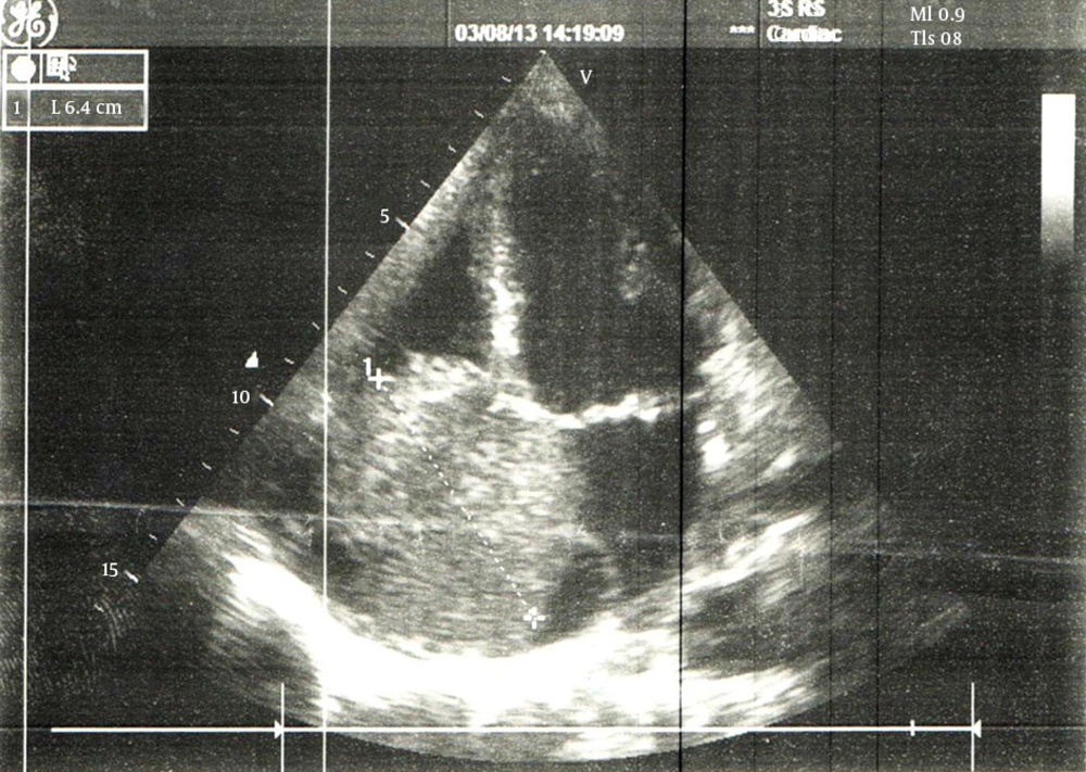
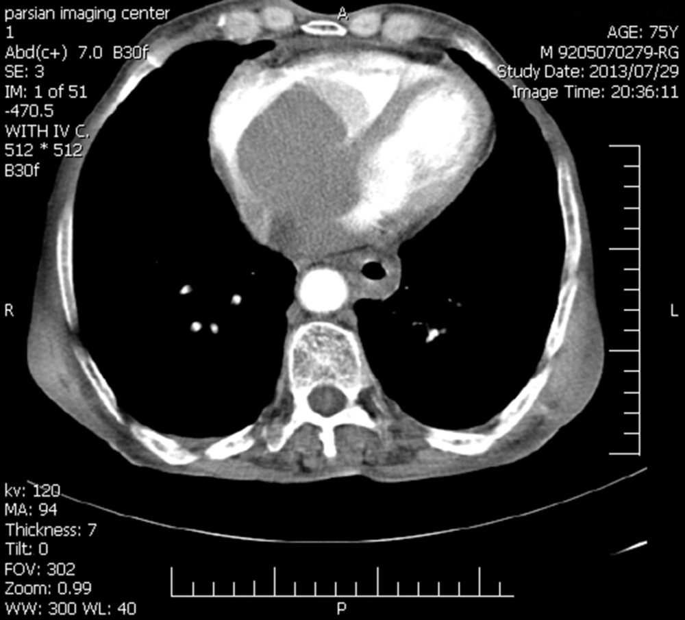
Surgery has recommended and in pre-operative evaluation he has referred to cardiologist. ECG had no significant and specific changes and in echocardiography, a large mass with diameter of 74 × 60 mm has seen in right atrium (Figure 1) and LV ejection fraction was 65%. Also, in thoracic sections of CT scan, an intra-cardiac mass has seen (Figure 2).
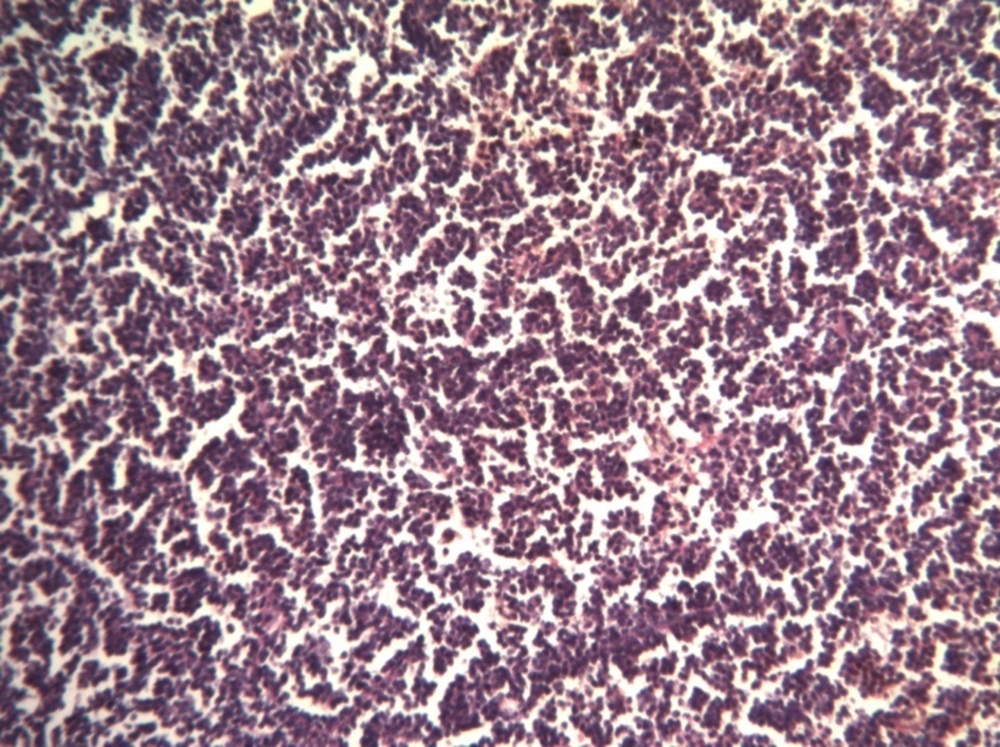
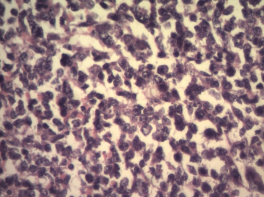
Bases on these new findings management of the patient has changed to cardiac surgery and open heart surgery has recommended and performed at August 2013. Pathologic examination of removed cardiac mass has reported as below after immunohistochemistry study: HMB 45 and CK: negative; CD20: positive. Compatible with non-Hodgkin lymphoma in favor of B-cell origin (Figures 3 and 4). He has referred to oncologist after recovery of heart surgery (September 2013).
In evaluation of medical history and further physical examination, he had no history of fever and sweating, but weight loss of 2 - 3 kg during recent weeks. Karnowsky performance status was 80%. At physical examination, he had an adenopathy of 2 × 1 cm at left jugulodigastric chain, and the another of 3 × 1 cm at left supraclavicular area and physical exam was normal otherwise.
In bone marrow biopsy, BM was hyper cellular, and involved with lymphoma infiltration. The patient has planned to treat by R-CHOP chemotherapy regimen (Rituximab-cyclophosphamide-Doxorubicin-vincristine-prednisolone).
He could not provide Rituximab due to economic problems, therefor he has received only CHOP regimen. After first cycle of treatment, cervical adenopathies have disappeared and at the end of seventh cycle, imaging of the neck , chest and abdominopelvic cavity by CT scan had no positive finding of disease but the patient has not satisfied to undergo BM biopsy for second time.
Patient treatment has completed after eight cycles of CHOP chemotherapy regimen at February 2014, but he was in good condition, without any evidences of disease after six months of follow up.
3. Discussion
Cardiac masses have been arising from the heart or pericardium, were potentially lethal whether defined as benign or malignant. Almost 75% of primary cardiac masses were benign. The most common primary malignant tumors were sarcomas and lymphomas (4).
Metastatic deposits have represented the vast majority of cardiac malignancies; the common primary malignancies sources have included cancers of lung, esophagus and breast as well as lymphoma, leukemia and melanoma (5). Cardiac involvement as an initial presentation of malignant lymphoma was a rare occurrence (2). Secondary involvement of the heart has seen in 8.7 - 27.2% of documented clinical case of lymphoma (2, 6, 7). Despite its life-threatening nature (8), the cardiac manifestations of lymphomatous involvement of the heart were often subclinical (9, 10). As in the present case there has been no cardiac symptom or sign. These symptoms and signs might include arrhythmias, pericardial effusion or tamponade, tumor embolization and obstruction of blood flow and valvular dysfunction. These symptoms have related on tumor location, size, growth rate, degree of invasion and friability.
In the present case and many other reports the majority of intracavital tumors have occurred on the right site of the heart, the reason for which has yet been to be found (1, 11). Plain chest radiographs lack sensitivity and specificity as an initial diagnostic tool but could demonstrate cardiomegaly or specific chamber enlargement. Echocardiography was the first non-invasive study for examining the chambers of the heart and pericardium (12) but trans esophageal echocardiography (TEE) was a more sensitive technique for assessing patients (13).
CT scan and MRI with gadolinium contrast injection were useful tools for demonstrating morphology, location, extension of disease, blood flow and cardiac function (14, 15). FDG-PET imaging has recently reported to reveal previously unsuspected cardiac involvement (16, 17).
Pathologic diagnosis has been essential in management of cardiac masses. Although traditionally this has required a thoracostomy, less invasive procedures has recently been available such as TEE-guided biopsy, endomyocardial biopsy, or percutaneous intracardiac biopsy with combined fluoroscopy and TEE or pericardial fluid sampling (18, 19).
Because of rarity, data has been lacking to produce definitive guidelines regarding management. The available literature suggested systemic chemotherapy was the only effective therapy (18) and the majority of cases have treated with combination chemotherapy with varying results (20, 21). There was an improvement in response and survival rates by adding of monoclonal therapies to chemotherapy (22). Radiation therapy has indicated in cardiac mass that progresses despite chemotherapy and its adverse side effects have been pericarditis, cardiomyopathy, diastolic dysfunction, conduction defects and coronary artery disease. Radical surgery has not advised.
Lymphomatous involvement of the heart has reported with greater frequency than the past. Cardiac involvement was rare as an initial presentation of malignant lymphoma and has often been subclinical. These tumors have seen more common in the right site of the heart and TEE, CT scan and MRI with Gd were effective tools in assessment of patients. Pathologic diagnosis would be essential to management of cardiac masses and the only effective treatment has been chemotherapy.


