1. Background
Breast cancer is the most widespread neoplasm in women worldwide (1, 2). So, early detection of this type of neoplasm is vital and helps the medical staff for choosing the best treatment strategy and planning (3, 4). Nowadays, mammography is the gold standard and best modality for diagnose or screening the breast cancer, which helps the physician for early detection of this type of cancer (2). Many studies have noted that, in addition to patient discomfort and the imposed radiation hazards, false negative reports of mammography may lead to misdiagnosis of breast cancer (5).
Trace element analysis is a common method for evaluating health state in living tissues (6, 7). As most of exertion and also toxic materials which lead to disorders are accumulated in body tissues such as hair. It seems that measuring the concentrations of these elements can be a useful method for diagnosis of such disease (6). Many studies have reported that this type of analysis can be helpful for diagnosis of neoplasms and cancers (8-10). In this regard, the trace element concentrations in epithelial (8), esophageal (9), prostate (10) cancers have been studied and in most of them, there was a significant relation between changes of health state and amount of trace elements (10).
Using hair samples to analyze the trace element concentrations is of interest among many researchers (6, 10). X-ray fluorescence (XRF) and X-ray diffraction (XRD) are the most common methods in studying the structure and concentration of elements of tissues and also crystalline materials, using low energy X-ray (3).
For breast tissue, because of the importance of disorders, researchers have published a considerable number of papers on using trace element analysis with the above mentioned modalities (11-13). There are various methods for analyzing trace elements and all of them have their own advantages and disadvantages. Therefore, comparative study between different methods has been done and consequently concentration levels in healthy individuals and cancer patients have been reported (14). Using Synchrotron Radiation X-ray Fluorescence (SRXFA), researchers reported that Selenium (Se) concentration in breast cancer patients is less than those suffering hyperplasia; however, concentration of Chrome (Cr) was higher than that in normal people (15). When comparing trace elements in male and female with Energy Dispersive X-ray Fluorescence (EDXRF), studies have revealed that Cr, Cu and Mo concentration in females is considerably higher than these elements in males, but for Mn, Ni and Zn the concentration is higher in males (16).
According to the literature, several published papers reported using X-ray Diffraction (XRD) analysis of healthy and also cancerous breast tissue (3, 17-21).
2. Objectives
In the present study, comparing the mean concentration ratio of Ca/Zn, Ca/Mg, Ca/P elements between healthy women and those with breast cancer patients was the main goal. In addition, the detection ability of tube based X-ray fluorescence method in the presence of early stages breast cancer was also evaluated and the results were compared with mammography. Finally, the sensitivity of tube based X-ray fluorescence method in distinguishing normal and abnormal breast tissues was also evaluated.
3. Materials and Methods
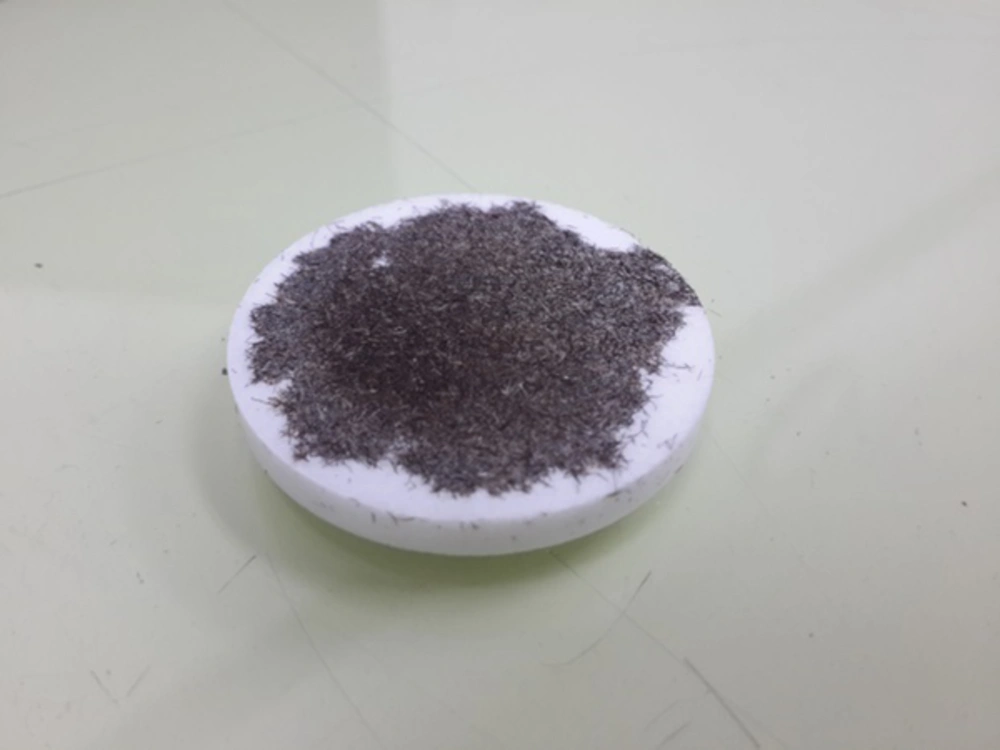
In this study, hair samples of 54 women (including 27 healthy and 27 cancer patients) who stayed in four cancer treatment centers in Tehran, Iran was analyzed. Healthy individuals were women who reported as healthy in physical examinations and mammography and cancer cases were among those patients that showed positive mammography and pathology reports. All of the donors were Iranian women aged between 33 - 79 years old (average 52.03 ± 11.44 years). The hair samples were obtained from women who had no colored hair for 6 weeks or more and hair samples of cancer cases were collected from patients who had not undertake chemotherapy and medicine therapy. Hair strands were cut from occipital and parietal region of skull and taken through sample preparation method introduced by International Atomic Energy Agency (22). For cleaning the samples from any kind of external contamination, coded samples were washed with ethanol and distilled water for three times and then dried in an oven for about 30 minutes at 80°C temperature. All samples were cut with surgical stainless steel scissors into fine pieces of about 2 mm and using a mortar, the samples became powdered and dried in the oven for about 10 hours at an 80°C temperature (23). Then, the amount of 0.8 g of each powdered sample had pressed on a boric acid (H3BO3, Merck Analysis grade, Germany) pellet with 4 cm diameter and 5 mm thickness using an automatic hydraulic press with 10,000 kg pressure (Herzog, Germany). Figure 1 shows hair sample mounted on boric acid.
For the measurements, Wave Length X-ray Fluorescence (WLXRF) method was used. Sample holder pellets were exposed to a PANalytical (formerly Philips), PW 2404 (PANalytical Inc., The Netherlands) with Rhodium anode X-ray tube. Each sample was filled in special sample holders of XRF spectrometer with a circular exposure window of 27 mm in diameter and exposed for 20 minutes.
Results of element concentration of healthy individuals and breast cancer patients were proceed and statistical parameters such as mean and standard deviation values of Ca, Zn, Mg, and P elements were analyzed with IBM SPSS (version 22.0, 2013) software. The Ca/Zn, Ca/Mg, and Ca/P ratios were also compared in healthy and patient groups.
The WLXRF results of coded hair samples were analyzed by XRF experts. Then, these results were matched with donors’ health state data. The results of healthy individuals and cancer patient groups were compared with mammography and pathology reports and based on the results, the sensitivity of the method versus mammography were also evaluated.
4. Results
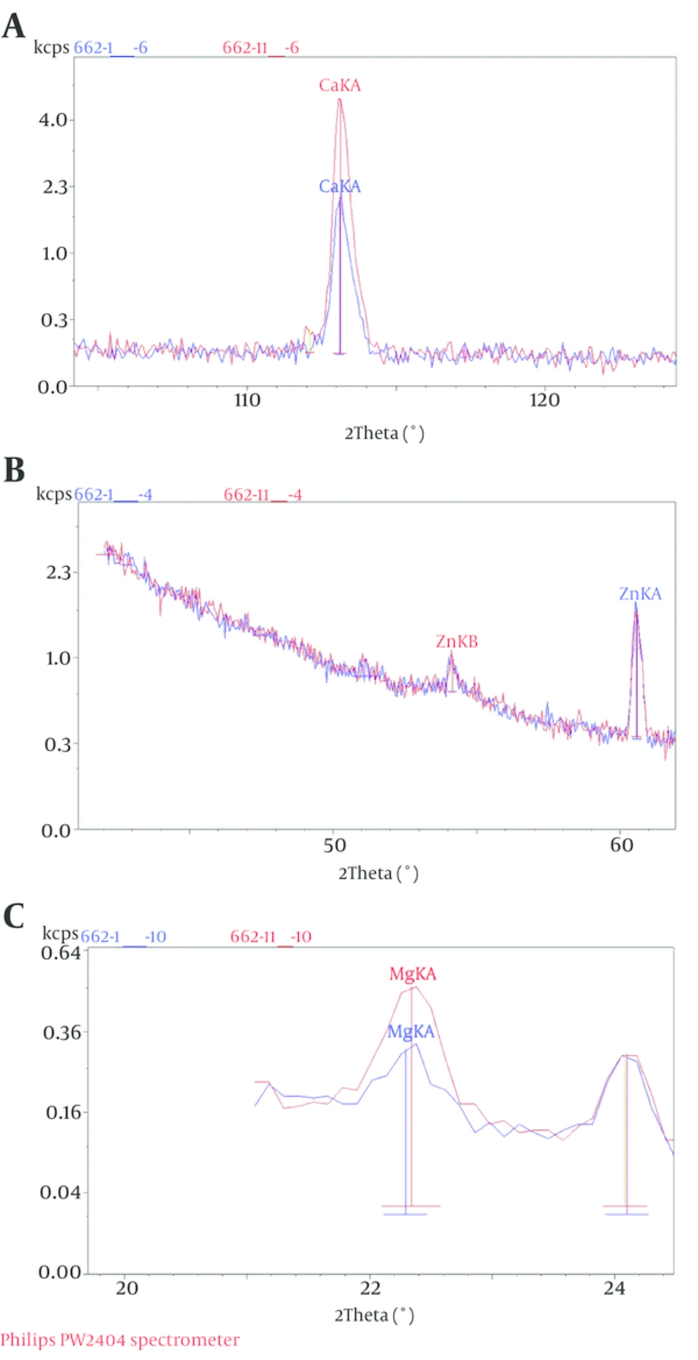
Results of WLXRF analysis of hair samples belong to healthy individuals and breast cancer patients including the trace element concentration, range, mean, and standard deviation values of Ca, Zn, Mg and P for healthy individuals and patients are shown in Table 1. Superimposed WLXRF curves (plots) of normal and patient related samples are also shown in Figure 2.
| Mean Concentration % | Range ± SD | Mean Healthy/Mean Cancer Concentration | |
|---|---|---|---|
| 2.430 | |||
| Healthy | 4.340 | 9.230 ± 1.948 | |
| Cancer | 1.763 | 5.756 ± 1.197 | |
| 1.514 | |||
| Healthy | 0.212 | 0.372 ± 0.105 | |
| Cancer | 0.140 | 0.818 ± 0.151 | |
| 1.430 | |||
| Healthy | 0.259 | 0.341 ± 0.097 | |
| Cancer | 0.181 | 0.371 ± 0.102 | |
| 1.249 | |||
| Healthy | 0.220 | 0.884 ± 0.158 | |
| Cancer | 0.177 | 0.146 ± 0.042 |
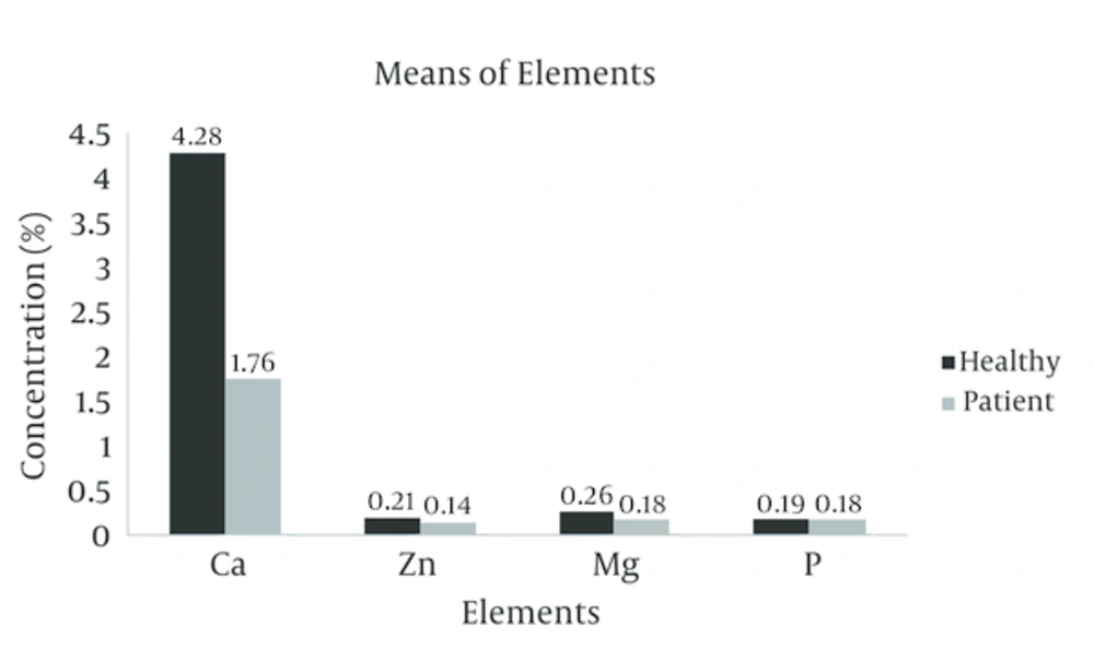
Figure 3 demonstrates the comparison of mean element concentration chart related to healthy individuals and cancer patients. In addition, in each group, the results were compared with mammography and pathology report of each individual, which are shown in Table 2.
| Number of Patients | WLXRF | Mammography | Pathology | ||||
|---|---|---|---|---|---|---|---|
| 27 | 2 | 25 | 4 | 23 | NA | 27 | |
| 27 | 26 | 1 | 21 | 6 | 27 | NA | |
| 96 | 92 | 77 | 85 | 100 | 100 | ||
Abbreviations: H; Healthy, NA; not available, P; Patient.
5. Discussion
The results showed that the concentrations of all elements in the scalp hair samples of healthy individuals were higher than cancer patients, indicated by the ratio of mean healthy to mean tumor concentrations (Figure 3). This result was compatible with previous studies that have been performed using synchrotron X-ray fluorescence (15). It seems that this evidence can be due to elemental increase in the tumor site, so there is less accumulation of elements far from the tumor (12).
As reported in Table 1, the mean concentration percentage level for each element in healthy group is higher than that of cancer group. The increase rate was the least for P and the highest for Ca. For each element, using T-test for comparing the mean concentration percentage level between healthy and cancer groups, the results showed significant differences for Ca, Zn, Mg, except for P with no significant difference.
When comparing the element accumulation in hair samples, as it can be seen from Figure 3, the highest amount of element concentration, which revealed accumulation, was related to Ca, Mg, Zn, and P, respectively.
Comparing with XRD procedures which have been previously done on the mentioned hair samples (3), the sensitivity of XRF (96%) was higher than the sensitivity of XRD (86%). But, from examination time point of view and the curve interpretation of the results, the method used here has longer time than that of XRD method. The sensitivity of the studied method is presented for both healthy and patient groups in Table 2. The sensitivity of WLXRF method was also higher than that of commonly used diagnosis method of mammography.
Therefore, in healthy state of breast tissue, XRF technique may be used as an alternative non-invasive and more sensitive method compared to other methods. It should be noted that mammography has discomfort and radiation hazard, compared with the modality used in the present study, but mammography has better detection. Efforts should be made to further study the importance of this useful and other relevant methods using more hair samples of breast cancer patients.


