1. Background
Lymphomas are divided into two main groups-Hodgkin and non-Hodgkin lymphomas. Hodgkin lymphoma (HL) commonly manifested as nodal involvement, while 40% of non-Hodgkin lymphomas (NHL) are extra-nodal (1). The head and neck area, particularly Waldeyer’s tonsillar ring (tonsils, pharynx and base of the tongue) are among the most commonly involved extra-nodal sites (2). The prevalence of NHL increases from the 5th to 7th decades of life and has a predilection for males. The most common clinical manifestation includes local swelling with or without ulceration (3). NHLs have several microscopic subgroups with different innate characteristics and variable responses to treatment.
Previous studies have shown significant differences in the prevalence rates of lymphoma worldwide (2). The CRC at Shahid Beheshti University of medical sciences is an accredited research center in Iran and information about cancer cases from all over the country is sent to this referral center. Therefore, the obtained statistics based on the data registered in this center would be highly reliable.
2. Objectives
The overall prevalence of oral lymphoma has not been previously assessed in Iran, this study aimed to assess the relative frequency of this lesion and its histopathological variations in Iran during a 6-year period. The results of this study can help determine the prevalence and dispersion of oral lymphoma in Iran. Moreover, the obtained statistics can be compared with those of other countries.
3. Materials and Methods
This multi-center, retrospective, cross-sectional study was conducted in two phases. In the first phase, cases of oral lymphoma registered in the CRC at Shahid Beheshti university were recorded and in the next phase, patient records and pathology reports of these cases were retrieved from the archives; then age and sex of the patients, location of the lesion and its microscopic type were recorded and classified in tables. This center has been collecting information on cancer patients from all major hospitals in Iran since 2003. The information includs identity and demographic information of patients, name of the respective physician, name of the hospital, site of biopsy, primary clinical diagnosis, and final pathological diagnosis. Data were statistically analyzed using descriptive statistics.
4. Results
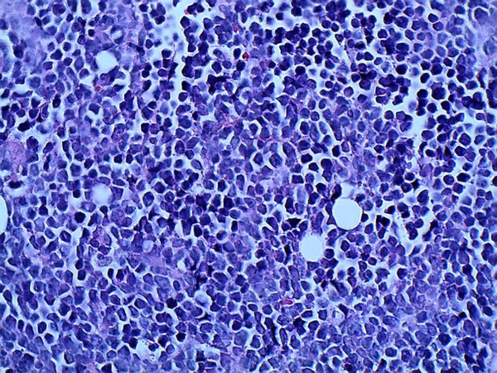
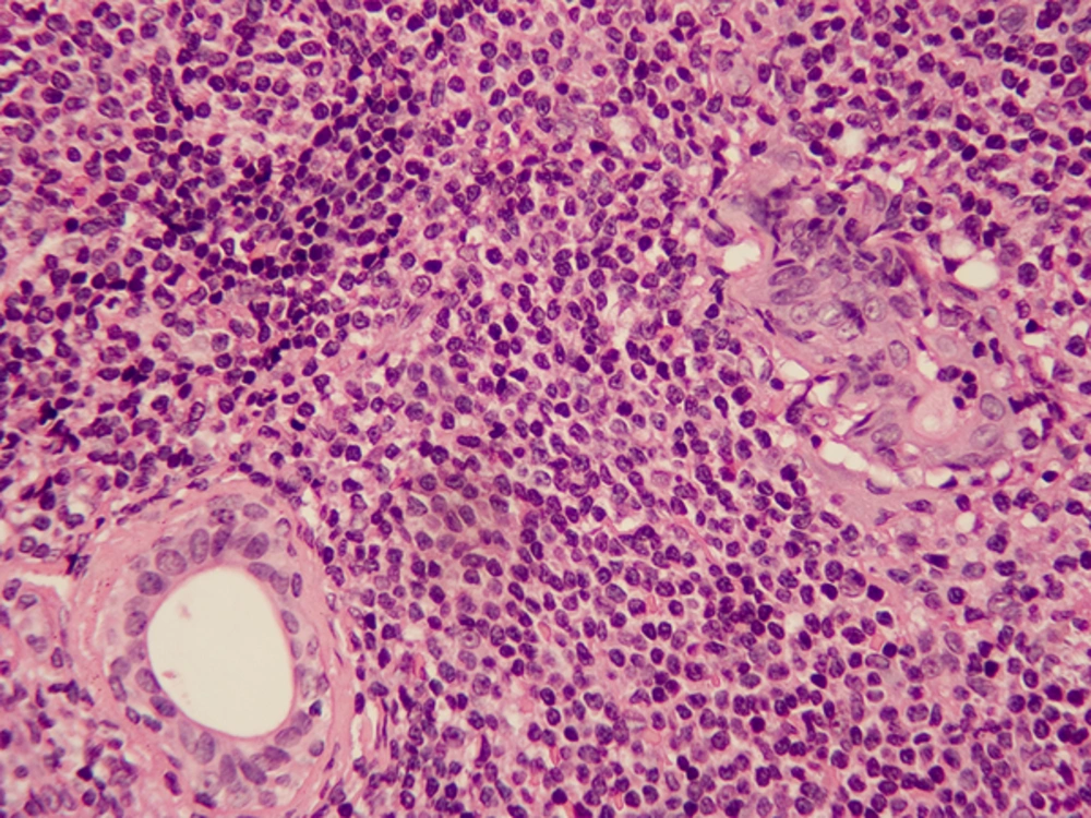
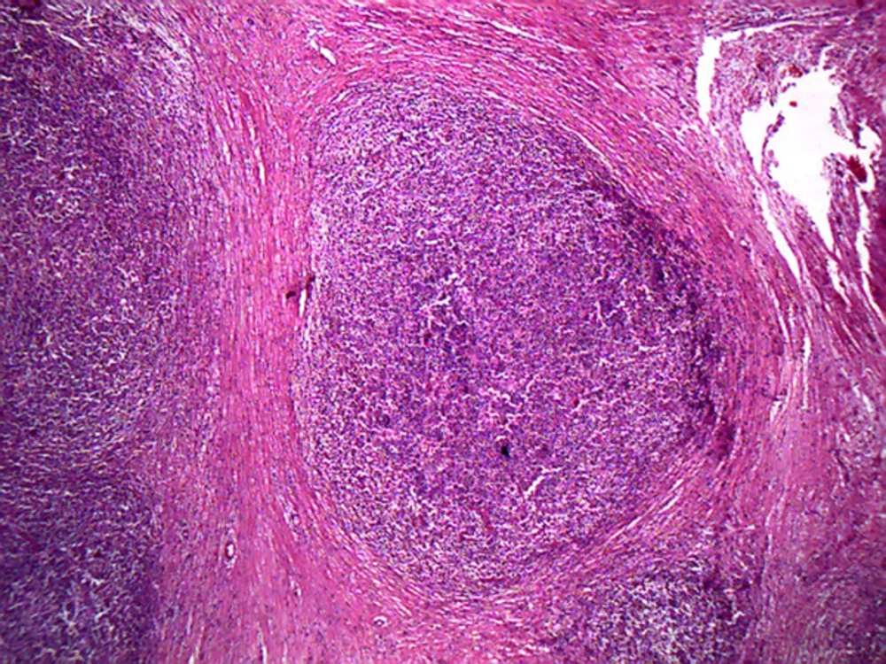
In this study, oral lymphoma accounted for 1.1% of head and neck malignancies and 8% of all lymphomas. During 2003 - 2008, 437 new cases of oral lymphoma were registered in the CRC. Most cases of oral lymphomas were of non-Hodgkin type (98%). Except for a descending incidence during 2005 - 2007, the overall trend of changes was ascending. Of 437 cases, 431 were B cell and the remaining were T cell lymphomas. Histopatholoic features of some oral lymphoma cases are illustrated in Figures 1 - 4. The most common form of oral lymphoma was diffuse large B cell lymphoma (DLBL) (240 cases, 54.9%) followed by small lymphocytic lymphoma (SLL) (54 cases, 12.4%) and follicular type (FL) (20 cases, 4.6%). Other types of lymphomas had a frequency of less than 5 cases. Oral lymphoma generally had the highest frequency in the 4th to 8th decades of life and the most commonly diagnosed cases were in the 6th and 7th decades of life. Burkitt’s lymphoma was an exception. Although only 6 cases of oral Burkitt’s lymphoma had been registered, 4 of them were younger than 20 years of age. The lowest age range was 0 to 9 years (8 cases) and the highest was over 80 years of age (34 cases).
A total of 283 (64.8%) cases of lymphomas had occurred in males and 154 (35.2%) in females. Thus, the male to female ratio was 1:84. All types of NHLs were more prevalent in males than in females, but the incidence ratio of males/females was variable for different types of lymphomas. The incidence of DLBL in males was 1.66 times that of females. This ratio was 18 for FL and 5.37 for SLL. On the other hand, in HL despite the scarcity of cases, rate of involvement in females was three times that of males. Composite Hodgkin and non-Hodgkin lymphoma were only seen in females. The tonsils were the most common site of involvement in the oral cavity (77.8%). Other areas ranked second with a significant difference (12.4%) followed by the palate (4.1%) and tongue (3.0%). Distribution of microscopic types based on age, sex and site of involvement is shown in Tables 1 to 3. In the current study, of 8 HL cases, 4 (50%) were classified under mixed cellularity subgroup, 3 (37.5%) were assigned to nodular lymphocyte predominant subgroup and 1 (12.5%) was classified under lymphocyte-rich subgroup.
| 0 - 9 | 10 - 19 | 20 - 29 | 30 - 39 | 40 - 49 | 50 - 59 | 60 - 69 | 70 - 79 | +80 | Unknown | |
|---|---|---|---|---|---|---|---|---|---|---|
| 8 | 20 | 27 | 51 | 67 | 72 | 72 | 62 | 32 | 13 | |
| 1 | 2 | 1 | 1 | 1 |
| Male | Female | |
|---|---|---|
| 57 | 43 | |
| 2 | ||
| 18 | 1 | |
| 1 | ||
| 148 | 89 | |
| 3 | ||
| 43 | 8 | |
| 5 | 2 | |
| 4 | 1 | |
| 1 | ||
| 281 | 145 | |
| 2 | 6 | |
| 0 | 3 |
| Base of Tongue | Gum | Palate | Tonsil | Other Ill-Defined in Lips, Oral Cavity & OroPharynx | Oropharynx | |
|---|---|---|---|---|---|---|
| 3 | 4 | 4 | 71 | 13 | 3 | |
| NA | NA | 1 | NA | NA | NA | |
| NA | NA | NA | 17 | 3 | NA | |
| NA | NA | NA | NA | 1 | NA | |
| 8 | NA | 7 | 198 | 23 | 4 | |
| NA | NA | 1 | NA | 1 | 1 | |
| 1 | 1 | 4 | 40 | 6 | 2 | |
| NA | NA | 1 | 4 | 1 | NA | |
| 1 | NA | NA | 3 | 1 | 1 | |
| NA | NA | NA | NA | 1 | NA | |
| 13 | 4 | 18 | 333 | 51 | 7 | |
| NA | NA | NA | 6 | 2 | NA | |
| NA | NA | NA | 2 | 1 | NA | |
| 13 | 5 | 18 | 340 | 54 | 7 |
Abbreviation: NA, not available.
5. Discussion
In the current study, oral lymphoma accounted for 1.1% of head and neck malignancies and 8% of all lymphomas. Evidence shows that lymphomas comprise 1.5% to 8.8% of the oral malignancies (4-6). The prevalence of oral lymphoma relative to all oropharyngeal malignancies was reported to be 3.4% in a study by Razavi et al. (6), in Isfahan, 8.8% in a study (5), in Gilan, 65.5%. According to a study conducted in Philadelphia, most cases of oral lymphomas were NHL and of B cell type with an estimated prevalence rate of 41% to 100% (7). In our study, B cell lymphomas comprised 98% of the cases of oral lymphomas. In contrast, a study in Japan reported a prevalence rate of 34% for B cell lymphoma and 28% for T cell lymphoma (8). An interesting point is that in lymphomas of the nasal cavity and paranasal sinuses, although close to the oral cavity, the prevalence of T cell lymphomas are very high and a previous study reported that 80% of the lymphomas of the nasal cavity and paranasal sinuses were T cell lymphomas (9). Another study conducted in Spain reported a prevalence rate of 44% for T cell lymphomas in the afore-mentioned areas (10). Thus, it may be concluded that B cell lymphomas are more prevalent in the oral cavity while T cell lymphomas more commonly occur in the nasal areas. In this study, the incidence of oral lymphoma was found to be 0.78 to 1.21 individuals per one million population from 2003 to 2008, respectively. The ascending trend of lymphoma was also reported by Mousavi et al. (5), in their study in 2009 on all lymphomas. The increased incidence may be due to several factors. First of all, it may be due to the more recent diagnostic techniques in the country enabling accurate diagnosis of lymphoma. Second of all, the new classification systems suggested for lymphoma may include malignancies that were not classified as a lymphoma previously. Thirdly, the quality of cancer registry has improved in the recent years.
In general, lymphoma is the eighth most common cancer in men and the tenth most common cancer in women. Although its incidence in less developed countries (4.2 in males, 2.8 in females per 100,000) is far less than that in developed countries (10.3 in males and 7 in females per 100,000), the morbidity and mortality due to this malignancy are much higher in less developed countries (11). In our study, the incidence of oral lymphoma in men was 1.84 times that of women. The incidence of lymphoma in Iran is 3.7 per 100,000 population in males and 2.3 per 100,000 population in females (5). In a study by Razavi et al. (6), the frequency of oral lymphoma in men was 1.29 times that in women. In other communities, the male/female lymphoma incidence ratio varied from 1/4 to 2/2 (12, 13). In most previous studies, men were more commonly affected than women. In contrast, in a study by Kemp et al. (1), number of affected women was only slightly higher than men (53%).
Different classifications have been offered for staging of lymphoma. In the current study, the most recent classification suggested by WHO for lymphoma was used. Most cases were NHL (97.5%). HL and the composite form only comprised 1.8% and 0.7% of all cases, respectively. Of the subgroup of NHLs, the type of lymphoma had not been specified in 21.7% of cases, while 54.9% of cases were DLBL. The prevalence of SLL and FL was 12.4% and 4.6%, respectively. Gnepp reported that DLBL was the most common type of lymphoma of Waldeyer’s tonsillar ring followed by peripheral T cell lymphoma, MALT (mucosa-associated lymphoid tissue) lymphoma and FL in a decreasing order of prevalence (14) In our study, MALT and peripheral T cell lymphoma were each seen in one case and follicular type was found in 20 cases. In a study by Mohtasham et al. (15), on 36 cases of oral lymphomas, DLBL (41.1%), low-grade B cell lymphoma (35.2%), peripheral T cell lymphoma (11.7%), Burkitt’s lymphoma (5.8%) and HL (5.8%) had the highest frequency in decreasing order. Although Mohtasham et al. (15) did not use the accredited WHO classification for lymphomas, DLBL was the most common subtype, which is in line with the results of the current study. In addition, Kemp et al. (1) reported that DLBL comprised 58% and B cell lymphoma comprised 98% of cases. Moreover, Epstein et al. (12), in their study on 361 cases of oral lymphoma demonstrated a frequency of 38% for large B cell lymphoma and 27.4% for small cell lymphoma. In their study, HL was only seen in 0.8% and Burkitt’s lymphoma in 1.6% of cases; which is in accordance with our findings. In a study (3) MALT was the most frequent microscopic subtype and DLBL ranked second, which is in contrast to our findings. This difference is explained by the fact that they included lymphomas of the salivary glands in their study.
In the current study, oral lymphoma had the highest frequency in the 4th to 8th decades of life and the diagnosed cases were mostly in their 60s or 70s. In other studies, the mean age of patients with oral lymphoma was 62.5 years and 71 years (1, 12). In contrast, Razavi et al. (6), in their study in Iran reported that more than half the cases of oral lymphomas had occurred in patients younger than 40 years of age. Considering the large sample size of our study, the reported age range in our study is more reliable than that of Razavi.
The tonsils are the most common site of occurrence of lymphoma in the oral cavity (77.8%). This finding is in accord with the results of other studies (12, 15, 16). Of 670 cases of oral lymphomas evaluated in a study by DePe-a et al. (17), one-third occurred in the tonsils and one-third in other parts of Waldeyer’s tonsillar ring. Kolokotronis et al. (16), also reported the tonsils to be the common site of involvement. In a study by Epstein et al. (12) 53.7% of cases had occurred in Waldeyer’s tonsillar ring (32.7% in the tonsils), 16.1% in the parotid gland and 13% in the pharynx. In a study by Kemp et al. (1), the maxilla and palate were the most common sites of oral lymphomas, which may be due to the exclusion of tonsils from their study. In a study by Etemad-Moghadam et al. (2) 22% of cases had occurred in the tonsils, 15% in the parotid and 13% in the pharynx; however, in our study only 1.6% of the lymphomas had occurred in the pharynx. This difference is probably due to the fact that we only included lymphomas of the oropharynx in our study, while in the above-mentioned study, lymphomas of the nasopharynx were also included. Moreover, we only evaluated cases of lymphomas in the oral cavity and excluded the major salivary glands, whereas another study (3), in their study in Greece reported the most common site of involvement to be the major salivary glands and Waldeyer’s ring ranked fourth. In our study, the base of the tongue, which is included in Waldeyer’s ring, was the fourth, and the palate was the third most common site of involvement, which is in line with the available literature. Unfortunately, data earlier than 2003 were not available in the archives and those available were not complete. Thus, the current study only evaluated a 6-year period. Approximately, 21.7% of lymphomas had been registered only as lymphoma with no specific information regarding histopathological subtype. If their histological subtype had been registered, our data would have been much more valuable. One limitation of the current study was insufficient data regarding the clinical condition of patients and stage of cancer. No information was available regarding the follow up of patients either; thus the survival rate could not be calculated. Complete registry of information of cancer patients in national registry systems can provide better data regarding the status of cancer in our country.
5.1. Conclusions
Oral lymphoma comprised 1.1% of all malignancies in the head and neck region and 8% of all lymphomas in Iranian population. The age of onset, site of involvement, sex of patients and histopathological subtype of oral lymphomas in Iran were similar to those of most other countries.


