1. Introduction
Wunderlich syndrome, which is a relatively rare condition, most often occurs due to benign causes, yet a significant number of cases are associated with malignancy (1). According to some previous studies, the most common causes of spontaneous renal hemorrhage are either benign or malignant neoplasms, with angiomyolipoma and renal cell carcinoma being the most prominent ones, respectively. Vascular conditions are the third most common reason for spontaneous renal hemorrhage with polyarteritis nodosa being the most frequent one (2).
RCC accounts for the highest proportion of malignant tumors of the kidney. However, spontaneous renal hemorrhage as the presentation of RCC is very uncommon (1).
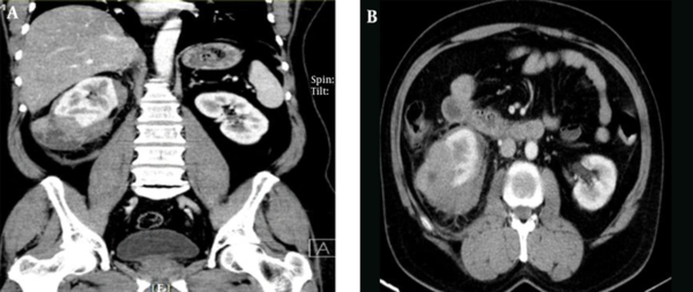
In this study we present a case with sudden right flank pain who was found to have a tumor mass measuring 75 mm with extracapsular extension and heterogeneous enhancement in the lower pole of the right kidney and perinephric hematoma secondary to rupture of capsular vessels. We performed radical nephrectomy and pathological evaluation of the specimen revealed choromophobe RCC.
2. Case Presentation
A 59-year-old man with complaint of sudden non-colicky right flank pain radiating to the back presented to our emergency department. He had no gross hematuria. Twenty years earlier he had a renal stone which was treated with extra-corporal shockwave lithotripsy (ESWL). The patient had no history of surgery or medical illness. On physical examination, he had no fever but diffuse tenderness was present over the right flank area. On ultrasound, increase in size and echogenicity of renal parenchyma and decrease in corticomedulary differentiation were reported. A hetero-echo exophytic mass measuring 47 × 63 mm with hyper-echoic components, probably angiomyolipoma, was also detected. Two stones measuring 4 - 6 mm and 6 - 7 mm were detected in upper and middle zone of the pyelocaleceal system, respectively, and there was no hydronephrosis present. Computed tomography (CT) showed a tumor mass measuring 75 mm with extracapsular extension and heterogeneous enhancement in the lower pole of the right kidney with perinephric hematoma secondary to rupture of capsular vessels. No evidence of fat density and angiomyolipoma were seen. A hypodense lesion measuring 10 mm was detected in segment VI of liver. Other organs were normal without lymphadenopathy. RCC is the most probable diagnosis (Figure 1). Serum creatinine level and bone scan showed normal results. We ordered a three-phase magnetic resonance imaging (MRI) and found that the hepatic lesion was a biliary cyst and not a metastatic one. The patient underwent radical nephrectomy of right kidney. A pathological study revealed chromophobe RCC with Fuhrman nuclear grade II and Tumor necrosis was seen. The tumor had invaded perinephric fat tissue. Ureter, renal vain, renal sinus, Gerota’s fascia and adrenal were tumor free. (Stage: PT3). This article involving human participant who signed the informed consent and we did not disclose her personal information.
3. Discussion
Nowadays, RCCs are mostly discovered as ‘incidentalomas’ in contrast to the past when they usually presented with classic presentations (1). Subcapsular and/or perinephric spontaneous bleeding were first described by Carl Reinhold August Wunderlich (2). Wunderlich syndrome is a relatively uncommon situation, with benign conditions as the most frequent causes (3). In one study, 70% of cases were due to benign causes, including vascular disease, infection, and benign neoplasm. Neoplastic causes accounted for 61.2% of these cases (4).
Based on the patient’s general condition, embolization or immediate surgery are the two options available (5, 6). Once the patient is stabilized, embolization is a wise choice, while immediate exploration also seems appropriate. In our case, the hematoma was not large and according to the imaging performed, the risk of malignancy was high; therefore, we decided to perform surgery immediately.
The most reliable modality in diagnosing retroperitoneal hemorrhage and RCC is contrast enhanced CT Scan (CECT) (7). However, the reliability of CT scan in diagnosing RCC at the time of bleeding is an area of controversy (8). Radical nephrectomy is the treatment of choice for tumors diagnosed as malignant by CT scan, and embolization could be the choice modality for benign conditions. But if malignancy is found on the follow-up CT scan, delayed surgery may be difficult due to fibrosis formation (9, 10).
3.1. Conclusions
Kidney neoplasms are the most common cause of spontaneous peri-nephric hemorrhage, Wunderlich syndrome, and approximately 50% of such neoplasms are malignant.
