1. Background
The malignancy of hepatocellular carcinoma (HCC) is reported as the third cause of cancer-derived death worldwide. HCC also leads to socio-economic problems in many countries, especially in Asian and African region countries. HCC is reported to affect approximately between 50 to150 individuals per 100,000 of population (1-3). HepG2 cells are a kind of human epithelial-like, originated from hepatocellular carcinoma patients. These cells were primarily derived from liver tissues of a 15-years-old male Caucasian patient, suffering from HCC. HCC is amongst the most frequent primary types of neoplastic malignancies, involving liver (4, 5). Almost all of the therapy protocols applied for treatment of HCC are associated with cytotoxicity effects. A particular treatment for HCC with a lesser extent of side effects is yet to be introduced. (5). In spite of attempts made regarding cancer treatment, there are still many unknown issues that deserve to be defined for getting the most appropriate therapy protocol for these malignancies (6). Activities of heterocyclic compounds and their side groups are essential for bio-functions of the living cells. The proline-derived compounds are widely present both in nature and in other pharmaceutical applications (7). Trans-4-hydroxy proline is a stereochemically rich and rapidly available molecule, containing several active sites for its functionality. These functional sites can bind various pharmacophores to make a diverse library of compounds to be employed in both biological and medical research fields. Amongst the proline based N-containing heterocycles, enalapril and captopril attracted more attention due to their extensive pharmaceutical activities, as angiotensin-converting enzyme (ACE) inhibitors (8, 9). The proline scaffold has already been shown in pharmacology and in nature to be a bioactive small organic molecule (10).
Apoptosis is described as a gene-regulated phenomenon of programmed cell death which is induced by most of the chemotherapeutic agents applied for therapy of malignancies (5). The induction of apoptosis in tumor cells is considered useful in cancer therapy and prevention. A broad range of natural and synthetic substances have been evidenced to display pro-apoptosis properties in various tumor cells. Therefore, the examination of the effects of apoptotic inducer reagents, either in their normal or modified form, is essential. The synthetic compounds with cytotoxic activities against tumor cells were also employed in many antitumor studies (11).
2. Objectives
Following synthesis of N-aryl-4-hydroxy-1-(2, 2, 2 trifluoroacetyl) pyrrolidine-2-carboxamide compound with a wide spectrum of biological activities. We examined the growth inhibitory effects of the pyrrolidine-2-carboxamide, a novel derivative of pyrrolidine on human hepatoma cancer (HepG2) cells, in present study by applying MTT (3-(4,5-dimethyl-2-thiazolyl)-2,5-diphenyl-2H-tetrazolium bromide) viability assay. In parallel, we also aimed to explore cellular apoptosis by detection of annexin V signal using flow cytometric analysis.
3. Methods
3.1. Materials
Some of biomaterials including fetal bovine serum (FBS), RPMI-1640 medium, trypsin enzyme and combined penicillin-streptomycin were all purchased from Gibco-BRL (Grand Island, NY, USA). We obtained MTT (3-(4,5-dimethyl-2-thiazolyl)-2,5-diphenyl-2H-tetrazolium bromide) and dimethyl sulfoxide (DMSO) from Roche (Germany). The RNA analysis materials such as RNA extraction Kit and Esay cDNA PCR reverse transcription kit were provided from (PARS Tous, Iran). Furthermore, the Annexin V- FITC apoptosis detection kit was purchased from eBioscience company (San Diego, CA).
3.2. Stock of Chemical Compound
Five mgs of chemical compound were dissolved in 1 mL of DMSO to achieve a main concentration of 5 mg/mL. The resultant stock solution was then gently filtered through a 0.45 micron filter, before each assay. Subsequently, 400 μL of the stock solution was gently mixed up and enriched with a volume of 600 μL of RPMI 1640 and other concentrations were provided by serial dilution.
3.3. Cell Culture
The HCC cell line (HepG2) which is defined as a liver cancer cell line was purchased from National Cell Bank of Iran (the Pasteur Institute of Iran, Tehran). Cells were seeded onto a 25 mL culture flask in presence of RPMI 1640 medium supplemented with 10% of FBS and 100 U/mL penicillin-streptomycin, at 37°C in a humidified atmosphere (90%) containing 95% O2, and 5% CO2. In parallel to malignant cells, nonmalignant cells (L929) were also cultured in RPMI 1640 medium contained 5% (v/v) of FBS and 100 units/mL penicillin-streptomycin. Cells were left overnight, and then incubated with various concentrations of pyrrolidin derivative (7.8 - 125 μg/mL) for 24, 48 and 72 h. For MTT assay, cells were seeded at a cellular concentration of 5000/well onto the 96-well culture plates. Along with each concentration as well as at different time points of the study, a control sample which remained untreated and received equal volume of medium was also established. All various treatments were carried out in triplicate.
3.4. Cell Viability Assay
The cell viability was determined by employing a modified MTT assay. In Brief, cells were seeded (5000/well) onto a flat-bottomed 96-well culture plate and allowed to grow for periods of 24, 48, and 72 hours subsequent to treatment with pyrrolidin derivative. At different time points, the medium was discarded, cells were labeled with MTT solution (5 mg/mL in PBS) for a duration of 4 hours and resulting formazan was solubilized with DMSO (100 μL). Finally, the optical density was read at 570 nm (620 nm as a reference) applying an ELISA reader.
3.5. Flow Cytometric Analysis of Apoptosis
The double staining method with annexin V-FITC and PI was carried out for flow cytometry analysis, using annexin V- FITC apoptosis detection kit. HepG2 cells were cultured for 48 hours and incubated with relative concentrations (0, 15.625, 31.5 and 62.5 μg/mL) of the compound. Consequently, both treated and untreated cells were harvested 24 hours following culture and were washed twice in PBS. Cells were then re-suspended in 200 μL of binding buffer (e.g. calcium buffer). Then 5 μL of annexin V- FITC was added to the cells and continued by the addition of 10 μL propidium iodide (PI). Samples were then incubated for a duration of 5 minutes in the dark at 4°C and were examined under a flow cytometer (BD FACS Calibur, BD Biosciences). BD CellQuest (BD Biosciences) version 1.0 was used for data analysis.
3.6. Statistical Analysis
The data distribution for each of separate trials met the requirements of normal distribution (one-sample kolmogrov-smirnov) and assumptions of variance homogeneity were checked with Levene’s test. All of the studied groups were compared via one-way ANOVA. Subsequently, the control group was statistically compared with the tested group using Dunnett’s test, following primitive confirmation (with F-test in variance analysis) of statistically significant differences in analyzed means. Kruskat-Wallis was tested for other variables due to data that were not normally distributed. Mann-Whitney U test was performed for the pair-wise Post-hoc comparison between the two groups. All tests were performed with the significance level of P < 0.05, using SPSS 20 software.
4. Results
4.1. Effects of Pyrrolidine-2-Carboxamide on Cell Viability
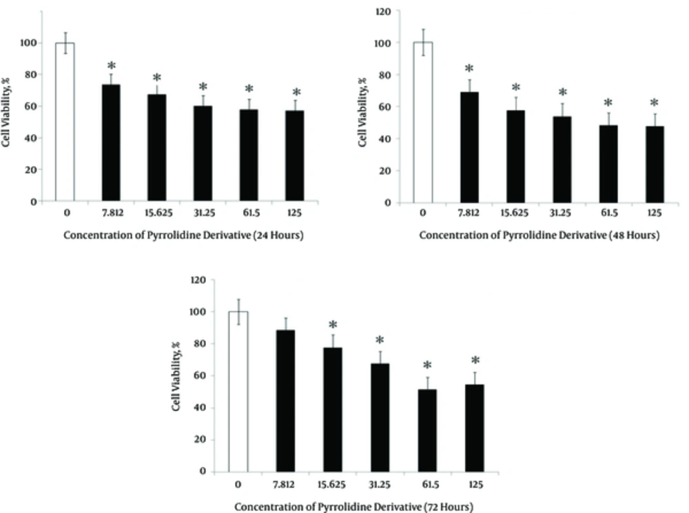
The cytotoxicity impacts of the one of pyrrolidine derivatives, (2R, 4S)-N- Aryl-4-hydroxy-1 - (2,2,2 trifluoroacetyl) pyrrolidine-2-carboxamide was examined on human HepG2 cells. Cells were treated with various doses (7.8125, 15.625, 31.25, 62.5 and 125 μg/mL) of the compound. Following periods of 24, 48 and 72 hours after treatment, the cell viability was evaluated employing the MTT assay. These findings demonstrated that the growth of treated HepG2 cells was inhibited, in comparison to control cells which were cultured along with treated cells. As clearly presented in Figure 1, the obtained IC50 values for pyrrolidine-2-carboxamide after 24 hours, 48 hours and 72 hours were 62.5, 125 and 62.5 μg/µL, respectively. We observed that at the highest concentration of the compound, the highest number of cells died and relatively the concentration of 62.5 µM and 125 µM (at 48 - 72 hours of treatment), while the minimum inhibition was found following 24 hours at the concentration of 7.812 μg/µL (Figure 1). Thus, pyrrolidine-2-carboxamide had inhibitory effects on the HepG2 cells growth (Table 1).
| Concentrations of Pyrrolidine Deivartis (μg/mL) | ||||||
|---|---|---|---|---|---|---|
| 0 | Control | 7.8125 | 15.625 | 31.25 | 62.5 | 125 |
| 24 | 0.756 ± 0.01 | 0.67 ± 0.086a | 0.67 ± 0.12 | 0.61 ± 0.14a | 0.589 ± 0.05a | 0.587 ± 0.05a |
| 48 | 0.734 ± 0.01 | 0.584 ± 0.057a | 0.572 ± 0.062a | 0.560 ± 0.003a | 0.551 ± 0.037a | 0.541 ± 0.80a |
| 72 | 0.716 ± 0.01 | 0.673 ± 0.086 | 0.671 ± 0.12 | 0.588 ± 0.05a | 0.574 ± 0.029a | 0.554 ± 0.12a |
Cell Proliferation After Different Concentrations of Pyrrolidine-2-Carboxamide ((2R, 4S)-N-(2, 5-Difluorophenyl)-4-Hydroxy-1-(2, 2, 2-Trifluoroacetyl) Pyrrolidine-2-Carboxamide) (x ± s, %) for 24, 48 and 72 h
4.2. Pyrrolidine-2-Carboxamide Induces Apoptosis and Necrosis in HepG2 cells
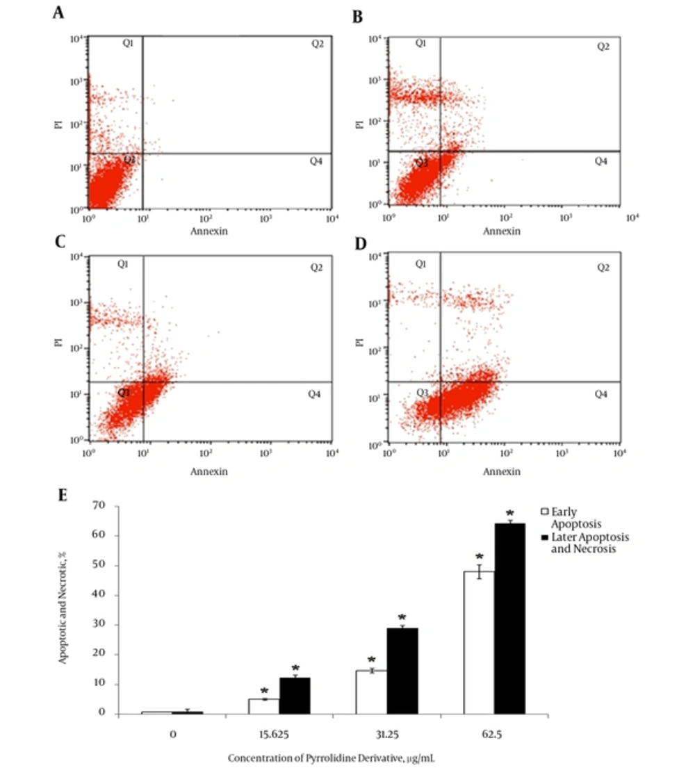
HepG2 cells have initially received various concentrations of pyrrolidine-2-carboxamide and the dose-dependent effects of the compound on apoptosis were explored. According to the cell viability analysis the maximum effective cytotoxic concentrations of Pyrrolidone 62.5 µM, the minimum cytotoxic concentration of 15.625 µM and the middle dose of 31.25 µM concentration were employed for determination of the apoptosis in HepG2 cells after 48 hours. Figure 2 demonstrates proof-of-principle data from the HepG2 dose response experiment as Annexin V FITC-A vs PI-A contour plots with quadrant gates showing four populations. In the untreated control sample, the highest proportion of cells (98.46%) were viable and non-apoptotic (Figure 2, Table 2), which by increasing the doses of pyrrolidine-2-carboxamide, an attenuated number of cells was observed in the Annexin V-PI- population in parallel with an increase in cells early apoptosis phase as well (Annexin V+PI-). Obviously, once cells were treated with three various concentrations of the compound 15.625, 31.25 and 62.5 µM for 48 hours, 65.24, 27.59 and 8.63 % of Annexin V-PI- cells were observed, respectively (Figure 2). Data obtained from four populations were further plotted against different concentrations of pyrrolidine-2-carboxamide. This also confirmed a dose-dependent fasion of enhancement in the Annexin V+PI- population and inversely a declined level of the Annexin-PI- population (Figure 2). A slight increase in the Annexin V+PI+ population was evidenced that indicating a late apoptosis has taken placed in cells at lower doses of pyrrolidine-2-carboxamide.
Cells were treated with different concentrations (A, control group; B, 15.625 µg/mL; C, 31.5 µg/mL; D, 62.5 µg/mL) of pyrrolidine-2-carboxamide for 48 h and stained with Annexin V fluorescein isothiocyanate (FITC) and propidium iodide (PI). Subsequently, apoptotic and necrotic cells were quantified by flow cytometry. The different subpopulations were defined as Q1, Annexin V/PI+, i.e. necrotic cells; Q2, Annexin V+/PI+, i.e. late apoptotic cells; Q3, Annexin V-/PI , i.e. normal live cells; and Q4, Annexin V+/PI , i.e. early apoptotic cells. E, comparison of induced early and late apoptosis as well as necrosis in treated HepG2 cells with pyrrolidine-2-carboxamide in different concentration. *P < 0.05 indicates a significant difference from the control cells.
Apoptosis Rates of HepG2 Cells Intervened by Different Concentrations of Pyrrolidine-2-Carboxamide ((2R, 4S)-N-(2, 5-Difluorophenyl)-4-Hydroxy-1-(2, 2, 2-Trifluoroacetyl) Pyrrolidine-2-Carboxamide) (x ± s, %)
5. Discussion
These days, issues of cancer prevention, detection and treatment have been the focus of several research teams, worldwide and achievements of these teams for the reduction of the risk of cancer is essential, as the world interest in this field (12). In the present study, the MTT assay confirmed the inhibitory effects of pyrrolidine-2-carboxamide on HepG2 perhaps through induction of the apoptosis phenomenon. As clearly shown in Figure 1, the cytotoxic effects observed for different concentrations of the pyrrolidine-2-carboxamide are following a dose dependent fashion. While a cell viability rate of more than 50% was observed following treatment with this compound, in a concentration below 125 µg/µL. These results revealed that when the concentration of pyrrolidone derivative was elevated, the proportion of dead cells was also induced in a dose-dependent fashion (Figure 1). The most populated portion of dead cells was seen in the concentration of 125 µM. In addition to the compound concentration, the duration of the incubation could also have a significant effect on the cytotoxicity of the compound (Table 1). Pyrrolidine structures with either an amine or alcoholic group at the fourth position have been widely applied as antiviral reagents, specifically for the treatment of influenza (11). In last few years, pyrrolidine derivatives have remarkably attracted research teams worldwide due to their applications as ACE inhibitor. In other words, it was proposed that either fluorine atom or fluorinated substituents play fundamental parts in natural bioactive compounds, through providing more structural complexity (3, 13). Fluorine is well evidenced to be an extremely important element in medicinal chemistry. Because it gives exclusive inter connective properties within organic molecules and in turn leading to changing the physical and biological characteristics. In particular, the trifluoromethyl group (CF3) is of the most frequently utilized fluorinated substituents in agricultural, medicinal and material sciences, due to its properties in offering simultaneous elevated electron density, high lipophilicity, and a steric demand similarly, as what observed in the isopropyl group (14). According to these bio-functional properties we therefore, synthesized some N-aryl-4-hydroxy-1-(2,2,2 trifluoroacetyl) pyrrolidine-2-carboxamide compounds which exhibited a range of biological activities. As an instance, the neuraminidase inhibitor capable of binding to the active site on the human cell systems that the neuraminidase binds to and hence inhibits the reproduction and propagation of the virus within the body (7).
To the best of our knowledge, this study is the first world report that addresses the fact that pyrrolidine-2-carboxamide induces apoptosis in HepG2 cells. Our findings revealed that this pyrrolidin derivative induced cytotoxic activity against HepG2 cells (Figure 2). The pyrrolidin-induced apoptosis was defined by distinct morphological features including chromatin condensation, cell and nuclear shrinkage, membrane blebbing and oligonucleosomal DNA fragmentation (15). As shown in Table 2, while the 62.5 mg/ml concentration of compound affected the viability of the HepG2 cells populations, apoptosis probably only partially contributed in this toxicity. It also may be concluded that non-apoptosis cell death programs are also involved in pyrrolidin derivative induced toxicity of these cells (2). Daniel and co-workers found that pyrrolidine dithiocarbamate complex has anti-proliferative effects on human breast cancer cells via dose and time dependent fashion, as well (16). Consistent with our findings, anti-tumoral property of dispirooxindole-pyrrolidine derivative was also reported as anticancer compound against A549 human lung adenocarcinoma cell line (17). Flow cytometric analysis shows a large percentage of apoptotic cells by treatment with pyrrolidine derivatives; however, in lower concentrations a small population of necrotic cells were also observed. Results obtained by this study indicated a dose dependent manner, so that the rate of apoptosis has increased with increasing the dose compound.
5.1. Conclusions
Overall, heterocyclics play major parts in biochemical processes and are also side groups of the most typical and essential constituents of living cells. According to the results obtained here, the pyrrolidine-2-carboxamide could possibly be considered as a potential candidate for production of drug for the treatment of human hepatocellular carcinoma in future.

