Visceral Leishmaniosis in Bone Marrow Aspiration

authors:

avatar Mohammad Naderisorki ORCID 1 , * , avatar Hossein Karami ORCID 1

Department of Pediatrics Hematology and Oncology, Thalassemia Research Center, Faculty of Medicine, Mazandaran University of Medical Sciences, Sari, Iran

how to cite: Naderisorki M, Karami H. Visceral Leishmaniosis in Bone Marrow Aspiration. Int J Infect. 2018;5(4):e83304. https://doi.org/10.5812/iji.83304.

Abstract

Introduction:

Visceral leishmaniosis, kala-azar, or black fever is a fatal disease caused by protozoan parasites of the genus Leishmania. Serological testing is more frequently used in endemic areas. The recombinant kinesin antigen (rK39) is a useful antigen in enzyme-linked immunosorbent assay (ELISA), also available in strip format as a rapid test, but the best way for the diagnosis of visceral leishmaniosis is histopathologic demonstration of the parasite by aspiration or biopsy of affected organs (usually, bone marrow or spleen aspirations).

Case Presentation:

Here, we present the case of a 5-year-old girl with fever, weight loss, weakness and massive hepatosplenomegaly. In lab data, pancytopenia was observed. ELISA rapid test was negative for leishmaniosis. Bone marrow aspiration showed amastigotes within macrophages and outside cells.

Conclusions:

The aim of this report was to emphasize that bone marrow aspiration is an accurate method for the diagnosis of visceral leishmaniosis.

1. Introduction

Visceral leishmaniosis is one of largest parasitic killers in the world, which is annually responsible for more than 200,000 infections worldwide (1). Fever, massive hepatomegaly, and splenomegaly are the most typical symptoms of the disease. The disease usually appears two to six months following infection (2). Without proper treatment the mortality rate for kala-azar is close to 100% (3).

Less invasive diagnostic methods are demonstration of specific antibodies, antigens, or parasite DNA in peripheral blood specimens (4). The direct agglutination test (DAT) has high sensitivity but relatively low specificity (5). The recombinant kinesin antigen (rK39) is a useful antigen in enzyme-linked immunosorbent assay (ELISA), also available in strip format as a rapid test (6). The direct agglutination test (DAT) requires less equipment than ELISA, thus, it is useful in developing settings (7).

The gold standard for the diagnosis of visceral leishmaniosis is the demonstration of parasite in tissue (usually bone marrow or spleen) (8). Bone marrow aspirates are generally safer than splenic aspirates and the sensitivity of bone marrow smear is about 60 to 85%.

2. Case Presentation

Our patient was a 5-year-old girl admitted due to prolonged fever, weight loss and weakness since 1 month ago. Physical examination showed massive hepatosplenomegaly and icter. After admission, laboratory tests revealed the following: red blood cells (RBCs), 3.66 × 10⁹ per Liter; hemoglobin, 8 g/L; white blood cells (WBCs), 3.2 × 10⁹ per Liter with 36% neutrophils, 54% lymphocytes, 6% monocytes, 2% eosinophils, and 1% basophils. A peripheral blood smear showed hypochromic-microcytic anemia. Blood chemistry analysis demonstrated total bilirubin of 4 mg/dL and direct bilirubin of 0.5 mg/dL. Aspartate transaminase (AST) was 134 (0 - 40) and alanine aminotransferase (ALT) 97 (0 - 55), and virology tests were negative. Chest X-ray was NL. Abdominal sonography showed only massive hepatosplenomegaly, and rapid ELISA test was negative for leishmaniosis.

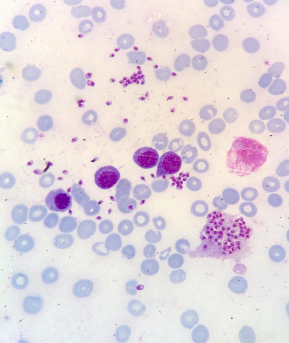
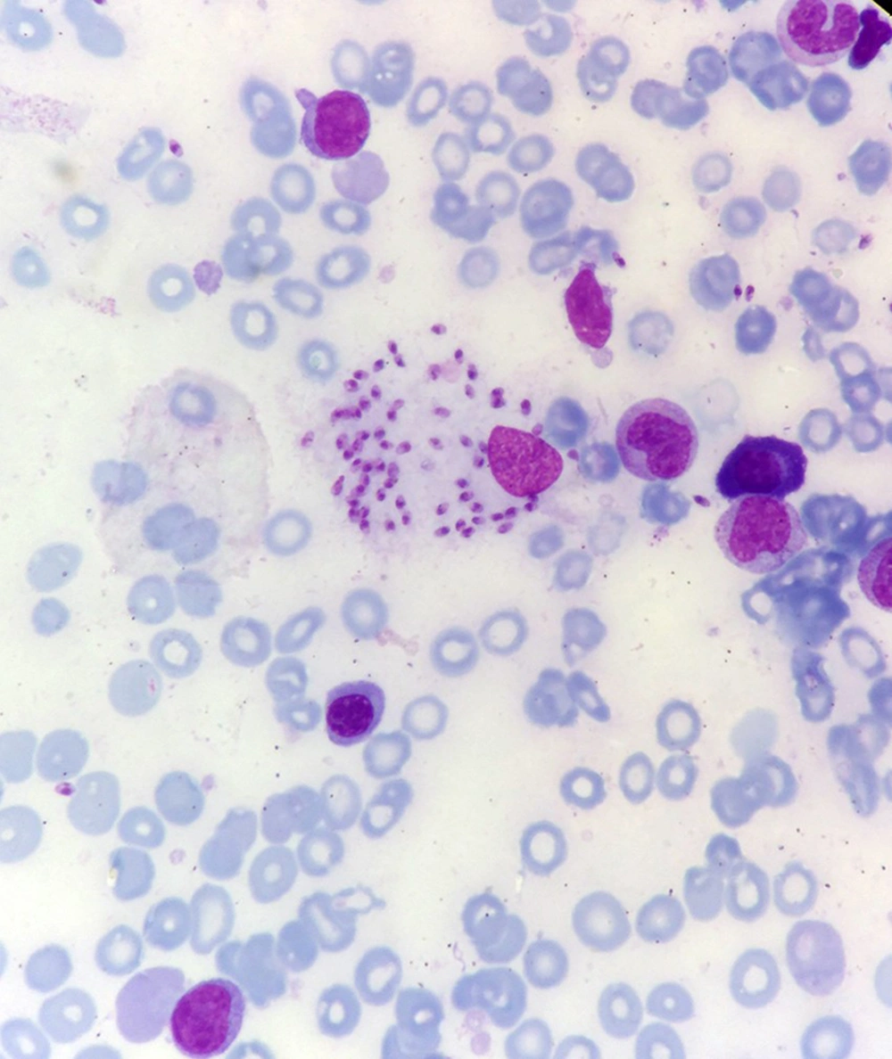
Bone marrow aspiration and bone marrow biopsy were performed, showing spherical or ovoid bodies, amastigotes, within macrophages and outside cells (Figures 1 and 2).

Leishman bodies within macrophages
Leishman bodies outside cells

3. Discussion

Although there are new diagnostic tools for visceral leishmaniosis, but the gold standard is visualization of the amastigotes in splenic or bone marrow aspirate. Montenegro skin test, a Leishman skin test, is negative in active visceral leishmaniosis and is no longer used (9).

The direct agglutination test (DAT), rK39 in ELISA, and indirect fluorescent antibody test (IFA) are not definitive proof of active visceral leishmaniosis, and in endemic areas positive antibody tests may be observed among asymptomatic individuals with subclinical infection (6, 10), and up to 32% of the healthy population may have a positive test but do not require treatment (11).

In our case, ELISA test was negative, and as noted in Figures 1 and 2, bone marrow aspiration showed Leishman bodies inside and outside macrophages in Wright-Giemsa stain.

References

  • 1.

    Desjeux P. The increase in risk factors for leishmaniasis worldwide. Trans R Soc Trop Med Hyg. 2001;95(3):239-43. [PubMed ID: 11490989]. https://doi.org/10.1016/S0035-9203(01)90223-8.

  • 2.

    Bern C, Jha SN, Das ML, Joshi AB, Bista MB, Hightower A, et al. Factors associated with visceral leishmaniasis in Nepal: Bed-net use is strongly protective. Am J Trop Med Hyg. 2000;63(3-4):184-8. [PubMed ID: 11388512]. https://doi.org/10.4269/ajtmh.2000.63.184.

  • 3.

    Desjeux P. Leishmaniasis. Public health aspects and control. Clin Dermatol. 1996;14(5):417-23. [PubMed ID: 8889319]. https://doi.org/10.1016/0738-081X(96)00057-0.

  • 4.

    Zijlstra EE, Ali MS, el-Hassan AM, el-Toum IA, Satti M, Ghalib HW, et al. Kala-azar: A comparative study of parasitological methods and the direct agglutination test in diagnosis. Trans R Soc Trop Med Hyg. 1992;86(5):505-7. [PubMed ID: 1475815]. https://doi.org/10.1016/0035-9203(92)90086-R.

  • 5.

    el Harith A, Kolk AH, Leeuwenburg J, Muigai R, Huigen E, Jelsma T, et al. Improvement of a direct agglutination test for field studies of visceral leishmaniasis. J Clin Microbiol. 1988;26(7):1321-5. [PubMed ID: 3410946]. [PubMed Central ID: PMC266601].

  • 6.

    Sundar S, Maurya R, Singh RK, Bharti K, Chakravarty J, Parekh A, et al. Rapid, noninvasive diagnosis of visceral leishmaniasis in India: Comparison of two immunochromatographic strip tests for detection of anti-K39 antibody. J Clin Microbiol. 2006;44(1):251-3. [PubMed ID: 16390983]. [PubMed Central ID: PMC1351954]. https://doi.org/10.1128/JCM.44.1.251-253.2006.

  • 7.

    Chappuis F, Rijal S, Soto A, Menten J, Boelaert M. A meta-analysis of the diagnostic performance of the direct agglutination test and rK39 dipstick for visceral leishmaniasis. BMJ. 2006;333(7571):723. [PubMed ID: 16882683]. [PubMed Central ID: PMC1592383]. https://doi.org/10.1136/bmj.38917.503056.7C.

  • 8.

    da Silva MR, Stewart JM, Costa CH. Sensitivity of bone marrow aspirates in the diagnosis of visceral leishmaniasis. Am J Trop Med Hyg. 2005;72(6):811-4. [PubMed ID: 15964968]. https://doi.org/10.4269/ajtmh.2005.72.811.

  • 9.

    Bern C, Amann J, Haque R, Chowdhury R, Ali M, Kurkjian KM, et al. Loss of leishmanin skin test antigen sensitivity and potency in a longitudinal study of visceral leishmaniasis in Bangladesh. Am J Trop Med Hyg. 2006;75(4):744-8. [PubMed ID: 17038705]. https://doi.org/10.4269/ajtmh.2006.75.744.

  • 10.

    Bern C, Chowdhury R, Breiman RF, Wahed MA, Wagatsuma Y, Secor WE, et al. The epidemiology of visceral leishmaniasis and asymptomatic leishmanial infection in a highly endemic Bangladeshi village. Am J Trop Med Hyg. 2007;76(5):909-14. [PubMed ID: 17488915]. https://doi.org/10.4269/ajtmh.2007.76.909.

  • 11.

    Boelaert M, Lynen L, Desjeux P, Van der Stuyft P. Cost-effectiveness of competing diagnostic-therapeutic strategies for visceral leishmaniasis. Bull World Health Organ. 1999;77(8):667-74. [PubMed ID: 10516788]. [PubMed Central ID: PMC2557711].